



Glycodeoxycholic acid
Based on 5 publication(s) in Google Scholar
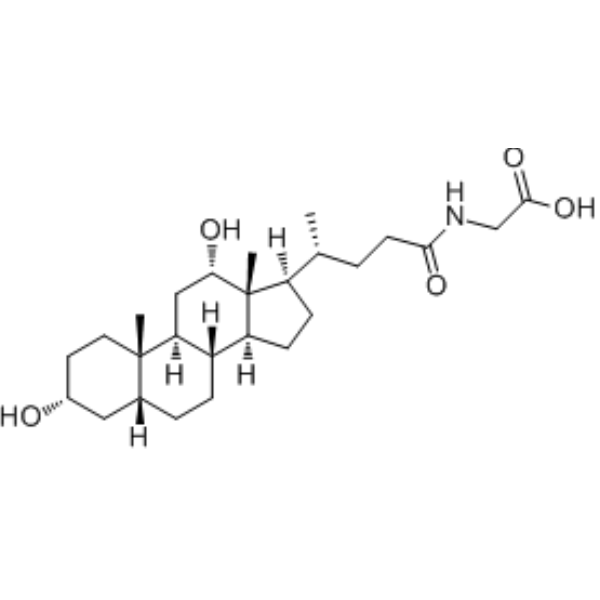
Glycodeoxycholic Acid is a natural product found in Streptomyces nigricans, Trypanosoma brucei and C. elegans. Glycodeoxycholic Acid induces hepatocyte necrosis and autophagy in patients with obstructive cholestasis.
For research use only. We do not sell to patients.
- Purity : 99.91%
- CAS No.: 360-65-6
- Formula: C26H43NO5
- Molecular Weight:449.62
-
Storage:Powder -20°C, 3 years , 4°C, 2 years ; In solvent -80°C, 2 years , -20°C, 1 year

To place orders, for customer services and technical support, please contact: MedChemExpress USA
Tel: 609-228-6898 E-mail: [email protected] [email protected]


-
 Biological Activity
-
Chemical Information
-
Solvent & Solubility
- Protocols
- Purity & Documentation
- References
-
 Help & FAQs


-
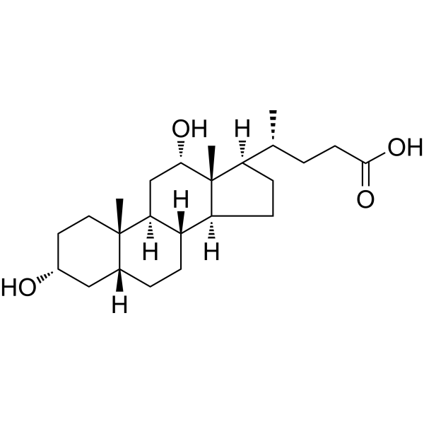
HY-N0593
Deoxycholic acidBile Acid Receptor Activator -
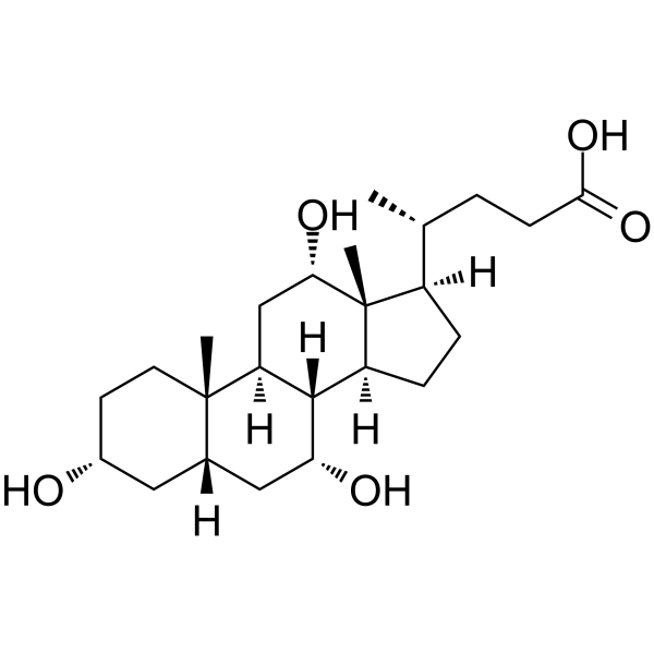
HY-N0324
Cholic acidEndogenous Metabolite -
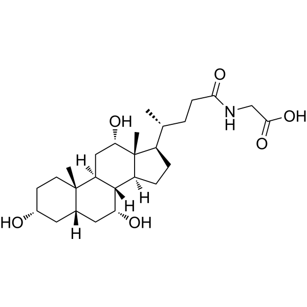
HY-N1423
Glycocholic acidBile Acid Derivative -
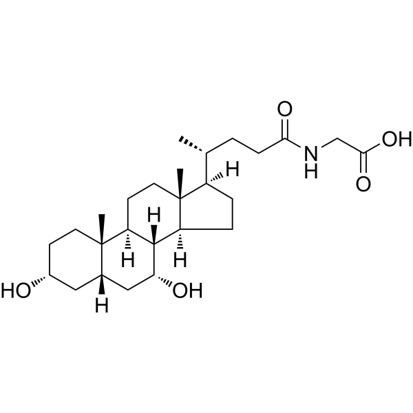
HY-N2334
Glycochenodeoxycholic acidEndogenous Metabolite -
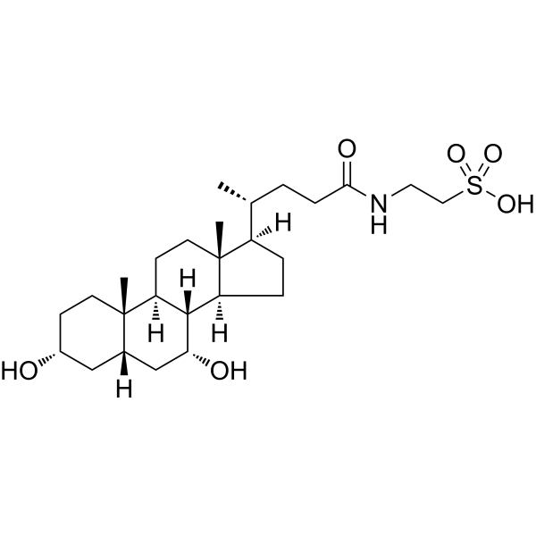
HY-N2027
Taurochenodeoxycholic acidEndogenous Metabolite -
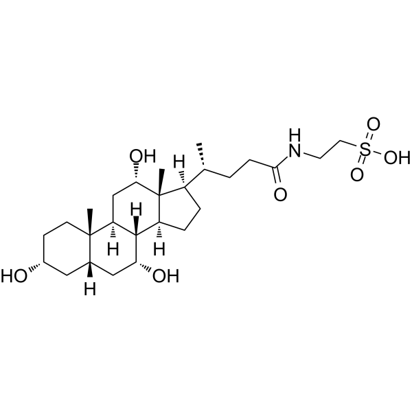
HY-B1788
Taurocholic acidImmunoregulation Agent -
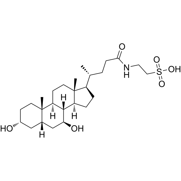
HY-19696
TauroursodeoxycholateEndoplasmic Reticulum Stress Inhibitor -
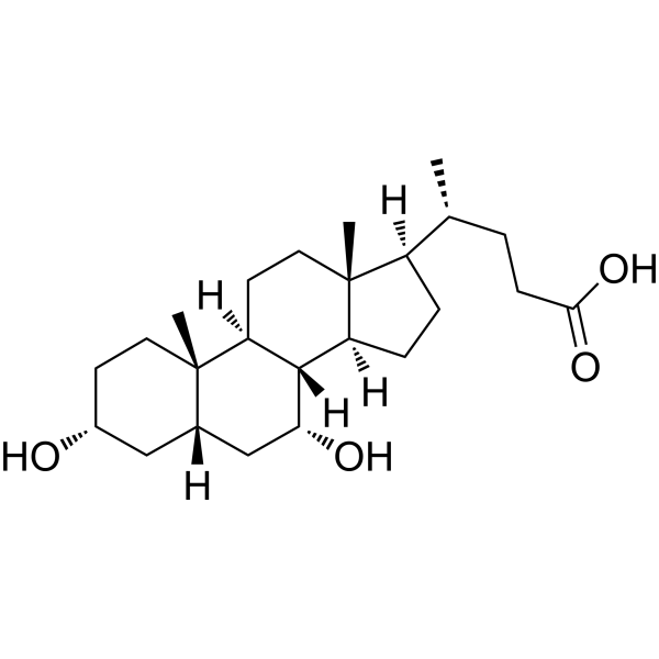
HY-76847
Chenodeoxycholic AcidEndogenous Metabolite -
HY-B0172
Lithocholic acidAutophagy Inducer -
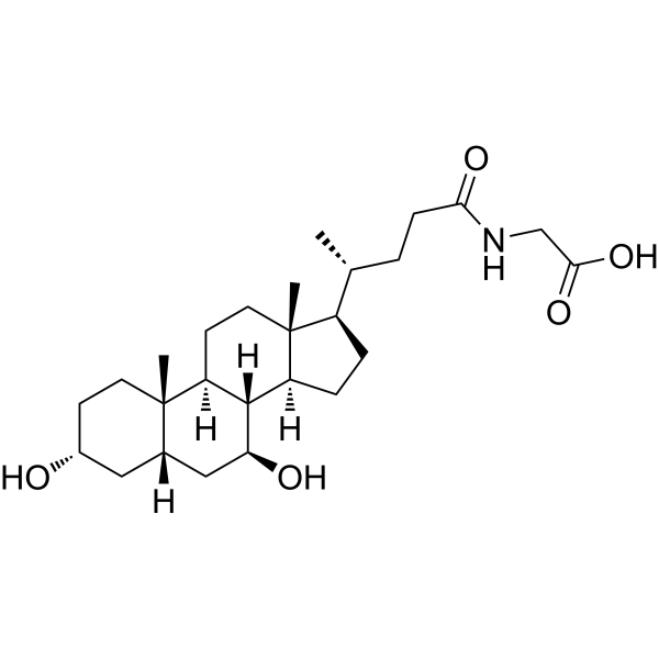
HY-N1424
Glycoursodeoxycholic acidEndogenous Metabolite -
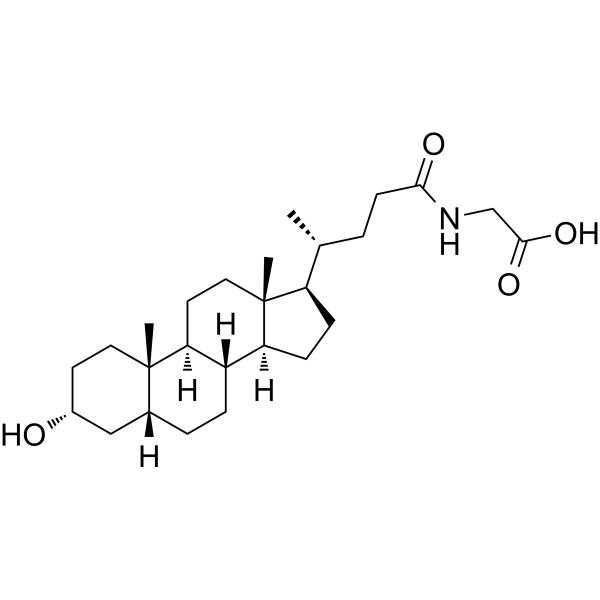
HY-116374
Glycolithocholic acidSecondary Bile Acid -
HY-B1899
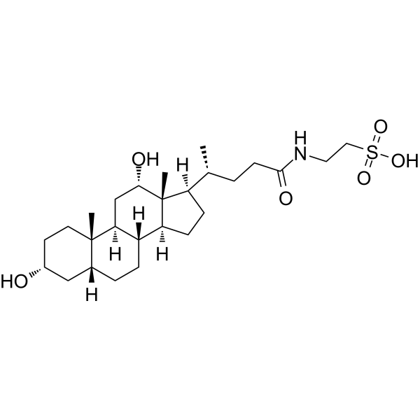
Taurodeoxycholic acidAnti-inflammatory Agent
Publications Citing Use of MedChemExpress (MCE) Glycodeoxycholic acid
More Customer Validation & Images

-
RT-PCR
All Endogenous Metabolite Isoforms
More

Biological Activity
Description
Cellular Effect
|
Cell Line

|
Type | Value | Description | References |
|---|---|---|---|---|
| CHO | EC50 |
1.18 μM
Compound: 7b, glyco
|
Agonist activity at human TGR5 expressed in CHO cells by luciferase assay
Agonist activity at human TGR5 expressed in CHO cells by luciferase assay
|
[PMID: 18307294] |
| HET-1A | CC50 |
1237 μM
Compound: 12, TDCA
|
Cytotoxicity against human HET-1A cells assessed as cell viability after 24 hrs by MTT assay
Cytotoxicity against human HET-1A cells assessed as cell viability after 24 hrs by MTT assay
|
[PMID: 20713311] |
In Vitro
Glycodeoxycholic Acid (200 μM, 24-48 h) induces stemness and chemotherapy resistance of hepatocellular carcinoma cells through STAT3 signaling pathway[1].
Glycodeoxycholic Acid (50 μM, pretreatment for 1 h) can eliminate UCB-induced cytochrome c oxidase inhibition, and significantly prevent oxidative stress, metabolic changes and cell death[2].
MedChemExpress (MCE) has not independently confirmed the accuracy of these methods. They are for reference only. Further protocols information, click here.
-
Cell Line:Huh7, LM3
-
Concentration:200 μM
-
Incubation Time:24, 48 h
-
Result:Increased cell viability treated with 5-FU and cisplatin.
-
Cell Line:Huh7, LM3
-
Concentration:200 μM
-
Incubation Time:24, 48 h
-
Result:Suppressed the expression of apoptotic genes and increased anti-apoptotic genes.
Promoted the expression of Sox2, Sox9, Nanog and CD133.
Down-regulated the level of E-cadherin and up-regulated vimentin.
Decreased the levels of SOCS2, SOCS5, PTPN1 and PTPN11.
In Vivo
MedChemExpress (MCE) has not independently confirmed the accuracy of these methods. They are for reference only.
-
Animal Model:Experimental macaque model[3]
-
Dosage:11.20 mg/kg
-
Administration:injected along the biliopancreatic duct
-
Result:Increased the levels of Serum amylase and lipase.
Elevated Blood pressure and heart rate.


Chemical Information
-
CAS No. 360-65-6
-
Appearance Solid
-
Molecular Weight 449.62
-
Formula C26H43NO5
-
Color White to off-white
-
SMILES
C[C@@H]([C@]1(CC[C@]2([C@@]3(CC[C@@]4(C[C@@H](CC[C@@]4([C@]3(C[C@@H]([C@@]21C)O)[H])C)O)[H])[H])[H])[H])CCC(NCC(O)=O)=O
-
Structure Classification
-
Initial Source
-
Shipping
Room temperature in continental US; may vary elsewhere.
-
Storage
Powder -20°C 3 years 4°C 2 years In solvent -80°C 2 years -20°C 1 year


Publications (5)
-
Journal Impact Factor
-
Most Recent
-
Nature
2025 Jul;643(8070):192-200. PMID: 39695227 -
Adv Sci (Weinh)
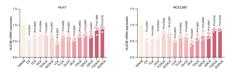
Targeting FDFT1 Reduces Cholesterol and Bile Acid Production and Delays Hepatocellular Carcinoma Progression Through the HNF4A/ALDOB/AKT1 Axis. [Abstract]2025 Mar;12(12):e2411719. PMID: 39899681
Glycodeoxycholic acid purchased from MedChemExpress. Usage Cited in: Adv Sci (Weinh). 2025 Mar;12(12):e2411719. [Abstract]
The mRNA levels of ALDOB in Huh7 or HCCLM3 cells treated with various bile acids (100 μM, 24 h). All data are presented as mean ± SD. Data were analyzed by one-way ANOVA with Bonferroni multiple-comparison correction. CA, cholic acid; TCA, taurocholic acid; GCA, glycocholic acid; TCDCA, taurochenodeoxycholic acid; GCDCA, glycochenodeoxycholic acid; LCA, lithocholic acid; TLCA, taurolithocholic acid; GLCA, glycolithocholic acid; DCA, deoxycholic acid; TDCA, taurodeoxycholic acid; GDCA, glycodeoxycholic acid; UDCA, ursodeoxycholic acid; TUDCA, tauroursodeoxycholic acid; GUDCA, glycoursodeoxycholic acid.
-
J Enzyme Inhib Med Chem
Natural product sennoside B disrupts liquid-liquid phase separation of SARS-CoV-2 nucleocapsid protein by inhibiting its RNA-binding activity. [Abstract]2025 Dec;40(1):2501743. PMID: 40371698 -
-
bioRxiv
2026 May 5:2026.04.30.721772. PMID: 42146479


Solvent & Solubility
In Vitro:
DMSO : 87.5 mg/mL (194.61 mM; ultrasonic and warming and heat to 60°C; Hygroscopic DMSO has a significant impact on the solubility of product, please use newly opened DMSO)
Please refer to the solubility information to select the appropriate solvent. Once prepared, please aliquot and store the solution to prevent product inactivation from repeated freeze-thaw cycles.
Storage method and period of stock solution: -80°C, 2 years; -20°C, 1 year. When stored at -80°C, please use it within 2 years. When stored at -20°C, please use it within 1 year.
Please refer to the solubility information to select the appropriate solvent. Once prepared, please aliquot and store the solution to prevent product inactivation from repeated freeze-thaw cycles.
Storage method and period of stock solution: -80°C, 2 years; -20°C, 1 year. When stored at -80°C, please use it within 2 years. When stored at -20°C, please use it within 1 year.
Concentration (start) × Volume (start) = Concentration (final) × Volume (final)
In Vivo:
Select the appropriate dissolution method based on your experimental animal and administration route.
- For the following dissolution methods, please ensure to first prepare a clear stock solution using an In Vitro approach and then sequentially add co-solvents:
- To ensure reliable experimental results, the clarified stock solution can be appropriately stored based on storage conditions. As for the working solution for In Vivo experiments, it is recommended to prepare freshly and use it on the same day.
- The percentages shown for the solvents indicate their volumetric ratio in the final prepared solution. If precipitation or phase separation occurs during preparation, heat and/or sonication can be used to aid dissolution.
Add each solvent one by one: 10% DMSO 40% PEG300 5% Tween-80 45% Saline
Solubility: ≥ 2.08 mg/mL (4.63 mM); Clear solution
This protocol yields a clear solution of ≥ 2.08 mg/mL (saturation unknown).
Taking 1 mL working solution as an example, add 100 μL DMSO stock solution (20.8 mg/mL) to 400 μL PEG300, and mix evenly; then add 50 μL Tween-80 and mix evenly; then add 450 μL Saline to adjust the volume to 1 mL.
Preparation of Saline: Dissolve 0.9 g sodium chloride in ddH₂O and dilute to 100 mL to obtain a clear Saline solution.
Add each solvent one by one: 10% DMSO 90% (20% SBE-β-CD in Saline)
Solubility: ≥ 2.08 mg/mL (4.63 mM); Clear solution
This protocol yields a clear solution of ≥ 2.08 mg/mL (saturation unknown).
Taking 1 mL working solution as an example, add 100 μL DMSO stock solution (20.8 mg/mL) to 900 μL 20% SBE-β-CD in Saline, and mix evenly.
Preparation of 20% SBE-β-CD in Saline (4°C, storage for one week): 2 g SBE-β-CD powder is dissolved in 10 mL Saline, completely dissolve until clear.
In Vivo Dissolution Calculator
Please enter the basic information of animal experiments:
-
-
-
-
Recommended: Prepare an additional quantity of animals to account for potential losses during experiments.
Please enter your animal formula composition:
-
%DMSO +
Recommended: Keep the proportion of DMSO in working solution below 2% if your animal is weak.
-
%+
-
+%Tween-80 + +
-
%Saline +
The co-solvents required include: DMSO, . All of co-solvents are available by MedChemExpress (MCE). , Tween 80. All of co-solvents are available by MedChemExpress (MCE).
Working solution concentration: 0.22 mg/mL
Method for preparing stock solution: mg drug dissolved in μL DMSO. Stock solution concentration: mg/mL.
1. Take μL DMSO stock solution;
2. Add μL .
μL , mix evenly;
3. Then add μL Tween 80, mix evenly;
4. Then add μL
Please ensure that the stock solution in the first step is dissolved to a clear state, and add co-solvents in sequence. You can use ultrasonic heating (ultrasonic cleaner, recommended frequency 20-40 kHz), vortexing, etc. to assist dissolution.

Protocols
-
Autophagy
Autophagy is a process in which eukaryotic cells use lysosomes to degrade their own cytoplasmic proteins and damaged organelles under the regulation of autophagy related gene (Atg). Microtubule-associated proteins light chain 3 (LC3) is recognized as autophagy marker, which transfers from cytoplasmic LC3 (LC3-I) to membrane type (LC3-II). LC3-II/I ratio could be detected by Western Blot and fluorescence microscopy.
-
Lysosome and acidic-vesicle live-cell staining
Lysosome and acidic-vesicle live-cell staining detects acidic intracellular compartments by using membrane-permeant acidotropic probes that accumulate in low-pH vesicles, including lysosomes, late endosomes, autolysosomes, and acidic phagosomes. LysoTracker staining is commonly used as an intensity-based readout of acidic lysosomal compartment abundance or enlargement, while acridine orange produces green fluorescence in less concentrated compartments and red fluorescence after concentration-dependent accumulation in acidic vesicular organelles. Loss or reduction of acridine-orange red signal can be used as a readout of lysosomal membrane permeabilization or reduced acidic-vesicle integrity. This protocol is designed for live cultured cells and can be adapted for fluorescence microscopy, high-content imaging, plate-reader readout, or flow cytometry when the selected literature supports the readout. Because these dyes report acidotropic accumulation rather than lysosome identity alone,
-
Macroautophagy Solutions
Macroautophagy is a conserved lysosome-dependent degradation pathway in which cytoplasmic material is sequestered into double-membrane autophagosomes and delivered to lysosomes for degradation and recycling. The pathway supports cellular homeostasis during nutrient limitation, organelle stress, protein-aggregate accumulation, infection, differentiation, and tissue remodeling by coupling cargo sequestration, autophagosome maturation, lysosomal fusion, and degradation of cargo-derived macromolecules. The core molecular sequence includes initiation by nutrient- and stress-regulated autophagy machinery, autophagosome nucleation, LC3/ATG8-family conjugation to autophagosomal membranes, cargo selection through receptors such as SQSTM1/p62, autophagosome-lysosome fusion, and lysosomal degradation. LC3 was identified as a mammalian homolog of yeast Atg8 that localizes to autophagosomal membranes after processing, and p62/SQSTM1 was shown to connect ubiquitinated cargo with autophagic degradati


Purity & Documentation
-
Data Sheet (278 KB)
-
SDS (394 KB)
- English - EN (394 KB)
- Français - FR (394 KB)
- Deutsch - DE (394 KB)
- Norwegian - NO (394 KB)
- Español - ES (394 KB)
- Swedish - SV (394 KB)
- Italian - IT (394 KB)
- Korean - KR (394 KB)
- Portuguese - PT (394 KB)
-
Handling Instructions (2659 KB)
References
[1]. Shi C, et al. Glycochenodeoxycholic acid induces stemness and chemoresistance via the STAT3 signaling pathway in hepatocellular carcinoma cells. Aging (Albany NY). 2020 Aug 3;12(15):15546-15555. [Content Brief]
[2]. Vaz AR, et al. Bilirubin selectively inhibits cytochrome c oxidase activity and induces apoptosis in immature cortical neurons: assessment of the protective effects of glycoursodeoxycholic acid. J Neurochem. 2010 Jan;112(1):56-65. [Content Brief]
[3]. Fauzi A, et al. Role of glycodeoxycholic acid to induce acute pancreatitis in Macaca nemestrina. J Med Primatol. 2022 Jun;51(3):134-142. doi: 10.1111/jmp.12577. Epub 2022 Mar 20. PMID: 35306662; PMCID: PMC9310849. [Content Brief]


Complete Stock Solution Preparation Table
Please refer to the solubility information to select the appropriate solvent. Once prepared, please aliquot and store the solution to prevent product inactivation from repeated freeze-thaw cycles.
Storage method and period of stock solution: -80°C, 2 years; -20°C, 1 year. When stored at -80°C, please use it within 2 years. When stored at -20°C, please use it within 1 year.
| Optional Solvent | Concentration Solvent Mass | 1 mg | 5 mg | 10 mg | 25 mg |
|---|---|---|---|---|---|
| DMSO | 1 mM | 2.2241 mL | 11.1205 mL | 22.2410 mL | 55.6025 mL |
| 5 mM | 0.4448 mL | 2.2241 mL | 4.4482 mL | 11.1205 mL | |
| 10 mM | 0.2224 mL | 1.1121 mL | 2.2241 mL | 5.5603 mL | |
| 15 mM | 0.1483 mL | 0.7414 mL | 1.4827 mL | 3.7068 mL | |
| 20 mM | 0.1112 mL | 0.5560 mL | 1.1121 mL | 2.7801 mL | |
| 25 mM | 0.0890 mL | 0.4448 mL | 0.8896 mL | 2.2241 mL | |
| 30 mM | 0.0741 mL | 0.3707 mL | 0.7414 mL | 1.8534 mL | |
| 40 mM | 0.0556 mL | 0.2780 mL | 0.5560 mL | 1.3901 mL | |
| 50 mM | 0.0445 mL | 0.2224 mL | 0.4448 mL | 1.1121 mL | |
| 60 mM | 0.0371 mL | 0.1853 mL | 0.3707 mL | 0.9267 mL | |
| 80 mM | 0.0278 mL | 0.1390 mL | 0.2780 mL | 0.6950 mL | |
| 100 mM | 0.0222 mL | 0.1112 mL | 0.2224 mL | 0.5560 mL |


Glycodeoxycholic acid Related Classifications
HY-125731 Related Classifications
 Powered by Bioz