



Heraclenin

Based on 1 Customer Validation
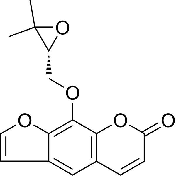
Heraclenin is a linear furanocoumarin natural product. Heraclenin binds to the type III effector protein XopQ and chitin deacetylase, exhibits antibacterial activity against Xanthomonas oryzae pv. oryzae, and displays antifungal activity against Colletotrichum lindemuthianum. Heraclenin stimulates Runx2 mRNA expression, induces osteoblast differentiation and mineralization. Heraclenin induces Chk1 phosphorylation, G2/M cell cycle arrest, DNA fragmentation, apoptosis, chromosomal aberrations, sister chromatid exchange, and inhibits proliferation. Heraclenin can be used for research on osteoporosis, melanoma, T-cell lymphoma, and leukemia.
Nur für Forschungszwecke. Wir verkaufen nicht an Patienten.
- CAS. Nr.: 2880-49-1
- Formel: C16H14O5
- Molecular Weight:286.28
-
Speicherung:
4°C, protect from light
* In solvent : -80°C, 6 months; -20°C, 1 month (protect from light)

To place orders, for customer services and technical support, please contact: MedChemExpress USA
Tel: 609-228-6898 E-mail: [email protected] [email protected]


-
 Biologische Aktivität
-
Chemical Information
-
Lösungsmittel & Löslichkeit
- Protokoll
- Reinheit & Dokumentation
- Verweise
-
 Help & FAQs





Biologische Aktivität
Beschreibung
Cellular Effect
|
Cell Line

|
Type | Value | Description | References |
|---|---|---|---|---|
| B16-F10 | IC50 |
1.01 μM
|
Inhibition of B16F10 melanoma cell proliferation under UVA irradiation (5 J cm^-2 for 64 min) assessed after 48 hrs by WST-1 assay.
Inhibition of B16F10 melanoma cell proliferation under UVA irradiation (5 J cm^-2 for 64 min) assessed after 48 hrs by WST-1 assay.
|
23802687 |
In Vitro
Heraclenin (3000-6000 ppm; 7 days) effectively inhibits the growth of Colletotrichum lindemuthianum in Potato Dextrose broth, reducing mycelial mat weight to 0.157 grams at 3000 ppm and 0.093 grams at 6000 ppm[2].
Heraclenin (increasing concentrations; 72 h) exhibits antiproliferative activity against both sensitive PAR and multidrug resistant MDR mouse T-cell lymphoma cell lines[5].
Heraclenin (24 h) demonstrates cytotoxic activity against both sensitive PAR and multidrug resistant MDR mouse T-cell lymphoma cell lines, as well as mild toxicity against normal murine NIH/3T3 fibroblasts[5].
Heraclenin (5-20 μM; 72 h) treatment up to 20 μM does not alter cell membrane integrity or reduce cell viability in C3H10T1/2 cells, confirming its non-toxic nature[3].
Heraclenin (5-20 μM; 7 days) promotes osteoblast differentiation and mineralization in C3H10T1/2 cells[3].
MedChemExpress (MCE) has not independently confirmed the accuracy of these methods. They are for reference only.
-
Cell Line:C3H10T1/2
-
Concentration:5, 10, 20 μM
-
Incubation Time:72 h
-
Result:Induced a significant dose-dependent increase in Runx2 mRNA expression in all treatment groups compared to the control.
Showed an optimum increase in Runx2 mRNA expression at 10 μM.
Showed decreased expression at 20 μM compared to 10 μM.
-
Cell Line:C3H10T1/2
-
Concentration:5, 10, 20 μM
-
Incubation Time:72 h
-
Result:Showed an average cell viability percentage of approximately 96% in control and treated groups.
Showed no change in the total cell number in all the tested groups with respect to the control.
-
Cell Line:C3H10T1/2
-
Concentration:5, 10, 20 μM
-
Incubation Time:7 days
-
Result:Showed an enhanced area of mineralization compared to the control group.
Confirmed enhanced mineralization by colorimetric quantification.
In Vivo
MedChemExpress (MCE) has not independently confirmed the accuracy of these methods. They are for reference only.
-
Animal Model:Hos:HRM (male, 5 weeks old, subcutaneous B16F10 melanoma model)[4]
-
Dosage:0.5 mg/kg; 1.0 mg/kg
-
Administration:i.p.; daily; 20 days
-
Result:Had no effect on tumor growth or final tumor weight at 0.5 or 1.0 mg/kg.
- RT-PCR
- Research Protocol for Infectious Diseases
- RNA extraction experimental
- Western Blot
- Annexin V plus membrane-impermeant dye apoptosis staining
- BrdU Incorporation Assay
- Protocol for Cell Cycle
- Bacterial live/dead nucleic-acid viability staining
- Real Time qPCR (Q-PCR)
- Cell Viability Determination by MTT Colorimetric Assay
- Kinase activity and phosphorylation assays
- Detection of Apoptosis
- TUNEL staining for apoptotic DNA fragmentation
- Flow cytometric DNA-content cell-cycle staining
- Mesenchymal stromal/stem cell osteogenic differentiation
- Apoptosis Solutions
- Protocol for Kinase activity and phosphorylation assays


Chemical Information
-
CAS. Nr. 2880-49-1
-
Appearance Solid
-
Molecular Weight 286.28
-
Formel C16H14O5
-
Color White to off-white
-
SMILES
CC1(C)[C@H](O1)COC2=C(OC=C3)C3=CC(C=C4)=C2OC4=O
-
Structure Classification
-
Versand
Room temperature in continental US; may vary elsewhere.
-
Speicherung
4°C, protect from light
* In solvent : -80°C, 6 months; -20°C, 1 month (protect from light)


Lösungsmittel & Löslichkeit
In Vitro:
DMSO : 100 mg/mL (349.31 mM; Need ultrasonic; Hygroscopic DMSO has a significant impact on the solubility of product, please use newly opened DMSO)
Please refer to the solubility information to select the appropriate solvent. Once prepared, please aliquot and store the solution to prevent product inactivation from repeated freeze-thaw cycles.
Storage method and period of stock solution: -80°C, 6 months; -20°C, 1 month (protect from light). When stored at -80°C, please use it within 6 months. When stored at -20°C, please use it within 1 month.
Please refer to the solubility information to select the appropriate solvent. Once prepared, please aliquot and store the solution to prevent product inactivation from repeated freeze-thaw cycles.
Storage method and period of stock solution: -80°C, 6 months; -20°C, 1 month (protect from light). When stored at -80°C, please use it within 6 months. When stored at -20°C, please use it within 1 month.
Konzentration (Stammlösung) × Volumen (Stammlösung) = Konzentration (Ziellösung) × Volumen (Ziellösung)
In Vivo:
Select the appropriate dissolution method based on your experimental animal and administration route.
- For the following dissolution methods, please ensure to first prepare a clear stock solution using an In Vitro approach and then sequentially add co-solvents:
- To ensure reliable experimental results, the clarified stock solution can be appropriately stored based on storage conditions. As for the working solution for In Vivo experiments, it is recommended to prepare freshly and use it on the same day.
- The percentages shown for the solvents indicate their volumetric ratio in the final prepared solution. If precipitation or phase separation occurs during preparation, heat and/or sonication can be used to aid dissolution.
Add each solvent one by one: 10% DMSO 40% PEG300 5% Tween-80 45% Saline
Solubility: 2.5 mg/mL (8.73 mM); Suspended solution; Need ultrasonic
This protocol yields a suspended solution of 2.5 mg/mL. Suspended solution can be used for oral and intraperitoneal injection.
Taking 1 mL working solution as an example, add 100 μL DMSO stock solution (25.0 mg/mL) to 400 μL PEG300, and mix evenly; then add 50 μL Tween-80 and mix evenly; then add 450 μL Saline to adjust the volume to 1 mL.
Preparation of Saline: Dissolve 0.9 g sodium chloride in ddH₂O and dilute to 100 mL to obtain a clear Saline solution.
Add each solvent one by one: 10% DMSO 90% (20% SBE-β-CD in Saline)
Solubility: ≥ 2.5 mg/mL (8.73 mM); Clear solution
This protocol yields a clear solution of ≥ 2.5 mg/mL (saturation unknown).
Taking 1 mL working solution as an example, add 100 μL DMSO stock solution (25.0 mg/mL) to 900 μL 20% SBE-β-CD in Saline, and mix evenly.
Preparation of 20% SBE-β-CD in Saline (4°C, storage for one week): 2 g SBE-β-CD powder is dissolved in 10 mL Saline, completely dissolve until clear.
In Vivo Dissolution Calculator
Please enter the basic information of animal experiments:
-
-
-
-
Recommended: Prepare an additional quantity of animals to account for potential losses during experiments.
Please enter your animal formula composition:
-
%DMSO +
Recommended: Keep the proportion of DMSO in working solution below 2% if your animal is weak.
-
%+
-
+%Tween-80 + +
-
%Saline +
The co-solvents required include: DMSO, . All of co-solvents are available by MedChemExpress (MCE). , Tween 80. All of co-solvents are available by MedChemExpress (MCE).
Working solution concentration: 0.22 mg/mL
Method for preparing stock solution: mg drug dissolved in μL DMSO. Stock solution concentration: mg/mL. * In solvent : -80°C, 6 months; -20°C, 1 month (protect from light)
1. Take μL DMSO stock solution;
2. Add μL .
μL , mix evenly;
3. Then add μL Tween 80, mix evenly;
4. Then add μL
Please ensure that the stock solution in the first step is dissolved to a clear state, and add co-solvents in sequence. You can use ultrasonic heating (ultrasonic cleaner, recommended frequency 20-40 kHz), vortexing, etc. to assist dissolution.


Reinheit & Dokumentation
-
Data Sheet (309 KB)
-
SDS (394 KB)
- English - EN (394 KB)
- Français - FR (394 KB)
- Deutsch - DE (394 KB)
- Norwegian - NO (394 KB)
- Español - ES (394 KB)
- Swedish - SV (394 KB)
- Italian - IT (394 KB)
- Korean - KR (394 KB)
- Portuguese - PT (394 KB)
-
Handling Instructions (2659 KB)
Verweise
[1]. Wu H, et al. Mechanism-based inactivation of CYP2D6 by imperatorin and drug-drug interaction in vitro and in vivo. Arch Toxicol. 2026 Jul;100(7):2885-2897. [Content Brief]
[3]. Shanmugam H, et al. Osteogenic stimulatory effect of heraclenin purified from bael in mouse mesenchymal stem cells in vitro. Chemico-biological interactions. 2019 Sep 01;310:108750. [Content Brief]
[4]. Kimura Y, et al. In vitro and in vivo antiproliferative effect of a combination of ultraviolet-A and alkoxy furocoumarins isolated from Umbelliferae medicinal plants, in melanoma cells. Photochemistry and photobiology. 2013;89(5):1216-25. [Content Brief]
[5]. Mottaghipisheh J, et al. Antiproliferative and cytotoxic activities of furocoumarins of Ducrosia anethifolia. Pharmaceutical biology. 2018 Dec;56(1):658-664. [Content Brief]
[6]. Appendino G, et al. Coumarins from Opopanax chironium. New dihydrofuranocoumarins and differential induction of apoptosis by imperatorin and heraclenin. Journal of natural products. 2004 Apr;67(4):532-6. [Content Brief]
[7]. Abel G, et al. Chromosome-damaging effects of heraclenin in human lymphocytes in vitro. Mutation research. 1986;169(1-2):51-4. [Content Brief]
[8]. Schimmer O, et al. Mutagenicity of a furocoumarin epoxide, heraclenin, in Chlamydomonas reinhardii. Mutation research. 1986;169(1-2):47-50. [Content Brief]


Complete Stock Solution Preparation Table
Please refer to the solubility information to select the appropriate solvent. Once prepared, please aliquot and store the solution to prevent product inactivation from repeated freeze-thaw cycles.
Storage method and period of stock solution: -80°C, 6 months; -20°C, 1 month (protect from light). When stored at -80°C, please use it within 6 months. When stored at -20°C, please use it within 1 month.
| Optional Solvent | Concentration Solvent Mass | 1 mg | 5 mg | 10 mg | 25 mg |
|---|---|---|---|---|---|
| DMSO | 1 mM | 3.4931 mL | 17.4654 mL | 34.9308 mL | 87.3271 mL |
| 5 mM | 0.6986 mL | 3.4931 mL | 6.9862 mL | 17.4654 mL | |
| 10 mM | 0.3493 mL | 1.7465 mL | 3.4931 mL | 8.7327 mL | |
| 15 mM | 0.2329 mL | 1.1644 mL | 2.3287 mL | 5.8218 mL | |
| 20 mM | 0.1747 mL | 0.8733 mL | 1.7465 mL | 4.3664 mL | |
| 25 mM | 0.1397 mL | 0.6986 mL | 1.3972 mL | 3.4931 mL | |
| 30 mM | 0.1164 mL | 0.5822 mL | 1.1644 mL | 2.9109 mL | |
| 40 mM | 0.0873 mL | 0.4366 mL | 0.8733 mL | 2.1832 mL | |
| 50 mM | 0.0699 mL | 0.3493 mL | 0.6986 mL | 1.7465 mL | |
| 60 mM | 0.0582 mL | 0.2911 mL | 0.5822 mL | 1.4555 mL | |
| 80 mM | 0.0437 mL | 0.2183 mL | 0.4366 mL | 1.0916 mL | |
| 100 mM | 0.0349 mL | 0.1747 mL | 0.3493 mL | 0.8733 mL |


Heraclenin Related Classifications
HY-N4053 Related Classifications
 Powered by Bioz