



NS1643
Based on 1 publication(s) in Google Scholar
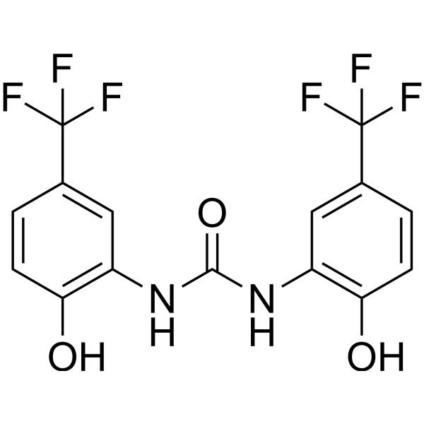
NS1643 is a partial agonist of human ether-a-go-go-related gene (hERG) K+ channels with an EC50 of 10.5 μM. NS1643 inhibits the growth of breast cancer tumors in TNBC mouse models. NS1643 inhibits cell migration and invasion of breast cancer cells.
For research use only. We do not sell to patients.
- Purity : 99.04%
- CAS No.: 448895-37-2
- Formula: C15H10F6N2O3
- Molecular Weight:380.24
-
Storage:Powder -20°C, 3 years , 4°C, 2 years ; In solvent -80°C, 2 years , -20°C, 1 year

To place orders, for customer services and technical support, please contact: MedChemExpress USA
Tel: 609-228-6898 E-mail: [email protected] [email protected]


-
 Biological Activity
-
Chemical Information
-
Solvent & Solubility
- Protocols
- Purity & Documentation
- References
-
 Help & FAQs


-
HY-15551
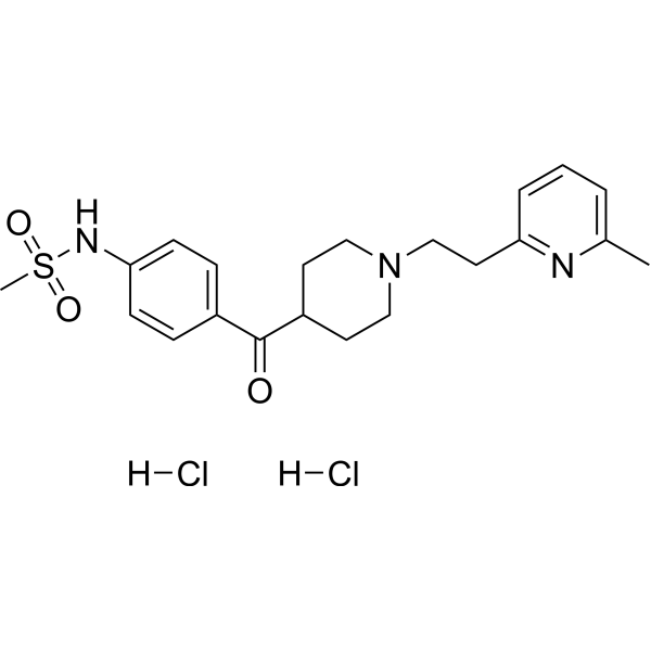
E-4031hERG Channels Blocker -
HY-B0232
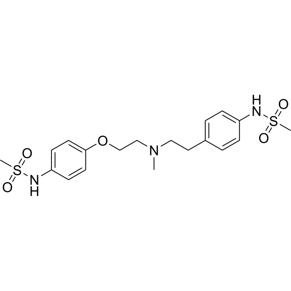
DofetilideIKr Blocker -
HY-108586
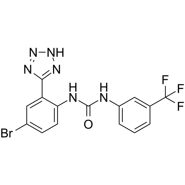
NS3623hERG1/IKr/Ito Activator -
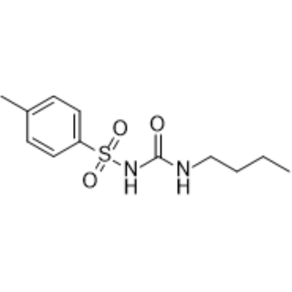
HY-B0401
TolbutamideKATP Inhibitor -

HY-B0401R
Tolbutamide (Standard)KATP Inhibitor -
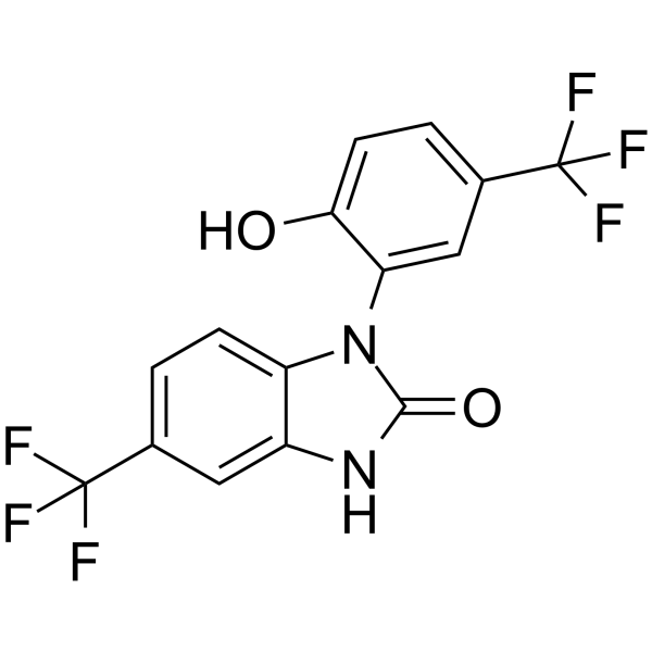
HY-12496
NS-1619BK Channel Opener -
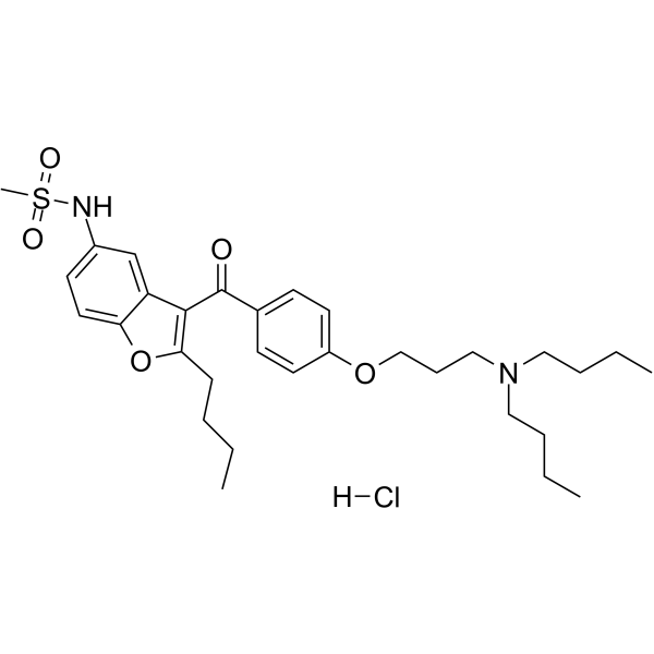
HY-75839
Dronedarone HydrochloridePotassium Channel Inhibitor -
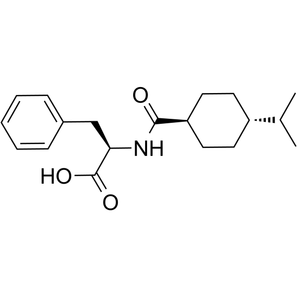
HY-B0422
NateglinideK+ Channel Inhibitor -
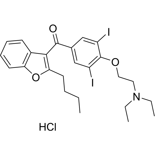
HY-14188
Amiodarone hydrochlorideAntiarrhythmic Agent -
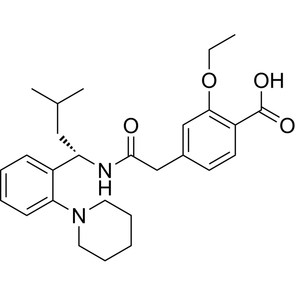
HY-15209
RepaglinideInsulin Secretagogue -
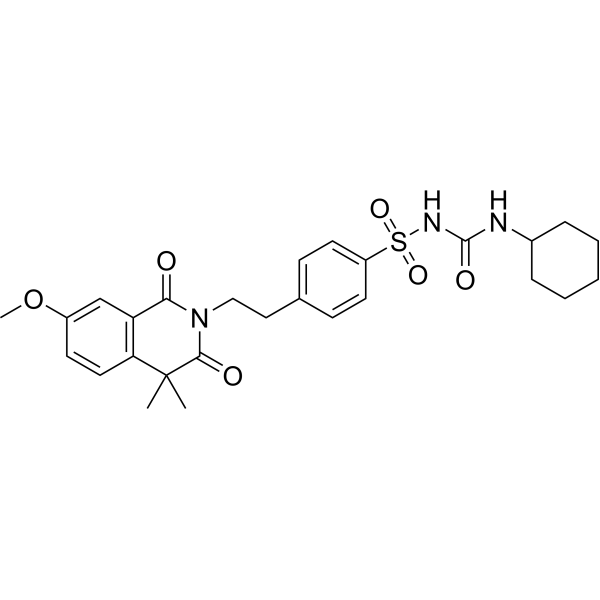
HY-B1114
GliquidoneAnti-Diabetic Drug
Publications Citing Use of MedChemExpress (MCE) NS1643
More

Biological Activity
Description
IC50 & Target
EC50: 10.5 μM (hERG) K+ channel[1]
In Vitro
NS1643 (0-100 μM) dose- and voltage-dependently increases hERG current in oocytes[1]. NS1643 (3, 10 and 30 μM) slows the rate of hERG inactivation throughout the testing voltage range and reduces the extent of hERG channel rectification[1]. NS1643 (10 and 50 μM) dose-dependently inhibits cell migration and invasion of MDA-MB-231 and SKBR3 cancer cells[2].
MedChemExpress (MCE) has not independently confirmed the accuracy of these methods. They are for reference only.
-
Cell Line:MDA-MB-231, SKBR3 and MCF7 breast cancer cell lines
-
Concentration:50 μM
-
Incubation Time:24 h
-
Result:Decreased Vimentin, N-cadherin and CD44 levels, and increased E-cadherin in breast cancer cell lines.
In Vivo
MedChemExpress (MCE) has not independently confirmed the accuracy of these methods. They are for reference only.
-
Animal Model:NSG mice with human-derived TNBC tumor xenografts[2]
-
Dosage:6 mg/kg
-
Administration:Intraperitoneal injection; 6 mg/kg; twice per week
-
Result:Significantly reduceed tumor growth and the metastatic liver tumors were significantly smaller than those in the control group. Decreased levels of human nuclear antigen (HNA).
- Scratch/Wound-Healing Migration Assay
- Cell migration
- Zigmond Chamber Chemotaxis Assay
- Under-Agarose Cell Migration Assay
- Neural Crest/Neuronal Cell Migration Explant Assay
- Transwell/Boyden Chamber Migration Assay
- 3D Collagen/Hydrogel Matrix Migration Assay
- Cell invasion
- Breast Cancer Modeling
- Cell-Exclusion Zone Migration Assay
- Dunn/Insall Chamber Chemotaxis Assay


Chemical Information
-
CAS No. 448895-37-2
-
Appearance Solid
-
Molecular Weight 380.24
-
Formula C15H10F6N2O3
-
Color White to off-white
-
SMILES
O=C(NC1=C(O)C=CC(C(F)(F)F)=C1)NC2=C(O)C=CC(C(F)(F)F)=C2
-
Shipping
Room temperature in continental US; may vary elsewhere.
-
Storage
Powder -20°C 3 years 4°C 2 years In solvent -80°C 2 years -20°C 1 year


Publications (1)
-
Journal Impact Factor
-
Most Recent
-
Int J Mol Sci
A Functional Pipeline of Genome-Wide Association Data Leads to Midostaurin as a Repurposed Drug for Alzheimer's Disease. [Abstract]2023 Jul 28;24(15):12079. PMID: 37569459


Solvent & Solubility
In Vitro:
DMSO : ≥ 100 mg/mL (262.99 mM; Hygroscopic DMSO has a significant impact on the solubility of product, please use newly opened DMSO)
* "≥" means soluble, but saturation unknown.
Please refer to the solubility information to select the appropriate solvent. Once prepared, please aliquot and store the solution to prevent product inactivation from repeated freeze-thaw cycles.
Storage method and period of stock solution: -80°C, 2 years; -20°C, 1 year. When stored at -80°C, please use it within 2 years. When stored at -20°C, please use it within 1 year.
Please refer to the solubility information to select the appropriate solvent. Once prepared, please aliquot and store the solution to prevent product inactivation from repeated freeze-thaw cycles.
Storage method and period of stock solution: -80°C, 2 years; -20°C, 1 year. When stored at -80°C, please use it within 2 years. When stored at -20°C, please use it within 1 year.
Concentration (start) × Volume (start) = Concentration (final) × Volume (final)
In Vivo:
Select the appropriate dissolution method based on your experimental animal and administration route.
- For the following dissolution methods, please ensure to first prepare a clear stock solution using an In Vitro approach and then sequentially add co-solvents:
- To ensure reliable experimental results, the clarified stock solution can be appropriately stored based on storage conditions. As for the working solution for In Vivo experiments, it is recommended to prepare freshly and use it on the same day.
- The percentages shown for the solvents indicate their volumetric ratio in the final prepared solution. If precipitation or phase separation occurs during preparation, heat and/or sonication can be used to aid dissolution.
Add each solvent one by one: 10% DMSO 40% PEG300 5% Tween-80 45% Saline
Solubility: ≥ 2.5 mg/mL (6.57 mM); Clear solution
This protocol yields a clear solution of ≥ 2.5 mg/mL (saturation unknown).
Taking 1 mL working solution as an example, add 100 μL DMSO stock solution (25.0 mg/mL) to 400 μL PEG300, and mix evenly; then add 50 μL Tween-80 and mix evenly; then add 450 μL Saline to adjust the volume to 1 mL.
Preparation of Saline: Dissolve 0.9 g sodium chloride in ddH₂O and dilute to 100 mL to obtain a clear Saline solution.
Add each solvent one by one: 10% DMSO 90% (20% SBE-β-CD in Saline)
Solubility: ≥ 2.5 mg/mL (6.57 mM); Clear solution
This protocol yields a clear solution of ≥ 2.5 mg/mL (saturation unknown).
Taking 1 mL working solution as an example, add 100 μL DMSO stock solution (25.0 mg/mL) to 900 μL 20% SBE-β-CD in Saline, and mix evenly.
Preparation of 20% SBE-β-CD in Saline (4°C, storage for one week): 2 g SBE-β-CD powder is dissolved in 10 mL Saline, completely dissolve until clear.
In Vivo Dissolution Calculator
Please enter the basic information of animal experiments:
-
-
-
-
Recommended: Prepare an additional quantity of animals to account for potential losses during experiments.
Please enter your animal formula composition:
-
%DMSO +
Recommended: Keep the proportion of DMSO in working solution below 2% if your animal is weak.
-
%+
-
+%Tween-80 + +
-
%Saline +
The co-solvents required include: DMSO, . All of co-solvents are available by MedChemExpress (MCE). , Tween 80. All of co-solvents are available by MedChemExpress (MCE).
Working solution concentration: 0.22 mg/mL
Method for preparing stock solution: mg drug dissolved in μL DMSO. Stock solution concentration: mg/mL.
1. Take μL DMSO stock solution;
2. Add μL .
μL , mix evenly;
3. Then add μL Tween 80, mix evenly;
4. Then add μL
Please ensure that the stock solution in the first step is dissolved to a clear state, and add co-solvents in sequence. You can use ultrasonic heating (ultrasonic cleaner, recommended frequency 20-40 kHz), vortexing, etc. to assist dissolution.


Purity & Documentation
-
Data Sheet (277 KB)
-
SDS (479 KB)
- English - EN (479 KB)
- Français - FR (479 KB)
- Deutsch - DE (479 KB)
- Norwegian - NO (479 KB)
- Español - ES (479 KB)
- Swedish - SV (479 KB)
- Italian - IT (479 KB)
- Korean - KR (479 KB)
- Portuguese - PT (479 KB)
-
Handling Instructions (2659 KB)
References
[1]. Casis O, et al. Mechanism of action of a novel human ether-a-go-go-related gene channel activator. Mol Pharmacol. 2006 Feb;69(2):658-65. Epub 2005 Nov 11. [Content Brief]
[2]. Breuer EK, et al. Potassium channel activity controls breast cancer metastasis by affecting β-catenin signaling. Cell Death Dis. 2019 Feb 21;10(3):180. [Content Brief]


Complete Stock Solution Preparation Table
Please refer to the solubility information to select the appropriate solvent. Once prepared, please aliquot and store the solution to prevent product inactivation from repeated freeze-thaw cycles.
Storage method and period of stock solution: -80°C, 2 years; -20°C, 1 year. When stored at -80°C, please use it within 2 years. When stored at -20°C, please use it within 1 year.
| Optional Solvent | Concentration Solvent Mass | 1 mg | 5 mg | 10 mg | 25 mg |
|---|---|---|---|---|---|
| DMSO | 1 mM | 2.6299 mL | 13.1496 mL | 26.2992 mL | 65.7480 mL |
| 5 mM | 0.5260 mL | 2.6299 mL | 5.2598 mL | 13.1496 mL | |
| 10 mM | 0.2630 mL | 1.3150 mL | 2.6299 mL | 6.5748 mL | |
| 15 mM | 0.1753 mL | 0.8766 mL | 1.7533 mL | 4.3832 mL | |
| 20 mM | 0.1315 mL | 0.6575 mL | 1.3150 mL | 3.2874 mL | |
| 25 mM | 0.1052 mL | 0.5260 mL | 1.0520 mL | 2.6299 mL | |
| 30 mM | 0.0877 mL | 0.4383 mL | 0.8766 mL | 2.1916 mL | |
| 40 mM | 0.0657 mL | 0.3287 mL | 0.6575 mL | 1.6437 mL | |
| 50 mM | 0.0526 mL | 0.2630 mL | 0.5260 mL | 1.3150 mL | |
| 60 mM | 0.0438 mL | 0.2192 mL | 0.4383 mL | 1.0958 mL | |
| 80 mM | 0.0329 mL | 0.1644 mL | 0.3287 mL | 0.8218 mL | |
| 100 mM | 0.0263 mL | 0.1315 mL | 0.2630 mL | 0.6575 mL |


NS1643 Related Classifications
HY-16916 Related Classifications
 Powered by Bioz