
ICT5040
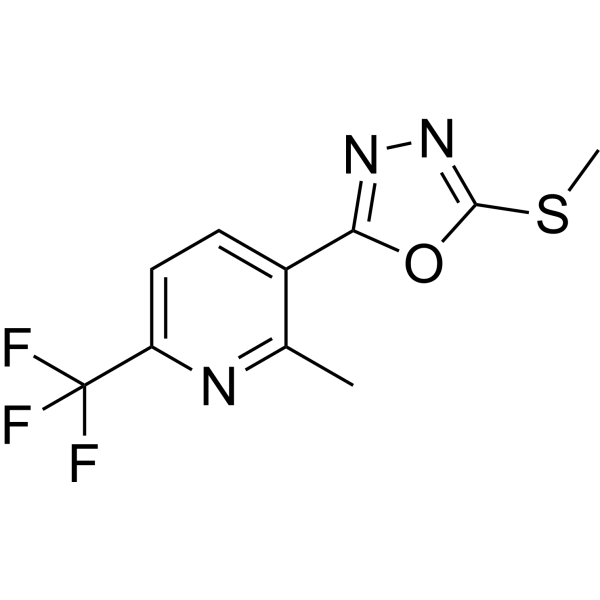
ICT5040 is a small molecule CXCR4 antagonist (IC50=3.8 μM). ICT5040 inhibits CXCL12-mediated proliferation and migration, and suppresses CXCL12-induced intracellular calcium mobilisation in U87 glioma cells.
For research use only. We do not sell to patients.
- CAS No.: 215655-21-3
- Formula: C10H8F3N3OS
- Molecular Weight:275.25
-
Storage:
Please store the product under the recommended conditions in the Certificate of Analysis.
Biological Activity
Description
IC50 & Target
[1]|
CXCR4 3.8 μM (IC50) |
In Vitro
ICT5040 (1-100 μM; 0-4 d) dose-dependently inhibits CXCL12-induced proliferation and migration of U87-glioma cells[1].
ICT5040 reduces CXCL12-induced intracellular calcium mobilisation in U87 glioma cells[1].
MedChemExpress (MCE) has not independently confirmed the accuracy of these methods. They are for reference only. Further protocols information, click here.
-
Cell Line:U87-glioma cells
-
Concentration:1 μM, 10 μM, 100 μM; with 100 ng/mL CXCL12
-
Incubation Time:0, 1, 2, 3, and 4 days
-
Result:Resulted reduction of CXCL12 induced proliferation and migration of U87 cells at day 3.
Chemical Information
-
CAS No. 215655-21-3
-
Molecular Weight 275.25
-
Formula C10H8F3N3OS
-
SMILES
FC(C1=CC=C(C2=NN=C(SC)O2)C(C)=N1)(F)F
-
Shipping
Room temperature in continental US; may vary elsewhere.
-
Storage
Please store the product under the recommended conditions in the Certificate of Analysis.
Protocols
-
Cell Cytotoxicity Assay
Cytotoxicity assays are usually based on the assessment of cell membrane damage, which can also be indirectly detected by measuring cell viability. Detection methods include MTT assay, CKK-8 assay, LDH assay and ATP assay, etc.
-
Cell migration
Cell migration is a method that plays an important role in wound healing, cell differentiation, embryonic development, etc.
-
Calcium Spark Assay
Calcium sparks are localized, transient increases in intracellular calcium concentration ([Ca2+]i) that occur in cardiac myocytes and represent elementary events underlying excitation-contraction coupling. These events are generated by the coordinated opening of clusters of ryanodine receptors (RyRs) on the sarcoplasmic reticulum membrane, leading to a brief release of Ca2+ into the cytosol. The detection and analysis of calcium sparks provide insights into the mechanisms of calcium handling and signaling in cardiac cells. Imaging techniques using fluorescent calcium indicators such as Fluo-3 are employed to visualize these subcellular calcium transients with high spatial and temporal resolution. The protocol is based on established methodologies described in primary literature for both experimental measurement and automated analysis of calcium sparks.
-
Ca2+ Staining Technique
Ca2+ staining is an experimental technique that utilizes specific fluorescent probes (such as Fluo-4 AM, Fura-2, etc.) to qualitatively or quantitatively detect dynamic changes in intracellular Ca2+ concentrations; this is achieved by monitoring the changes in fluorescent signals generated when these probes bind to free intracellular calcium ions. The underlying principle relies primarily on the presence of chelating groups within the probe's molecular structure that possess high affinity for calcium ions.
-
Cell Viability Determination by MTT Colorimetric Assay
The following protocol uses the MTT colorimetric assay as a classic literature-established method for assessing cell viability/metabolic activity in cultured mammalian cells. MTT[3-(4,5-dimethylthiazol-2-yl)-2,5-diphenyltetrazolium bromide] is reduced by metabolically active cells to a colored formazan product; the amount of formazan is quantified spectrophotometrically and provides an indirect measure of metabolically active viable cells. Importantly, MTT reduction reflects cellular oxidoreductase/metabolic activity rather than an absolute direct count of living cells, so changes in cellular metabolism can alter the signal independently of cell number.
Purity & Documentation
References
Calculators
Concentration (start) × Volume (start) = Concentration (final) × Volume (final)

