



Girinimbine

Based on 1 Customer Validation
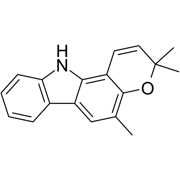
Girinimbine (Girinimbin) is a carbazole alkaloid with a variety of biological effects. Girinimbine can induce apoptosis, and has antitrypanosomal, antiplatelet activity, antibacterial activity, anti-inflammatory, antioxidant and antitumor activities.
商品は「研究用試薬」です。人や動物の医療用・臨床診断用・食品用の製品ではありません。
研究用途以外に使用した場合、当社は一切の責任を負いかねます。
- 純度 : 99.23%
- CAS 番号: 23095-44-5
- 分子式: C18H17NO
- 分子量:263.33
-
保管条件:
4°C, sealed storage, away from moisture and light
* In solvent : -80°C, 6 months; -20°C, 1 month (sealed storage, away from moisture and light)

To place orders, for customer services and technical support, please contact: MedChemExpress USA
Tel: 609-228-6898 E-mail: [email protected] [email protected]


-
 生物活性
生物活性
-
化学情報
- プロトコル
- 純度とドキュメンテーション
- 参考文献
-
 Help & FAQs


Parasite アイソフォーム固有の製品をすべて表示
More

生物活性
製品説明
Cellular Effect
|
Cell Line

|
Type | Value | Description | References |
|---|---|---|---|---|
| A2780 | IC50 |
>50 μM
Compound: Girinimbine
|
Anticancer activity against human A2780 cells by MTT assay
Anticancer activity against human A2780 cells by MTT assay
|
[PMID: 33316408] |
| A-431 | IC50 |
>50 μM
Compound: Girinimbine
|
Anticancer activity against human A431 cells by MTT assay
Anticancer activity against human A431 cells by MTT assay
|
[PMID: 33316408] |
| Bel-7402 | IC50 |
>50 μM
Compound: Girinimbine
|
Anticancer activity against human H7402 cells by MTT assay
Anticancer activity against human H7402 cells by MTT assay
|
[PMID: 33316408] |
| BGC-823 | IC50 |
>50 μM
Compound: Girinimbine
|
Anticancer activity against human BGC-823 cells by MTT assay
Anticancer activity against human BGC-823 cells by MTT assay
|
[PMID: 33316408] |
| HCT-8 | IC50 |
34.69 μM
Compound: Girinimbine
|
Anticancer activity against human HCT-8 cells by MTT assay
Anticancer activity against human HCT-8 cells by MTT assay
|
[PMID: 33316408] |
| HeLa | IC50 |
>50 μM
Compound: Girinimbine
|
Anticancer activity against human HeLa cells by MTT assay
Anticancer activity against human HeLa cells by MTT assay
|
[PMID: 33316408] |
| HepG2 | IC50 |
>160 μM
Compound: 7
|
Growth inhibition of human HepG2 cells after 48 hrs by WST-8 based CCK8 assay
Growth inhibition of human HepG2 cells after 48 hrs by WST-8 based CCK8 assay
|
[PMID: 22093759] |
| KB | IC50 |
263.1 μM
Compound: 7
|
Cytotoxicity against human KB cells by resazurin reduction assay
Cytotoxicity against human KB cells by resazurin reduction assay
|
[PMID: 22482432] |
| KB | IC50 |
7.02 μM
Compound: 5
|
Cytotoxicity against human KB cells by resazurin microplate assay
Cytotoxicity against human KB cells by resazurin microplate assay
|
[PMID: 21302964] |
| KETR3 | IC50 |
>50 μM
Compound: Girinimbine
|
Anticancer activity against human KETR3 cells by MTT assay
Anticancer activity against human KETR3 cells by MTT assay
|
[PMID: 33316408] |
| MCF7 | IC50 |
28.8 μM
Compound: 7
|
Cytotoxicity against human MCF7 cells by resazurin reduction assay
Cytotoxicity against human MCF7 cells by resazurin reduction assay
|
[PMID: 22482432] |
| MCF7 | IC50 |
49.76 μM
Compound: Girinimbine
|
Anticancer activity against human MCF7 cells by MTT assay
Anticancer activity against human MCF7 cells by MTT assay
|
[PMID: 33316408] |
| NCI-H187 | IC50 |
22.37 μM
Compound: 5
|
Cytotoxicity against human NCI-H187 by resazurin microplate assay
Cytotoxicity against human NCI-H187 by resazurin microplate assay
|
[PMID: 21302964] |
| NCI-H187 | IC50 |
58.9 μM
Compound: 7
|
Cytotoxicity against human NCI-H187 cells by resazurin reduction assay
Cytotoxicity against human NCI-H187 cells by resazurin reduction assay
|
[PMID: 22482432] |
| NCI-H460 | IC50 |
27.01 μM
Compound: Girinimbine
|
Anticancer activity against human H460 cells by MTT assay
Anticancer activity against human H460 cells by MTT assay
|
[PMID: 33316408] |
| SW1990 | IC50 |
>50 μM
Compound: Girinimbine
|
Anticancer activity against human SW1990 cells by MTT assay
Anticancer activity against human SW1990 cells by MTT assay
|
[PMID: 33316408] |
| Vero | IC50 |
>200 μM
Compound: 5
|
Cytotoxicity against african green monkey Vero cells by green fluorescent protein microplate assay
Cytotoxicity against african green monkey Vero cells by green fluorescent protein microplate assay
|
[PMID: 21302964] |
体外実験
Girinimbine (1-400 µM; 24-72 h) decreases the viability of HepG2 cells in 24, 48 and 72 h with IC50 values of 61 µM, 56 µM, and 40 µM respectively. Girinimbine (10-100 µM; 24-48 h) increase of LDH leakage in both concentration- and time-dependent manner in HepG2 cells[1].
Girinimbine (56 µM; 24-48 h) treatment results in DNA fragmentation and elevates levels of caspase-3 in HepG2 cells[1].
HepG2 cells[1].
Girinimbine (56 µM; 12-48 h) treatment also displays a time-dependent accumulation of the Sub-G0/G1 peak (hypodiploid) and caused G0/G1-phase arrest[1].
Girinimbine shows a potent antitrypanosomal activitywith an IC50 value of 10.16 µg/mL[3].
MedChemExpress (MCE) has not independently confirmed the accuracy of these methods. They are for reference only.
-
Cell Line:HepG2 cells
-
Concentration:1 µM, 10 µM, 50 µM, 100 µM, 200 µM
-
Incubation Time:24 h, 48 h and 72 h
-
Result:Inhibited the proliferation of HepG2 cells in vitro in a dose- and time-dependent manner.
-
Cell Line:HepG2 cells
-
Concentration:56 µM
-
Incubation Time:24 h, 48 h
-
Result:Showed typical morphological features of apoptosis.
-
Cell Line:HepG2 cells
-
Concentration:56 µM
-
Incubation Time:12 h, 24 h, 48 h
-
Result:Induced G0/G1-phase arrest in HepG2 cells.
体内実験
In vivo in zebrafish embryos, Girinimbine (20 μg/mL; 24 hours) shows significant distribution of apoptotic cells in embryos[2].
MedChemExpress (MCE) has not independently confirmed the accuracy of these methods. They are for reference only.
-
Animal Model:Male ICR mice (25-35 g) treated with carrageenan[2]
-
Dosage:10 mg/kg, 30 mg/kg, and 100 mg/kg
-
Administration:Orally gavage; once
-
Result:Helped limit total leukocyte migration, and reduced pro-inflammatory cytokine levels in the peritoneal fluid.


化学情報
-
CAS 番号 23095-44-5
-
性状 Solid
-
分子量 263.33
-
分子式 C18H17NO
-
Color Off-white to light yellow
-
SMILES
CC(OC1=C(C)C=C23)(C)C=CC1=C3NC4=C2C=CC=C4
-
別名
Girinimbin
-
Structure Classification
-
Initial Source
-
輸送条件
Room temperature in continental US; may vary elsewhere.
-
保管条件
4°C, sealed storage, away from moisture and light
* In solvent : -80°C, 6 months; -20°C, 1 month (sealed storage, away from moisture and light)


純度とドキュメンテーション
-
データシート (277 KB)
-
SDS (252 KB)
- English - EN (252 KB)
- Français - FR (252 KB)
- Deutsch - DE (252 KB)
- Norwegian - NO (252 KB)
- Español - ES (252 KB)
- Swedish - SV (252 KB)
- Italian - IT (252 KB)
- Korean - KR (252 KB)
- Portuguese - PT (252 KB)
-
取扱説明書 (2659 KB)
参考文献
[1]. Suvitha Syam, et al. The growth suppressing effects of girinimbine on HepG2 involve induction of apoptosis and cell cycle arrest. Molecules. 2011 Aug 23;16(8):7155-70. [Content Brief]
[2]. Venoos Iman, et al. Anticancer and anti-inflammatory activities of girinimbine isolated from Murraya koenigii. Drug Des Devel Ther. 2016 Dec 28;11:103-121. [Content Brief]
[3]. H O Dyary, et al. Antitrypanosomal and cytotoxic activities of botanical extracts from Murraya koenigii (L.) and Alpinia mutica Roxb. Trop Biomed. 2019 Mar 1;36(1):94-102. [Content Brief]

Calculators
濃度 (開始) × 体積 (開始) = 濃度 (終了) × 体積 (終了)


Girinimbine 関連分類
HY-N9488 関連分類
 Powered by Bioz