



Sertaconazole
Based on 2 publication(s) in Google Scholar
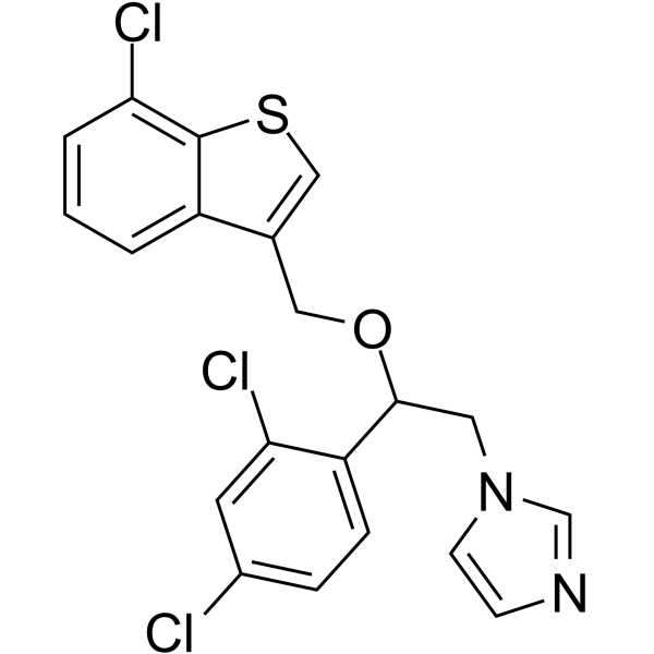
Sertaconazole (FI7056 free base) is a broad-spectrum topical antifungal agent, exhibits anti-inflammatory activity via activation of a p38-COX-2-PGE2 pathway. Sertaconazole is also a microtubule inhibitor, shows antiproliferative effect, induces apoptosis and autophagy, and can also inhibit the migration of cells.
商品は「研究用試薬」です。人や動物の医療用・臨床診断用・食品用の製品ではありません。
研究用途以外に使用した場合、当社は一切の責任を負いかねます。
- 純度 : 99.42%
- CAS 番号: 99592-32-2
- 分子式: C20H15Cl3N2OS
- 分子量:437.77
-
保管条件:Powder -20°C, 3 years , 4°C, 2 years ; In solvent -80°C, 6 months , -20°C, 1 month

To place orders, for customer services and technical support, please contact: MedChemExpress USA
Tel: 609-228-6898 E-mail: [email protected] [email protected]


-
 生物活性
生物活性
-
化学情報
-
溶剤 & 溶解度
- プロトコル
- 純度とドキュメンテーション
- 参考文献
-
 Help & FAQs


MedChemExpress(MCE)の使用を引用している文献 Sertaconazole
More

生物活性
製品説明
体外実験
Sertaconazole (0.03-40 µg/mL; 24 h) inhibits 150 strains of yeasts which includes six Candida species with arithmetic mean MIC of 0.77 µg/mL[1].
Sertaconazole (1 µg/mL; 5, 10, 30, 60 min) activates p38 MAP kinase in a time-dependent manner[2].
Sertaconazole (1, 2 µg/mL; 6, 8, or 24 h) increases a twofold release of PGE2 via COX-2 in keratinocytes, which is dependent on p38 activation[2].
Cetaconazole (10, 20, 30, 40 µM; 24 h) induces strong mitotic arrest by depolymerizing interphase and spindle microtubules, thereby inducing chromosome aggregation defects and causing anti-proliferation effect[3].
Sertaconazole (20, 40 µM; 24 h) induces apoptosis through p53 pathway in HeLa cells[3].
Sertaconazole (20, 30 µM; 24, 48, and 72 h) inhibits the migration of HeLa cells in a concentration-dependent manner[3].
Sertaconazole (15, 30 µM; 24 h) induces autophagy in A549, H460 cells[4].
MedChemExpress (MCE) has not independently confirmed the accuracy of these methods. They are for reference only.
-
Cell Line:C. albicans, C. guilliermondii, C. krusei, C. parapsilosi, C. tropicalis, C. glabrata
-
Concentration:0.03-40 µg/m
-
Incubation Time:24 h
-
Result:Againsted 150 strains of yeasts (six Candida species) which included C. albicans, C. guilliermondii, C. krusei, C. parapsilosi, C. tropicalis, C. glabrata species with arithmetic mean MIC values of 1.02, 0.51, 0.38, 0.31, 1.67 and 0.78 µg/mL, respectively.
-
Cell Line:HaCaT cells
-
Concentration:1 µg/mL
-
Incubation Time:5, 10, 30, 60 min
-
Result:Showed activity of activating p38 MAP kinase and Hsp27 in a time-dependent manner.
-
Cell Line:HaCaT cells
-
Concentration:1, 2 µg/mL
-
Incubation Time:6 or 8 h
-
Result:Induced 50% expression of COX-2 and resulted in a twofold increased in PGE2 release.
-
Cell Line:siRNA-transfected HaCaT cells (without p38 MAP kinase expression)
-
Concentration:1 µg/mL
-
Incubation Time:24 h
-
Result:Mediated induction of PGE2 was dependent on p38 activation.
-
Cell Line:HeLa, HEK-293, MCF-7, A549 cells
-
Concentration:0-100 µM
-
Incubation Time:24 h
-
Result:Showed antiproliferation activity with IC50s of 38, 45.1, 41.5, and 40.8 μM for HeLa, HEK-293, A549, and MCF-7 cells, respectively.
Exhibited mitotic block activity and induced cell death at concentration above 30 μM, but no significant increased in the number of mitotic cells.
Depolymerized interphase and spindle microtubules inducing defect in chromosomal congression.
-
Cell Line:HeLa cells
-
Concentration:10, 20, 40 µM
-
Incubation Time:24 h
-
Result:Induced approximately 5%, 10%, and 21% cells apoptotic at concentrations of 10, 20 and 40 μM, respectively.
-
Cell Line:A549 cells
-
Concentration:20, 40 µM
-
Incubation Time:24 h
-
Result:Induced apoptosis through p53 pathway that the expression of p53 from 30% to 50% and 95% and p21 from 11 to 39% and 40% respectively.
Resulted in Noxa and Puma, two direct transcriptional targets of p53 to be overexpressed.
-
Cell Line:HeLa cells
-
Concentration:20, 30 µM
-
Incubation Time:24, 48, and 72 h
-
Result:Inhibited the migration of HeLa cells at concentrations lesser than its IC50, which in a concentration-dependent manner.
-
Cell Line:A549, H460 cells
-
Concentration:15, 30 µM
-
Incubation Time:24 h
-
Result:Increased endogenous LC3 puncta and LC3 intensity, which indicated induction of autophagy in A549 and H460 cells.
体内実験
MedChemExpress (MCE) has not independently confirmed the accuracy of these methods. They are for reference only.
-
Animal Model:CD-1 mice (TPA-induced ear edema model)[2].
-
Dosage:1% (w/v)
-
Administration:Apply to the left ear, once.
-
Result:Exhibited a significant reduction of inflammation in mice by mediating PGE2 release.
- Detection of Apoptosis
- TUNEL staining for apoptotic DNA fragmentation
- Annexin V plus membrane-impermeant dye apoptosis staining
- Cotton Pellet Granuloma
- Cell migration
- Carrageenan-Induced Paw Edema
- Autophagy
- Apoptosis Solutions
- Research Protocol for Inflammation-related Diseases
- Lysosome and acidic-vesicle live-cell staining
- Macroautophagy Solutions
- Cell Viability Determination by MTT Colorimetric Assay
臨床実験
| NCT Number | Sponsor | Condition | Start Date |
Phase

|
|---|---|---|---|---|
| NCT01329991 | Plexxikon| | 2011-05 | PHASE1 |


化学情報
-
CAS 番号 99592-32-2
-
性状 Solid
-
分子量 437.77
-
分子式 C20H15Cl3N2OS
-
Color White to off-white
-
SMILES
ClC1=CC=C(C(OCC2=CSC3=C(Cl)C=CC=C32)CN4C=CN=C4)C(Cl)=C1
-
別名
FI7056 free base
-
輸送条件
Room temperature in continental US; may vary elsewhere.
-
保管条件
Powder -20°C 3 years 4°C 2 years In solvent -80°C 6 months -20°C 1 month


Publications (2)
-
Journal Impact Factor
-
Most Recent
-
J Exp Clin Cancer Res
Sertaconazole-repurposed nanoplatform enhances lung cancer therapy via CD44-targeted drug delivery. [Abstract]2023 Jul 29;42(1):188. PMID: 37507782 -
MedComm (2020)
Sertaconazole provokes proapoptotic autophagy via stabilizing TRADD in nonsmall cell lung cancer cells. [Abstract]2021 Dec 16;2(4):821-837. PMID: 34977879


溶剤 & 溶解度
体外:
DMSO : 25 mg/mL (57.11 mM; Need ultrasonic; Hygroscopic DMSO has a significant impact on the solubility of product, please use newly opened DMSO)
Please refer to the solubility information to select the appropriate solvent. Once prepared, please aliquot and store the solution to prevent product inactivation from repeated freeze-thaw cycles.
Storage method and period of stock solution: -80°C, 6 months; -20°C, 1 month. When stored at -80°C, please use it within 6 months. When stored at -20°C, please use it within 1 month.
Please refer to the solubility information to select the appropriate solvent. Once prepared, please aliquot and store the solution to prevent product inactivation from repeated freeze-thaw cycles.
Storage method and period of stock solution: -80°C, 6 months; -20°C, 1 month. When stored at -80°C, please use it within 6 months. When stored at -20°C, please use it within 1 month.
濃度 (開始) × 体積 (開始) = 濃度 (終了) × 体積 (終了)
体内:
Select the appropriate dissolution method based on your experimental animal and administration route.
- For the following dissolution methods, please ensure to first prepare a clear stock solution using an In Vitro approach and then sequentially add co-solvents:
- To ensure reliable experimental results, the clarified stock solution can be appropriately stored based on storage conditions. As for the working solution for In Vivo experiments, it is recommended to prepare freshly and use it on the same day.
- The percentages shown for the solvents indicate their volumetric ratio in the final prepared solution. If precipitation or phase separation occurs during preparation, heat and/or sonication can be used to aid dissolution.
Add each solvent one by one: 10% DMSO 40% PEG300 5% Tween-80 45% Saline
Solubility: ≥ 2.5 mg/mL (5.71 mM); Clear solution
This protocol yields a clear solution of ≥ 2.5 mg/mL (saturation unknown).
Taking 1 mL working solution as an example, add 100 μL DMSO stock solution (25.0 mg/mL) to 400 μL PEG300, and mix evenly; then add 50 μL Tween-80 and mix evenly; then add 450 μL Saline to adjust the volume to 1 mL.
Preparation of Saline: Dissolve 0.9 g sodium chloride in ddH₂O and dilute to 100 mL to obtain a clear Saline solution.
Add each solvent one by one: 10% DMSO 90% (20% SBE-β-CD in Saline)
Solubility: 2.5 mg/mL (5.71 mM); Suspended solution; Need ultrasonic
This protocol yields a suspended solution of 2.5 mg/mL. Suspended solution can be used for oral and intraperitoneal injection.
Taking 1 mL working solution as an example, add 100 μL DMSO stock solution (25.0 mg/mL) to 900 μL 20% SBE-β-CD in Saline, and mix evenly.
Preparation of 20% SBE-β-CD in Saline (4°C, storage for one week): 2 g SBE-β-CD powder is dissolved in 10 mL Saline, completely dissolve until clear.
In Vivo Dissolution Calculator
Please enter the basic information of animal experiments:
-
-
-
-
Recommended: Prepare an additional quantity of animals to account for potential losses during experiments.
Please enter your animal formula composition:
-
%DMSO +
Recommended: Keep the proportion of DMSO in working solution below 2% if your animal is weak.
-
%+
-
+%Tween-80 + +
-
%Saline +
The co-solvents required include: DMSO, . All of co-solvents are available by MedChemExpress (MCE). , Tween 80. All of co-solvents are available by MedChemExpress (MCE).
Working solution concentration: 0.22 mg/mL
Method for preparing stock solution: mg drug dissolved in μL DMSO. Stock solution concentration: mg/mL.
1. Take μL DMSO stock solution;
2. Add μL .
μL , mix evenly;
3. Then add μL Tween 80, mix evenly;
4. Then add μL
Please ensure that the stock solution in the first step is dissolved to a clear state, and add co-solvents in sequence. You can use ultrasonic heating (ultrasonic cleaner, recommended frequency 20-40 kHz), vortexing, etc. to assist dissolution.


純度とドキュメンテーション
-
データシート (290 KB)
-
SDS (394 KB)
- English - EN (394 KB)
- Français - FR (394 KB)
- Deutsch - DE (394 KB)
- Norwegian - NO (394 KB)
- Español - ES (394 KB)
- Swedish - SV (394 KB)
- Italian - IT (394 KB)
- Korean - KR (394 KB)
- Portuguese - PT (394 KB)
-
取扱説明書 (2659 KB)
参考文献
[1]. Carrillo-Muñoz AJ, et al. In-vitro antifungal activity of sertaconazole, econazole, and bifonazole against Candida spp. J Antimicrob Chemother. 1995 Oct;36(4):713-6. [Content Brief]
[2]. Sur R, et al. Anti-inflammatory activity of sertaconazole nitrate is mediated via activation of a p38-COX-2-PGE2 pathway. J Invest Dermatol. 2008 Feb;128(2):336-44. [Content Brief]
[3]. Sebastian J, et al. Sertaconazole induced toxicity in HeLa cells through mitotic arrest and inhibition of microtubule assembly. Naunyn Schmiedebergs Arch Pharmacol. 2021 Jun;394(6):1231-1249. [Content Brief]
[4]. Zhang W, et al. Sertaconazole provokes proapoptotic autophagy via stabilizing TRADD in nonsmall cell lung cancer cells. MedComm (2020). 2021 Dec 16;2(4):821-837. [Content Brief]


Complete Stock Solution Preparation Table
Please refer to the solubility information to select the appropriate solvent. Once prepared, please aliquot and store the solution to prevent product inactivation from repeated freeze-thaw cycles.
Storage method and period of stock solution: -80°C, 6 months; -20°C, 1 month. When stored at -80°C, please use it within 6 months. When stored at -20°C, please use it within 1 month.
| Optional Solvent | Concentration Solvent Mass | 1 mg | 5 mg | 10 mg | 25 mg |
|---|---|---|---|---|---|
| DMSO | 1 mM | 2.2843 mL | 11.4215 mL | 22.8430 mL | 57.1076 mL |
| 5 mM | 0.4569 mL | 2.2843 mL | 4.5686 mL | 11.4215 mL | |
| 10 mM | 0.2284 mL | 1.1422 mL | 2.2843 mL | 5.7108 mL | |
| 15 mM | 0.1523 mL | 0.7614 mL | 1.5229 mL | 3.8072 mL | |
| 20 mM | 0.1142 mL | 0.5711 mL | 1.1422 mL | 2.8554 mL | |
| 25 mM | 0.0914 mL | 0.4569 mL | 0.9137 mL | 2.2843 mL | |
| 30 mM | 0.0761 mL | 0.3807 mL | 0.7614 mL | 1.9036 mL | |
| 40 mM | 0.0571 mL | 0.2855 mL | 0.5711 mL | 1.4277 mL | |
| 50 mM | 0.0457 mL | 0.2284 mL | 0.4569 mL | 1.1422 mL |


Sertaconazole 関連分類
HY-B0736 関連分類
 Powered by Bioz