



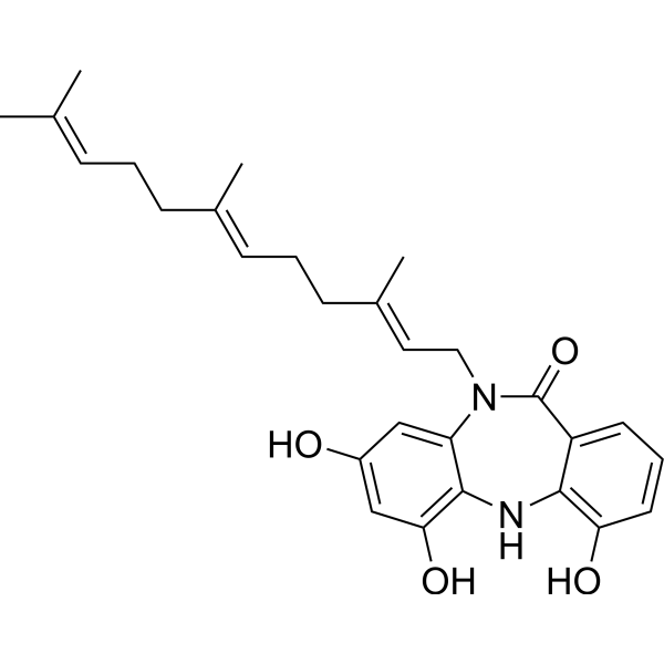
Diazepinomicin
Diazepinomicin (ECO-4601) is an anticancer and antibacterial agent. Diazepinomicin can be produced by a Micromonospora strain. Diazepinomicin induces Apoptosis. Diazepinomicin inhibits the proteases Rhodesain and Cathepsin L at an IC50 of 70-90 μM. Diazepinomicin possesses anti-inflammatory and anti-tumor activity. Diazepinomicin has demonstrated activity against hepatocellular carcinoma. Diazepinomicin shows antiparasitic activity against trypomastigote forms of Trypanosoma brucei with an IC50 of 13.5 μM. Diazepinomicin exhibits moderate antibacterial activity against specific Gram-positive bacteria, with an MIC of approximately 32 μg/mL.
For research use only. We do not sell to patients.
- CAS No.: 733035-26-2
- Formula: C28H34N2O4
- Molecular Weight:462.58
-
Storage:
-20°C, protect from light, stored under nitrogen
* In solvent : -80°C, 6 months; -20°C, 1 month (protect from light, stored under nitrogen)

To place orders, for customer services and technical support, please contact: MedChemExpress USA
Tel: 609-228-6898 E-mail: [email protected] [email protected]


-
 Biological Activity
-
Chemical Information
- Protocols
- Purity & Documentation
- References
-
 Help & FAQs


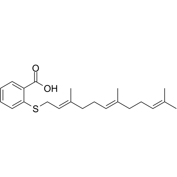
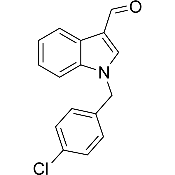
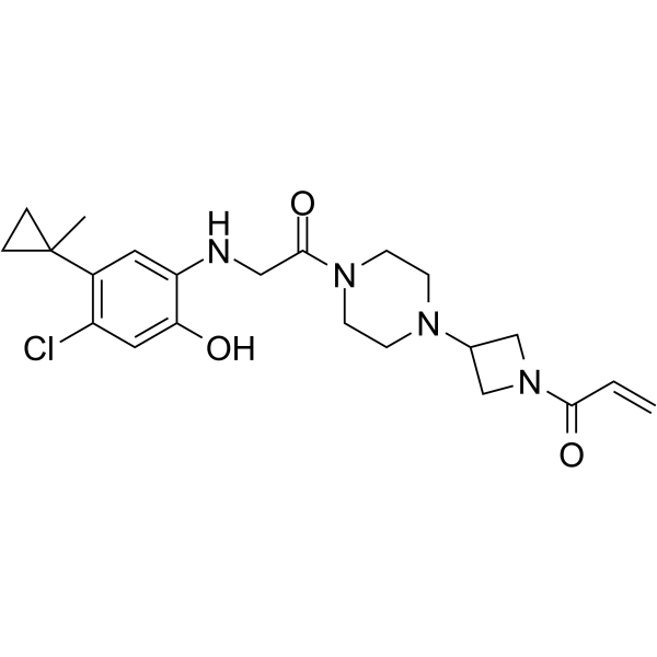


Biological Activity
Description
IC50 & Target
[1]|
Microbial Metabolite |
Trypanosoma |
cathepsin L 70-90 μM (IC50) |
Cellular Effect
|
Cell Line

|
Type | Value | Description | References |
|---|---|---|---|---|
| HUVEC | IC50 |
0.43 μg/mL
Compound: 1, BU-4664L
|
Antiinvasive activity against HUVEC assessed as inhibition of cell migration after 6 hrs by Matrigel assay
Antiinvasive activity against HUVEC assessed as inhibition of cell migration after 6 hrs by Matrigel assay
|
[PMID: 20056543] |
| HUVEC | IC50 |
0.72 μg/mL
Compound: 1, BU-4664L
|
Antiangiogenic activity against HUVEC cells assessed as inhibition of vascular formation after 6 hrs by wound healing assay
Antiangiogenic activity against HUVEC cells assessed as inhibition of vascular formation after 6 hrs by wound healing assay
|
[PMID: 20056543] |
| HUVEC | IC50 |
2 μg/mL
Compound: 1, BU-4664L
|
Cytotoxicity against HUVEC cells
Cytotoxicity against HUVEC cells
|
[PMID: 20056543] |
| NCI-H460 | GI50 |
13.6 μM
Compound: 1; BU-4664L
|
Antiproliferative activity against human NCI-H460 cells assessed as cell growth inhibition by MTT assay
Antiproliferative activity against human NCI-H460 cells assessed as cell growth inhibition by MTT assay
|
[PMID: 34838651] |
| PC-3 | GI50 |
1.95 μM
Compound: 1; BU-4664L
|
Antiproliferative activity against human PC-3 cells assessed as cell growth inhibition by MTT assay
Antiproliferative activity against human PC-3 cells assessed as cell growth inhibition by MTT assay
|
[PMID: 34838651] |
| Renca | IC50 |
0.78 μg/mL
Compound: 1, BU-4664L
|
Antiinvasive activity against mouse Renca by Matrigel assay
Antiinvasive activity against mouse Renca by Matrigel assay
|
[PMID: 20056543] |
| Renca | IC50 |
8.3 μg/mL
Compound: 1, BU-4664L
|
Cytotoxicity against mouse Renca cells
Cytotoxicity against mouse Renca cells
|
[PMID: 20056543] |
In Vitro
Diazepinomicin (1-25 μM; 24-72 h) induces dose-dependent antiproliferative activity in hepatocellular carcinoma HepG2 cells[1].
Diazepinomicin exhibits moderate antibacterial activity against specific Gram-positive bacteria, with an MIC of approximately 32 μg/mL[2].
MedChemExpress (MCE) has not independently confirmed the accuracy of these methods. They are for reference only. Further protocols information, click here.
-
Cell Line:human hepatoma HepG2 cells
-
Concentration:10-15 μM
-
Incubation Time:24 h
-
Result:Induced clumping of cells with slight distortion, as well as very condensed and fragmented nuclei (hallmark features of apoptosis). Induced condensed nuclei and apoptotic bodies, characteristic of apoptotic cells. Induced DNA fragmentation, visualized as characteristic DNA ladder bands on agarose gel electrophoresis.
Clinical Trial
| NCT Number | Sponsor | Condition | Start Date |
Phase
|
|---|---|---|---|---|
| NCT01329991 | Plexxikon| | 2011-05 | PHASE1 |


Chemical Information
-
CAS No. 733035-26-2
-
Appearance Solid
-
Molecular Weight 462.58
-
Formula C28H34N2O4
-
Color Off-white to gray
-
SMILES
O=C1C2=CC=CC(O)=C2NC3=C(O)C=C(O)C=C3N1C/C=C(C)/CC/C=C(C)/CC/C=C(C)\C
-
Synonyms
ECO-4601; TLN-4601; BU 4664L
-
Shipping
Room temperature in continental US; may vary elsewhere.
-
Storage
-20°C, protect from light, stored under nitrogen
* In solvent : -80°C, 6 months; -20°C, 1 month (protect from light, stored under nitrogen)

Protocols
-
Research Protocol for Infectious Diseases
Infectious-disease experiments test how pathogens interact with host barriers, innate immune receptors, inflammatory signaling, pathogen replication, and tissue injury; pattern-recognition receptors such as TLRs, RIG-I-like receptors, NOD-like receptors, and inflammasomes detect microbial molecules and activate NF-κB, interferon, and cytokine responses. The central hypothesis is that infection severity reflects the balance between pathogen burden and host response: protective inflammation restricts pathogen growth, whereas excessive or mislocalized inflammation contributes to tissue damage and disease phenotype. Unresolved questions include which host pathways are protective versus pathogenic, why some infection models fail to translate to human disease, and which combined readouts best predict clinically relevant infection outcomes.
-
Apoptosis
Apoptosis, also called programmed cell death, is generally characterized by distinct morphological characteristics.
-
TUNEL staining for apoptotic DNA fragmentation
TUNEL staining detects DNA strand breaks by using terminal deoxynucleotidyl transferase to add labeled nucleotides to exposed 3′-OH DNA termini, generating either microscopic staining in fixed cells or tissue sections, or fluorescence/cytometric signal in cell suspensions. TUNEL positivity reflects DNA fragmentation but should not be interpreted alone as definitive apoptosis, because TUNEL can also label necrotic, autolytic, mechanically damaged, or DNA-repair-associated DNA breaks.
-
Cell Cytotoxicity Assay
Cytotoxicity assays are usually based on the assessment of cell membrane damage, which can also be indirectly detected by measuring cell viability. Detection methods include MTT assay, CKK-8 assay, LDH assay and ATP assay, etc.
-
Annexin V plus membrane-impermeant dye apoptosis staining
Annexin V-based apoptosis assays rely on the detection of phosphatidylserine (PS) externalization from the inner leaflet of the plasma membrane to the outer leaflet, an early biochemical hallmark of apoptosis. Fluorescently labeled Annexin V binds PS in a calcium-dependent manner, enabling identification of early apoptotic cells by flow cytometry or fluorescence microscopy. When combined with a membrane-impermeant DNA-binding dye (e. g. , propidium iodide), this approach allows discrimination between viable (Annexin V−/dye−), early apoptotic (Annexin V+/dye−), and late apoptotic or necrotic (Annexin V+/dye+) cell populations by assessing membrane integrity and PS exposure.
-
Apoptosis Solutions
Apoptosis is a regulated, generally non-lytic cell-death pathway that removes unwanted, damaged, infected, or abnormal cells through coordinated morphological changes, caspase activation, DNA fragmentation, and membrane remodeling. The intrinsic apoptosis pathway is controlled mainly by mitochondrial outer membrane permeabilization, BCL-2 family proteins, cytochrome c release, apoptosome formation, caspase-9 activation, and downstream executioner caspase-3/7 activation. The extrinsic apoptosis pathway is initiated by death receptors such as Fas, TNFR, and TRAIL receptors, which recruit adaptor proteins and activate caspase-8 before engaging executioner caspases or mitochondrial amplification through BID cleavage. Apoptosis is linked to many phenotypes, including cancer cell killing, tissue homeostasis, immune regulation, neurodegeneration, infection response, and treatment-induced cytotoxicity; unresolved questions include how apoptosis interacts with necroptosis, pyroptosis, ferroptos
-
Research Protocol for Inflammation-related Diseases
The NLRP3 inflammasome is a cytosolic innate immune signaling platform that integrates priming signals and danger-signal activation to promote caspase-1 activation, maturation of IL-1β and IL-18, and gasdermin D-mediated pyroptotic cell death. The core experimental logic is to determine whether inflammatory disease phenotypes are driven by increased NLRP3 expression, ASC-containing inflammasome assembly, caspase-1 cleavage, GSDMD cleavage, and extracellular release of IL-1β/IL-18 rather than by nonspecific cell injury alone. The pathway is strongly linked to inflammation-related disease phenotypes because monosodium urate crystals activate NALP3/NLRP3 inflammasome signaling in gout-like crystal inflammation, cholesterol crystals activate NLRP3 inflammasomes in atherogenesis models, and DSS-induced intestinal inflammation has been reported to involve NLRP3 inflammasome activity. However, experimental colitis studies also show context-dependent protective effects of NLRP3 inflammasome co
-
Liver Cancer Modeling
Liver cancer can be classified into primary liver cancer and secondary liver cancer. Secondary liver cancer is the metastatic liver cancer. Primary liver cancer includes hepatocellular carcinoma (HCC), intrahepatic cholangiocarcinoma (ICC) and fibrolamellar HCC, of which HCC is the most common form, accounting for approximately 90% of primary liver cancers[1]. HCC mouse models include chemical agent-induced models, transplanted tumor models, and genetic engineered models.
-
Gram Staining of Tissue Sections
Gram staining of tissue sections is a histochemical technique used to differentiate Gram-positive and Gram-negative bacteria within histological specimens based on differences in bacterial cell wall structure and dye retention, adapted from classical bacteriological Gram staining into tissue-compatible “histological Gram stain” variants. In tissue applications, modifications of the Brown-Hopps and Brown-Brenn methods are commonly used to improve differentiation of microorganisms embedded within host connective tissue and to reduce overstaining or loss of Gram-negative signal, which are known limitations of earlier approaches. The principle relies on crystal violet-iodine complex retention in Gram-positive organisms and subsequent decolorization and counterstaining steps that allow contrast visualization of Gram-negative organisms against tissue background.
-
Bacterial live/dead nucleic-acid viability staining
The LIVE/DEAD bacterial viability staining method is based on differential permeability of nucleic-acid-binding fluorescent dyes, most commonly SYTO 9 and propidium iodide (PI), which enables discrimination of bacterial populations with intact versus compromised cytoplasmic membranes. SYTO 9 penetrates both intact and damaged bacterial membranes and binds nucleic acids to produce green fluorescence, whereas propidium iodide penetrates only cells with compromised membranes and fluoresces red while also reducing SYTO 9 signal through competitive binding and fluorescence interactions. The resulting fluorescence pattern is interpreted as a proxy for membrane integrity, which is widely used as an indicator of bacterial viability in microscopy, flow cytometry, and spectroscopic platforms. However, mechanistic studies show that SYTO 9 and PI interactions involve displacement and fluorescence resonance energy transfer effects, which can influence signal interpretation depending on dye ratios a


Purity & Documentation
-
Data Sheet (273 KB)
-
SDS (252 KB)
- English - EN (252 KB)
- Français - FR (252 KB)
- Deutsch - DE (252 KB)
- Norwegian - NO (252 KB)
- Español - ES (252 KB)
- Swedish - SV (252 KB)
- Italian - IT (252 KB)
- Korean - KR (252 KB)
- Portuguese - PT (252 KB)
-
Handling Instructions (2659 KB)
References

Calculators
Concentration (start) × Volume (start) = Concentration (final) × Volume (final)


Diazepinomicin Related Classifications
HY-N6674 Related Classifications
 Powered by Bioz
Keywords
- Diazepinomicin
- 733035-26-2
- ECO-4601
- TLN-4601
- BU 4664L
- ECO4601
- ECO 4601
- TLN4601
- TLN 4601
- TLN-4601
- Apoptosis
- Bacterial
- Cathepsin
- Parasite
- Endogenous Metabolite
- human hepatoma HepG2 cells
- Bcl2 protein
- BAX protein
- Gram-positive bacteria
- apoptotic nuclear condensation
- apoptotic body formation
- apoptosis
- DNA fragmentation
- human hepatoma cells
- hepatocellular carcinoma
- Inhibitor
- inhibitor
- inhibit