



NF157

Based on 1 Customer Validation
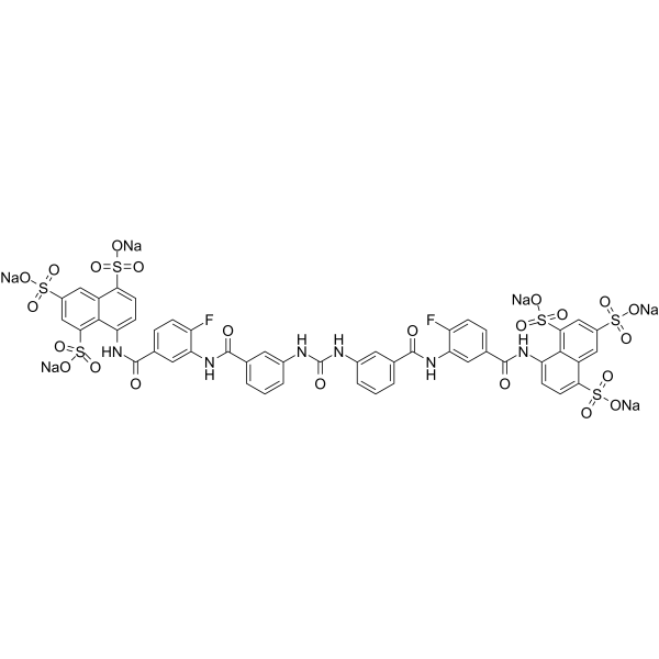
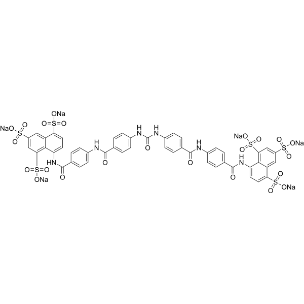
NF157 is a highly selective nanomolar P2Y11 antagonist with a pKi of 7.35. The IC50s are 463 nM, 1811 µM, 170 µM for P2Y11 (Ki=44.3 nM), P2Y1 (Ki=187 µM), P2Y2 (Ki=28.9 µM), respectively. NF157, significantly reduces expression of metalloproteinase (MMP)-3, MMP-13, can be used in the treatment of osteoarthritis (OA).
연구목적의 판매만을 진행합니다. 환자를 대상으로 한 판매는 하지 않습니다.
- Purity : 99.13%
- CAS No.: 104869-26-3
- 화학식: C49H28F2N6Na6O23S6
- 분자량:1437.10
-
보관:
4°C, sealed storage, away from moisture
* In solvent : -80°C, 6 months; -20°C, 1 month (sealed storage, away from moisture)

To place orders, for customer services and technical support, please contact: MedChemExpress USA
Tel: 609-228-6898 E-mail: [email protected] [email protected]


-
 Biological Activity
-
Chemical Information
-
용액&용해도
- Protocol
- 순도&문서
- References
-
 Help & FAQs


-
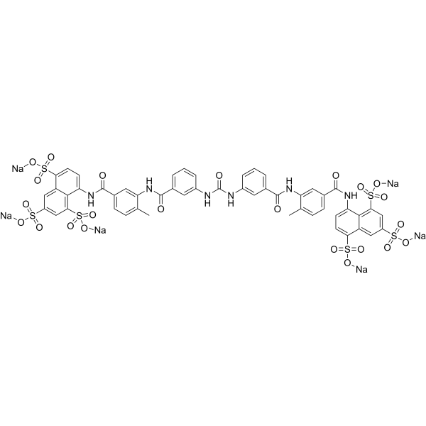
HY-B0879A
Suramin sodium saltPTPases Inhibitor -
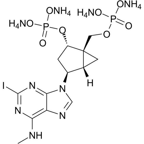
HY-108658
MRS2500 tetraammoniumP2Y1 Receptor Antagonist -
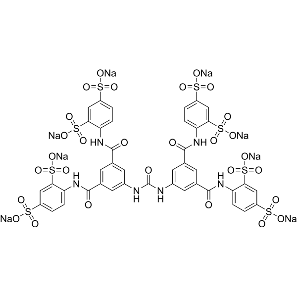
HY-112461A
NF449 octasodiumP2X1 Receptor Antagonist -
HY-15310
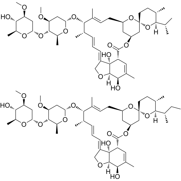
IvermectinAnti-parasite Agent -
HY-15568
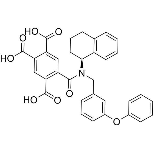
A-317491P2X3/P2X2/3 Antagonist -
HY-101044
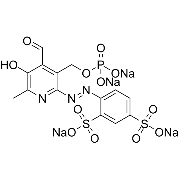
PPADS tetrasodiumP2X Receptor Antagonist -
HY-110126
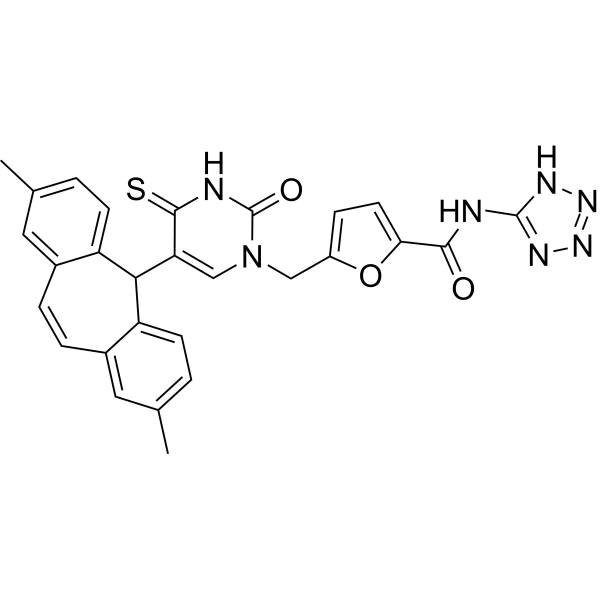
AR-C118925XXP2Y2 Receptor Antagonist -
HY-108661A
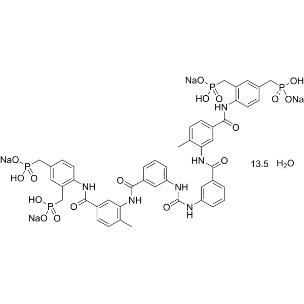
NF546 hydrateP2Y11 Agonist -
HY-108676
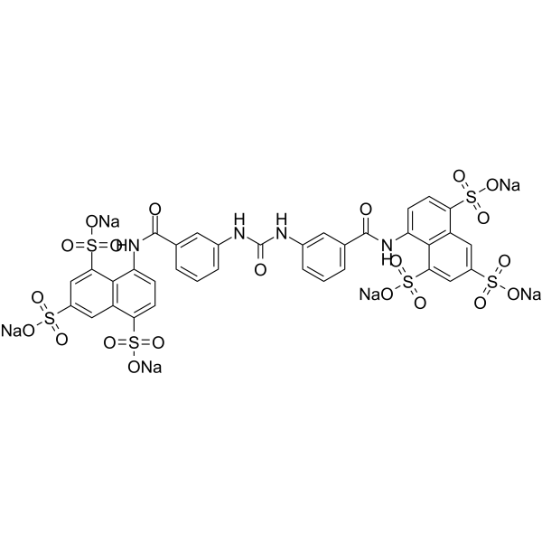
NF023 hexasodiumP2X1 Receptor Antagonist -
HY-13104
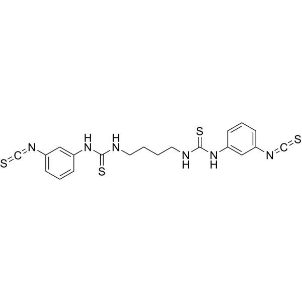
MRS 2578Apoptosis Inducer -
HY-110322
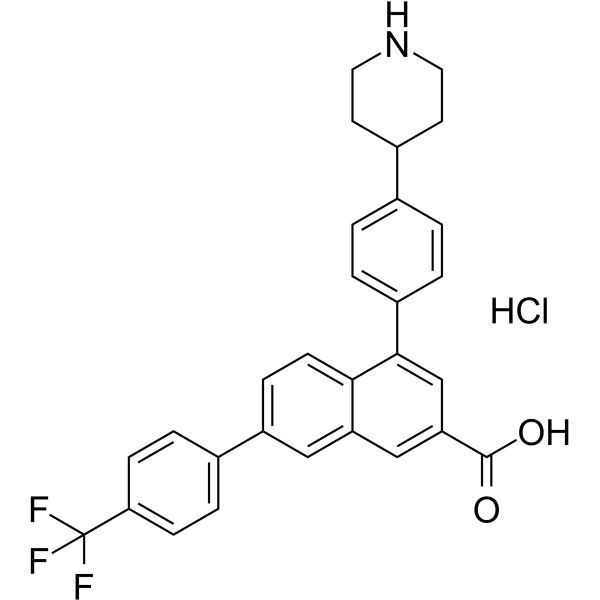
PPTN hydrochlorideP2Y14-R Antagonist -
HY-D0976
NF279P2X1 Receptor Antagonist
All P2Y Receptor Isoforms
More

Biological Activity
제품 설명
IC50 & Target
pKi: 7.35 (P2Y11)[1]
IC50: 463 nM (P2Y11), 1811 µM (P2Y1), 170 µM (P2Y2)[1]
Ki: 44.3 nM (P2Y11), 187 µM (P2Y1), 28.9 µM (P2Y2)[1]
In Vitro
NF157 displays selectivity for P2Y11 over P2Y1 (>650-fold), P2Y2 (>650-fold), P2X2 (3-fold), P2X3 (8-fold), P2X4 (>22-fold), and P2X7 (>67-fold) but no selectivity over P2X1[1].
NF157 (30 and 60 µM; 24 hours) causes a significant reduction in degradation of type II collagen in a dose-dependent manner. 60 µM NF157 nearly completely rescues type II collagen from degradation induced by TNF-α (10 ng/mL)[2].
NF157 (30 and 60 µM; 24 hours) almost fully restores nuclear translocation of p65 triggered by TNF-α (10 ng/mL) and significantly reduces the luciferase activity of NF-κB[2].
MedChemExpress (MCE) has not independently confirmed the accuracy of these methods. They are for reference only.
-
Cell Line:SW1353 cells
-
Concentration:30 and 60 µM
-
Incubation Time:24 hours
-
Result:Ameliorated TNF-α-induced degradation of type II collagen.
-
Cell Line:SW1353 cells
-
Concentration:30 and 60 µM
-
Incubation Time:24 hours
-
Result:Reduced TNF-α-induced activation of NF-κB.


Chemical Information
-
CAS No. 104869-26-3
-
Appearance Solid
-
분자량 1437.10
-
화학식 C49H28F2N6Na6O23S6
-
Color Off-white to pink
-
SMILES
O=C(NC1=CC(C(NC2=CC(C(NC3=CC=C(S(=O)(O[Na])=O)C4=CC(S(=O)(O[Na])=O)=CC(S(=O)(O[Na])=O)=C34)=O)=CC=C2F)=O)=CC=C1)NC5=CC(C(NC6=CC(C(NC7=CC=C(S(=O)(O[Na])=O)C8=CC(S(=O)(O[Na])=O)=CC(S(=O)(O[Na])=O)=C78)=O)=CC=C6F)=O)=CC=C5
-
선적
Room temperature in continental US; may vary elsewhere.
-
보관
4°C, sealed storage, away from moisture
* In solvent : -80°C, 6 months; -20°C, 1 month (sealed storage, away from moisture)


용액&용해도
In Vitro:
DMSO : 33.33 mg/mL (23.19 mM; Need ultrasonic; Hygroscopic DMSO has a significant impact on the solubility of product, please use newly opened DMSO)
H2O : 1 mg/mL (0.70 mM; ultrasonic and warming and heat to 60°C)
Please refer to the solubility information to select the appropriate solvent. Once prepared, please aliquot and store the solution to prevent product inactivation from repeated freeze-thaw cycles.
Storage method and period of stock solution: -80°C, 6 months; -20°C, 1 month (sealed storage, away from moisture). When stored at -80°C, please use it within 6 months. When stored at -20°C, please use it within 1 month.
* Note: If you choose water as the stock solution, please dilute it to the working solution, then filter and sterilize it with a 0.22 μm filter before use.
Please refer to the solubility information to select the appropriate solvent. Once prepared, please aliquot and store the solution to prevent product inactivation from repeated freeze-thaw cycles.
Storage method and period of stock solution: -80°C, 6 months; -20°C, 1 month (sealed storage, away from moisture). When stored at -80°C, please use it within 6 months. When stored at -20°C, please use it within 1 month.
* Note: If you choose water as the stock solution, please dilute it to the working solution, then filter and sterilize it with a 0.22 μm filter before use.
Concentration (start) × Volume (start) = Concentration (final) × Volume (final)
In Vivo:
Select the appropriate dissolution method based on your experimental animal and administration route.
- For the following dissolution methods, please ensure to first prepare a clear stock solution using an In Vitro approach and then sequentially add co-solvents:
- To ensure reliable experimental results, the clarified stock solution can be appropriately stored based on storage conditions. As for the working solution for In Vivo experiments, it is recommended to prepare freshly and use it on the same day.
- The percentages shown for the solvents indicate their volumetric ratio in the final prepared solution. If precipitation or phase separation occurs during preparation, heat and/or sonication can be used to aid dissolution.
Add each solvent one by one: 10% DMSO 90% (20% SBE-β-CD in Saline)
Solubility: ≥ 2.5 mg/mL (1.74 mM); Clear solution
This protocol yields a clear solution of ≥ 2.5 mg/mL (saturation unknown).
Taking 1 mL working solution as an example, add 100 μL DMSO stock solution (25.0 mg/mL) to 900 μL 20% SBE-β-CD in Saline, and mix evenly.
Preparation of 20% SBE-β-CD in Saline (4°C, storage for one week): 2 g SBE-β-CD powder is dissolved in 10 mL Saline, completely dissolve until clear.
In Vivo Dissolution Calculator
Please enter the basic information of animal experiments:
-
-
-
-
Recommended: Prepare an additional quantity of animals to account for potential losses during experiments.
Please enter your animal formula composition:
-
%DMSO +
Recommended: Keep the proportion of DMSO in working solution below 2% if your animal is weak.
-
%+
-
+%Tween-80 + +
-
%Saline +
The co-solvents required include: DMSO, . All of co-solvents are available by MedChemExpress (MCE). , Tween 80. All of co-solvents are available by MedChemExpress (MCE).
Working solution concentration: 0.22 mg/mL
Method for preparing stock solution: mg drug dissolved in μL DMSO. Stock solution concentration: mg/mL. * In solvent : -80°C, 6 months; -20°C, 1 month (sealed storage, away from moisture)
1. Take μL DMSO stock solution;
2. Add μL .
μL , mix evenly;
3. Then add μL Tween 80, mix evenly;
4. Then add μL
Please ensure that the stock solution in the first step is dissolved to a clear state, and add co-solvents in sequence. You can use ultrasonic heating (ultrasonic cleaner, recommended frequency 20-40 kHz), vortexing, etc. to assist dissolution.


순도&문서
-
Data Sheet (275 KB)
-
SDS (252 KB)
- English - EN (252 KB)
- Français - FR (252 KB)
- Deutsch - DE (252 KB)
- Norwegian - NO (252 KB)
- Español - ES (252 KB)
- Swedish - SV (252 KB)
- Italian - IT (252 KB)
- Korean - KR (252 KB)
- Portuguese - PT (252 KB)
-
Handling Instructions (2659 KB)
References
[1]. Ullmann H, et al. Synthesis and structure-activity relationships of suramin-derived P2Y11 receptor antagonists with nanomolar potency. J Med Chem. 2005 Nov 3;48(22):7040-8. [Content Brief]
[2]. Wang D, et al. Inhibition of P2Y11R ameliorated TNF-α-induced degradation of extracellular matrix in human chondrocytic SW1353 cells. Am J Transl Res. 2019 Apr 15;11(4):2108-2116. [Content Brief]


Complete Stock Solution Preparation Table
Please refer to the solubility information to select the appropriate solvent. Once prepared, please aliquot and store the solution to prevent product inactivation from repeated freeze-thaw cycles.
Storage method and period of stock solution: -80°C, 6 months; -20°C, 1 month (sealed storage, away from moisture). When stored at -80°C, please use it within 6 months. When stored at -20°C, please use it within 1 month.
| Optional Solvent | Concentration Solvent Mass | 1 mg | 5 mg | 10 mg | 25 mg |
|---|---|---|---|---|---|
| DMSO | 1 mM | 0.6958 mL | 3.4792 mL | 6.9585 mL | 17.3961 mL |
| 5 mM | 0.1392 mL | 0.6958 mL | 1.3917 mL | 3.4792 mL | |
| 10 mM | 0.0696 mL | 0.3479 mL | 0.6958 mL | 1.7396 mL | |
| 15 mM | 0.0464 mL | 0.2319 mL | 0.4639 mL | 1.1597 mL | |
| 20 mM | 0.0348 mL | 0.1740 mL | 0.3479 mL | 0.8698 mL |
* Note: If you choose water as the stock solution, please dilute it to the working solution, then filter and sterilize it with a 0.22 μm filter before use.


NF157 Related Classifications
HY-108672 Related Classifications
 Powered by Bioz