



M3541

Based on 1 Customer Validation
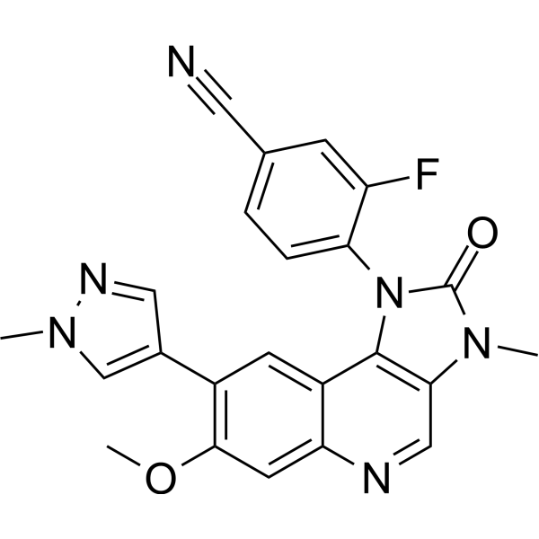
M3541 is an orally effective DNA-dependent protein kinase (DNA-PK) inhibitor. M3541 inhibits ionizing radiation-induced ATM autophosphorylation and phosphorylation of downstream substrates (KAP1, CHK2, p53), blocks DSB end resection and homologous recombination repair, and simultaneously induces non-homologous end joining, G2-M phase arrest, polyploidization, and cell death. M3541 can be used for research on solid tumors, acute myeloid leukemia, non-small cell lung cancer, triple-negative breast cancer, etc..
For research use only. We do not sell to patients.
- Purity : 99.28%
- CAS No.: 1360628-91-6
- Formula: C23H17FN6O2
- Molecular Weight:428.42
-
Storage:Powder -20°C, 3 years , 4°C, 2 years ; In solvent -80°C, 6 months , -20°C, 1 month

To place orders, for customer services and technical support, please contact: MedChemExpress USA
Tel: 609-228-6898 E-mail: [email protected] [email protected]


-
 Biological Activity
-
Chemical Information
-
Solvent & Solubility
- Protocols
- Purity & Documentation
- References
-
 Help & FAQs


-
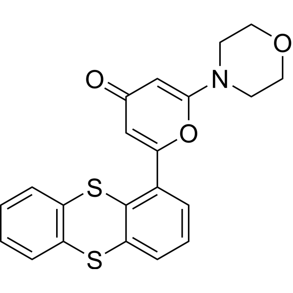
HY-12016
KU-55933ATM Inhibitor -
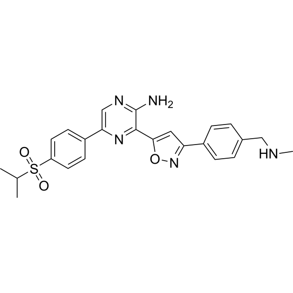
HY-13902
BerzosertibATR Inhibitor -
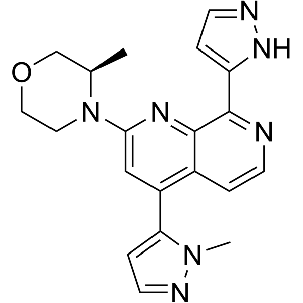
HY-101566
ElimusertibATR Inhibitor -
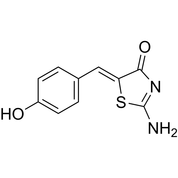
HY-19959
MirinMre11-Rad50-Nbs1 Complex Inhibitor -
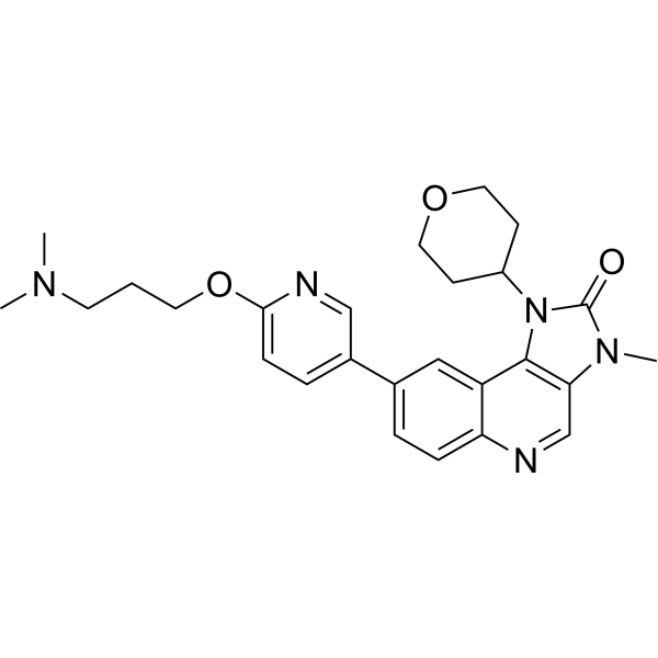
HY-100016
AZD0156ATM Inhibitor -
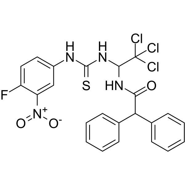
HY-15520
CGK733ATM/ATR Inhibitor -
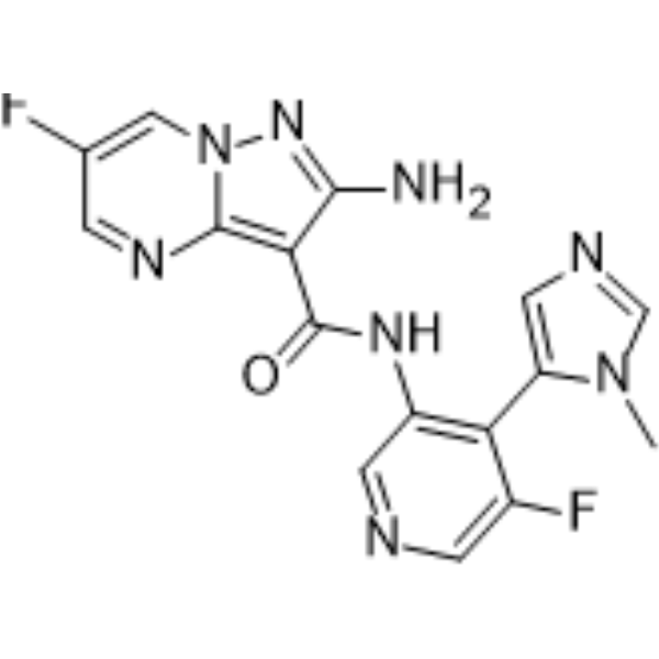
HY-111451
TuvusertibATR Inhibitor -
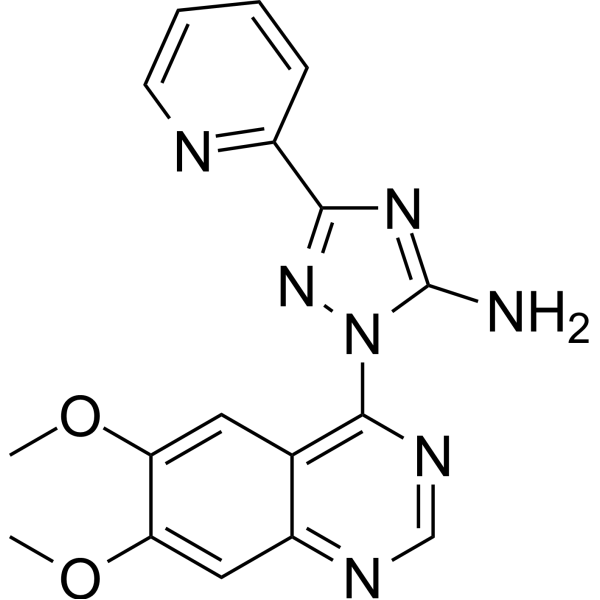
HY-11002
CP-466722ATM/ATR Inhibitor -

HY-101566R
Elimusertib (Standard)ATR Inhibitor -

HY-100016R
AZD0156 (Standard)ATM Inhibitor


Biological Activity
Description
|
ATM 025 μM (IC50) |
DNA-PK |
Chk2 |
KAP1 |
p53 |
In Vitro
M3541 (1 μM; 1 h) significantly impairs DSB repair in A549 cells, leading to the accumulation of unrepaired DSBs[1].
M3541 (1 μM; 1 h) combined with IR (5 Gy) inhibits the growth of A549 cells[1].
M3541 (1 μM; 1 h) exhibits radiosensitizing effects in A549 and 13 other cancer cell lines, significantly reducing clonogenic survival[1].
M3541 (1 µM; 6-96 h) inhibits early p53 activation induced by the combination of Calicheamicin (HY-19609)/M3814 (Nedisertib) (HY-101570) in MV4-11 cells, supporting that enhanced p53 activity is a downstream effect of M3814-induced ATM hyperactivation[2].
M3541 alone (up to 1.0 μM) shows minimal toxicity in A549 and NCI-H460 cells but sensitizes these cells to ionizing radiation[3].
M3541 (1 μM; 24 h) inhibition of ATM impairs RAD51 and RPA foci formation and increases DNA damage in FA-deficient HeLa and U2OS cells[3].
M3541 further impairs DNA end resection in FANCD2-deficient U2OS cells[3].
M3541 (1 μM; 24 h) further reduces DNA end resection in FANCD2-knockout U2OS cells[3].
M3541 (1 μM; 72 h) further increases NHEJ activity in U2OS cells with knockdown of FA/BRCA pathway components[3].
M3541 (1 μM; 72 h) reduces HR activity in FA/BRCA pathway-knockdown U2OS cells[3].
M3541 selectively inhibits the growth of PL11, HS766T, and TOV21G cancer cell lines with FA pathway defects[3].
M3541 (1 μM; 24 h) slightly decreases phospho-ATM levels below baseline and significantly reverses M6620 (Berzosertib) (HY-13902)-induced phospho-ATM levels in A549 cancer cells[5].
M3541 (1 μM; 24 h) alone has minimal effect on ATM/p53 signaling proteins, but in A549 cells it inhibits M6620-induced activation of these proteins[5].
M3541 (5 days) alone has minimal effect on the cell viability of A549 parental cells and ATM-deficient cells, but exhibits a synergistic effect with M6620 in A549 parental cells[5].
M3541 (1 μM; 1 h) abolishes the elevation of p53 induced by DNA-PK inhibitors in irradiated HCT116 and RKO cells, indicating that this event occurs downstream of the ATM pathway[6].
M3541 sensitizes multiple tumor cell lines to ionizing radiation and a topoisomerase inhibitor in vitro[7].
M3541 (1 μM; 1 h) effectively inhibits the catalytic activity of ATM and its downstream signaling in irradiated A549, A375, and RKO cancer cells, with 1 μM causing over 90% inhibition[9].
M3541 (1 μM; 1 h) initially suppresses IR-induced γH2AX foci but leads to the accumulation of numerous unrepaired DSBs at 24 h in A549 cells[9].
M3541 (1 h) significantly enhances radiation-induced cell death and inhibits the growth of HeLa cells[9].
M3541 (7 days) significantly enhances radiation-induced chromosome misalignment, missegregation, and prolonged metaphase in HeLa cells[9].
M3541 (1 μM; 1 h) promotes total chromosomal aberrations in irradiated A549 cells[9].
M3541 strongly enhances structural damage and micronucleus formation in irradiated A549 cells[9].
M3541 strongly enhances inflammatory signaling in irradiated A549 cells[9].
M3541 is a highly potent ATP-competitive ATM kinase inhibitor with an IC50 < 1 nM[10].
M3541 (1 μM; 24 h) alone does not affect cell cycle distribution, but partially reverses the G1 phase arrest induced by M6620 in A549, A375, and H460 cancer cells[5].
M3541 (1 μM) prevents complete cell cycle arrest induced by DNA-PK inhibition in irradiated A375 cells, confirming the role of ATM in this process[6].
M3541 (1 μM; 1 h) inhibits the function of ATM in cell cycle checkpoint control in irradiated A549 cells, causing cell cycle arrest to shift primarily toward G2/M[9].
The inhibitory activity of M3541 requires wild-type ATM kinase and functional signaling, with an average IC50 of 71 nM in wild-type ATM cell lines and 670 nM in mutant ATM cell lines[1].
M3541 (1 μM) alone causes growth inhibition in IMR90 and WI38 fibroblast cell lines, but induces only slight cell death[5].
MedChemExpress (MCE) has not independently confirmed the accuracy of these methods. They are for reference only.
-
Cell Line:A549, HT29, HCT116, PC3
-
Concentration:0.125-3 μM
-
Incubation Time:1 hour pre-treatment before 5 Gy IR
-
Result:Completely suppressed radiotherapy-induced ATM phosphorylation in A549 cells at 1 μM or higher.
Did not affect ATR-dependent CHK1 phosphorylation in HT29 cells or DNA-PK autophosphorylation in HCT116 cells.
Did not affect PI3K pathway activity measured by pAKT levels in PC3 cells.
-
Cell Line:A549
-
Concentration:1 μM
-
Incubation Time:1 hour pre-treatment before 5 Gy IR
-
Result:Showed a substantial increase in the H2AX foci fraction, indicating significantly impaired DSB repair and accumulation of unrepaired DSBs.
Showed only a minor increase in foci number in unirradiated cells treated with the inhibitor alone.
-
Cell Line:A549 NucLight
-
Concentration:1 μM
-
Incubation Time:1 hour pre-treatment before 5 Gy IR; monitored for 6 days
-
Result:Treatment alone showed a slight growth retardation.
IR plus M3541 combination halted cell growth during the 6-day observation period.
-
Cell Line:A549
-
Concentration:1 μM
-
Incubation Time:1 hour pre-treatment before 5 Gy IR
-
Result:IR plus M3541 combination induced a predominant G2-M phase arrest.
Showed increased sub-G1 and polyploid populations in the next two days, suggesting aberrant mitotic transition.
Cell-cycle profile at day 5 reflected a mix of cells with increasing ploidy as well as cells undergoing cell death.
-
Cell Line:MV4-11
-
Concentration:1 µM
-
Incubation Time:6, 24, 48, and 96 h
-
Result:Abrogated the enhancing effect of M3814 on the p53 targets p21, Mdm2, and Puma, bringing their levels close to untreated levels at 6 h and still significantly reduced relative to the calicheamicin/M3814 combination at 24 h.
-
Cell Line:HeLa (FANCA-KO) and U2OS (FANCD2-KO) cells
-
Concentration:1 μM
-
Incubation Time:24 h
-
Result:FANCA or FANCD2 knockout resulted in increased γ-H2AX foci, which was further enhanced by M3541.
In FANCA-KO HeLa cells, M3541 further reduced RAD51 foci and RPA foci, while no changes were observed in 53BP1 foci.
In FANCD2-KO U2OS cells, M3541 further reduced RAD51 foci, while no changes were observed in 53BP1 foci.
-
Cell Line:A549
-
Concentration:1 μM
-
Incubation Time:24 h
-
Result:Decreased phospho-ATM levels slightly below baseline.
Dramatically reversed M6620-induced phospho-ATM levels.
-
Cell Line:A549
-
Concentration:1 μM
-
Incubation Time:24 h
-
Result:Did not show significant effects on p-KAP1, p-CHK2, p-p53, and p21 levels compared to DMSO.
Suppressed M6620-induced p-KAP1, p-CHK2, p-p53, and p21 levels.
Maintained p-CHK1 levels previously lowered by ATRi.
-
Cell Line:A549, A375, H460
-
Concentration:1 μM
-
Incubation Time:24 h
-
Result:Did not have any impact on the cell-cycle profiles of A549, A375, or H460 cells.
Partially abrogated the M6620-induced G1 checkpoint and promoted cycling in all three cell lines.
-
Cell Line:A549 parental, p53-null, and ATM-null
-
Concentration:1 μM
-
Incubation Time:6 days
-
Result:Did not impact the parental A549 cells.
Further enhanced the effect on cell viability observed with M6620 alone in the A549 p53-null cells.
Had no additional effect in the A549 ATM-null cells.
-
Cell Line:HCT116 and RKO
-
Concentration:1 μM
-
Incubation Time:1 h
-
Result:Blocked the p53 boost induced by the DNA-PK inhibitor M3814 in irradiated HCT116 and RKO cells.
-
Cell Line:A549, A375, and RKO cancer cell lines
-
Concentration:1 μM
-
Incubation Time:1 h (pre-treatment)
-
Result:Suppressed ATM autophosphorylation at serine 1981 concentration-dependently.
Caused over 90% ATM inhibition at 1 μM compared to IR alone.
Reduced phospho-CHK2T68 and phospho-p53Ser15 levels.
Inhibited IR-induced p53 transcriptional target p21.
Increased levels of phospho-ATRT1989, phospho-CHK1S317, and phospho-CHK1S345.
-
Cell Line:A549 cancer cells
-
Concentration:1 μM
-
Incubation Time:1 h (pre-treatment); 0.5, 4, and 24 h post-radiation
-
Result:Dramatically reduced γH2AX foci at 30 minutes compared to IR alone.
Increased γH2AX foci to >10 in ~90% of cell population at 24 hours, compared to ~10% for IR or M3541 alone.
-
Cell Line:A549 cancer cells
-
Concentration:1 μM
-
Incubation Time:1 h (pre-treatment); 24 h post-IR
-
Result:Did not affect cell cycle distribution alone compared to vehicle.
Induced a cell cycle shift from G1 to predominantly G2/M phase arrest in irradiated cells.
In Vivo
M3541 (100 mg/kg; p.o.; in combination with fractionated radiotherapy (30 × 2 Gy over 6 weeks)) achieves complete and durable tumor regressions in 3 out of 4 xenograft models for 140-160 days[1].
The triple combination of M3541 (100 mg/kg; p.o.; 10 minutes before each IR fraction (2 Gy, 5 days on/2 days off for 2 weeks)), cisplatin (3 mg/kg), and fractionated radiotherapy (20 Gy total) demonstrates enhanced antitumor activity and progression-free survival in the FaDu xenograft model with reversible body weight effects[1].
The combination of M4076 (50-100 mg/kg; p.o.; once daily) and M4344 results in complete tumor growth inhibition in the MiaPaCa2 model and almost complete tumor regression in the MV4.11 model[5].
M4076 (50 mg/kg; p.o.; twice daily) potentiates the efficacy of ATRis in multiple TNBC PDX cancer models, achieving a tumor control rate of approximately 42% and an objective response rate of approximately 20% in combination with M4344[5].
M3541 (p.o.) strongly enhances the antitumor activity of fractionated radiation in human tumor xenograft-bearing nude mice, leading to complete tumor regression[7].
MedChemExpress (MCE) has not independently confirmed the accuracy of these methods. They are for reference only.
-
Animal Model:NMRI mice (female, immunodeficient, 7-9 weeks old, FaDu xenograft)[1]
-
Dosage:100 mg/kg (PD); 10, 50, or 200 mg/kg (efficacy)
-
Administration:p.o.; single dose 10 min before 2 Gy IR (PD); for duration of fractionated radiotherapy (efficacy)
-
Result:Inhibited CHK2 phosphorylation with strongest effects corresponding to the highest plasma concentration of M3541, demonstrating exposure-dependent inhibition of IR-induced CHK2 phosphorylation.
Inhibited tumor growth dose-dependently in FaDu xenografts at 10, 50, or 200 mg/kg.
Achieved complete regression on day 30 in the 200 mg/kg group, but most tumors re-grew by day 70.
-
Animal Model:NMRI mice (female, immunodeficient, 7-9 weeks old, FaDu xenograft); CD1 mice (female, immunodeficient, 7-9 weeks old, Capan-1 and NCI-H1975 xenografts)[1]
-
Dosage:100 mg/kg
-
Administration:p.o.; in combination with fractionated radiotherapy (30 × 2 Gy over 6 weeks)
-
Result:Strongly enhanced IR efficacy in all four models.
Achieved complete and durable tumor regressions in 3 out of 4 models (FaDu, NCI-H1975, Capan-1) with tumors not re-growing for 140-160 days.
Partially regressed IR-resistant NCI-H460 tumors during the treatment period.
Resulted in moderate body weight loss during treatment but no dermatitis or visible skin reaction.
-
Animal Model:NMRI mice (female, immunodeficient, 7-9 weeks old, FaDu xenograft)[1]
-
Dosage:100 mg/kg
-
Administration:p.o.; 10 minutes before each IR fraction (2 Gy, 5 days on/2 days off for 2 weeks)
-
Result:Demonstrated stronger antitumor activity and progression-free survival with limited effect on body weight.
Body weight returned to the weight of vehicle-treated mice after treatment stop.
-
Animal Model:H2dRag2 (C;129P2-H2d-Rag2 tm1Fwa IL2rgtm1) (female, 6- to 8-week-old, subcutaneous MiaPaCa2 pancreatic cancer and MV4.11 AML xenograft models)[5]
-
Dosage:50, 100 mg/kg
-
Administration:p.o.; once daily
-
Result:Inhibited tumor growth in the MiaPaCa2 model with a complete tumor growth inhibition in combination with M4344.
Inhibited tumor growth in the MV4.11 model with an almost complete tumor regression in combination with M4344.
-
Animal Model:athymic nude (Foxn1nu ENVIGO) (female, 5-week-old, subcutaneous 26 patient-derived triple-negative breast cancer xenograft models)[5]
-
Dosage:50 mg/kg
-
Administration:p.o.; twice daily
-
Result:Improved the single-agent activity of M4344 in most models with a mean RTV of 120% for the combination versus 310% for M4344 alone.
Achieved a tumor control rate of approximately 42% (11/26) and an objective response rate of approximately 20% (5/26) in combination with M4344.
Clinical Trial
| NCT Number | Sponsor | Condition | Start Date |
Phase

|
|---|---|---|---|---|
| NCT01329991 | Plexxikon| | 2011-05 | PHASE1 |


Chemical Information
-
CAS No. 1360628-91-6
-
Appearance Solid
-
Molecular Weight 428.42
-
Formula C23H17FN6O2
-
Color White to light yellow
-
SMILES
N#CC1=CC=C(N(C2=C3C=NC4=CC(OC)=C(C5=CN(C)N=C5)C=C24)C(N3C)=O)C(F)=C1
-
Shipping
Room temperature in continental US; may vary elsewhere.
-
Storage
Powder -20°C 3 years 4°C 2 years In solvent -80°C 6 months -20°C 1 month


Solvent & Solubility
In Vitro:
DMSO : 4.55 mg/mL (10.62 mM; ultrasonic and warming and adjust pH to 3 with 1 M HCL and heat to 60°C; Hygroscopic DMSO has a significant impact on the solubility of product, please use newly opened DMSO)
Please refer to the solubility information to select the appropriate solvent. Once prepared, please aliquot and store the solution to prevent product inactivation from repeated freeze-thaw cycles.
Storage method and period of stock solution: -80°C, 6 months; -20°C, 1 month. When stored at -80°C, please use it within 6 months. When stored at -20°C, please use it within 1 month.
Please refer to the solubility information to select the appropriate solvent. Once prepared, please aliquot and store the solution to prevent product inactivation from repeated freeze-thaw cycles.
Storage method and period of stock solution: -80°C, 6 months; -20°C, 1 month. When stored at -80°C, please use it within 6 months. When stored at -20°C, please use it within 1 month.
Concentration (start) × Volume (start) = Concentration (final) × Volume (final)
In Vivo:
Select the appropriate dissolution method based on your experimental animal and administration route.
- For the following dissolution methods, please ensure to first prepare a clear stock solution using an In Vitro approach and then sequentially add co-solvents:
- To ensure reliable experimental results, the clarified stock solution can be appropriately stored based on storage conditions. As for the working solution for In Vivo experiments, it is recommended to prepare freshly and use it on the same day.
- The percentages shown for the solvents indicate their volumetric ratio in the final prepared solution. If precipitation or phase separation occurs during preparation, heat and/or sonication can be used to aid dissolution.
Add each solvent one by one: 10% DMSO 90% (20% SBE-β-CD in Saline)
Solubility: ≥ 0.46 mg/mL (1.07 mM); Clear solution
This protocol yields a clear solution of ≥ 0.46 mg/mL (saturation unknown).
Taking 1 mL working solution as an example, add 100 μL DMSO stock solution (4.6 mg/mL) to 900 μL 20% SBE-β-CD in Saline, and mix evenly.
Preparation of 20% SBE-β-CD in Saline (4°C, storage for one week): 2 g SBE-β-CD powder is dissolved in 10 mL Saline, completely dissolve until clear.
Add each solvent one by one: 10% DMSO 40% PEG300 5% Tween-80 45% Saline
Solubility: ≥ 0.45 mg/mL (1.05 mM); Clear solution
This protocol yields a clear solution of ≥ 0.45 mg/mL (saturation unknown).
Taking 1 mL working solution as an example, add 100 μL DMSO stock solution (4.5 mg/mL) to 400 μL PEG300, and mix evenly; then add 50 μL Tween-80 and mix evenly; then add 450 μL Saline to adjust the volume to 1 mL.
Preparation of Saline: Dissolve 0.9 g sodium chloride in ddH₂O and dilute to 100 mL to obtain a clear Saline solution.
In Vivo Dissolution Calculator
Please enter the basic information of animal experiments:
-
-
-
-
Recommended: Prepare an additional quantity of animals to account for potential losses during experiments.
Please enter your animal formula composition:
-
%DMSO +
Recommended: Keep the proportion of DMSO in working solution below 2% if your animal is weak.
-
%+
-
+%Tween-80 + +
-
%Saline +
The co-solvents required include: DMSO, . All of co-solvents are available by MedChemExpress (MCE). , Tween 80. All of co-solvents are available by MedChemExpress (MCE).
Working solution concentration: 0.22 mg/mL
Method for preparing stock solution: mg drug dissolved in μL DMSO. Stock solution concentration: mg/mL.
1. Take μL DMSO stock solution;
2. Add μL .
μL , mix evenly;
3. Then add μL Tween 80, mix evenly;
4. Then add μL
Please ensure that the stock solution in the first step is dissolved to a clear state, and add co-solvents in sequence. You can use ultrasonic heating (ultrasonic cleaner, recommended frequency 20-40 kHz), vortexing, etc. to assist dissolution.


Purity & Documentation
-
Data Sheet (326 KB)
-
SDS (251 KB)
- English - EN (251 KB)
- Français - FR (251 KB)
- Deutsch - DE (251 KB)
- Norwegian - NO (251 KB)
- Español - ES (251 KB)
- Swedish - SV (251 KB)
- Italian - IT (251 KB)
- Korean - KR (251 KB)
- Portuguese - PT (251 KB)
-
Handling Instructions (2659 KB)
References
[1]. Zimmermann A, et al. A New Class of Selective ATM Inhibitors as Combination Partners of DNA Double-Strand Break Inducing Cancer Therapies. Molecular cancer therapeutics. 2022 Jun 01;21(6):859-870. [Content Brief]
[8]. Fuchss T, et al. Imidazolylchinoline [Patent]. DE 102010035744 A1. Germany; 2012‑03‑01.


Complete Stock Solution Preparation Table
Please refer to the solubility information to select the appropriate solvent. Once prepared, please aliquot and store the solution to prevent product inactivation from repeated freeze-thaw cycles.
Storage method and period of stock solution: -80°C, 6 months; -20°C, 1 month. When stored at -80°C, please use it within 6 months. When stored at -20°C, please use it within 1 month.
| Optional Solvent | Concentration Solvent Mass | 1 mg | 5 mg | 10 mg | 25 mg |
|---|---|---|---|---|---|
| DMSO | 1 mM | 2.3342 mL | 11.6708 mL | 23.3416 mL | 58.3539 mL |
| 5 mM | 0.4668 mL | 2.3342 mL | 4.6683 mL | 11.6708 mL | |
| 10 mM | 0.2334 mL | 1.1671 mL | 2.3342 mL | 5.8354 mL |


M3541 Related Classifications
HY-160096 Related Classifications
 Powered by Bioz
Keywords
- M3541
- 1360628-91-6
- M 3541
- M-3541
- ATM/ATR
- DNA-PK
- Checkpoint Kinase (Chk)
- MDM-2/p53
- ATM serine/threonine kinase
- triple-negative breast cancer
- ionizing radiation
- non-homologous end joining
- A549
- DNA double-strand break repair
- homologous recombination repair
- acute myeloid leukemia
- FA/BRCA pathway
- non-small cell lung cancer
- Inhibitor
- inhibitor
- inhibit