



Mancozeb

Based on 1 Customer Validation
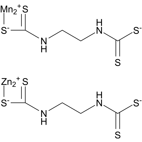
Mancozeb is a widely used fungicide that is effective against fungal diseases in most cereals, vegetables, fruits and ornamental plants. In addition, Mancozeb can cause liver damage in mice by activating the Keap1/Nrf2 signaling pathway. Mancozeb upregulates lactate dehydrogenase and cytochrome c to alter cell metabolism and induce cell death. Mancozeb has reproductive toxicity and can induce apoptosis in ovarian cells.
For research use only. We do not sell to patients.
- Purity : 85.50%
- CAS No.: 8018-01-7
- Formula: C8H12Mn2N4S8Zn22-
- Molecular Weight:661.36
-
Storage:Powder -20°C, 3 years , 4°C, 2 years ; In solvent -80°C, 2 years , -20°C, 1 year

To place orders, for customer services and technical support, please contact: MedChemExpress USA
Tel: 609-228-6898 E-mail: [email protected] [email protected]


-
 Biological Activity
-
Chemical Information
-
Solvent & Solubility
- Protocols
- Purity & Documentation
- References
-
 Help & FAQs


-
HY-13755
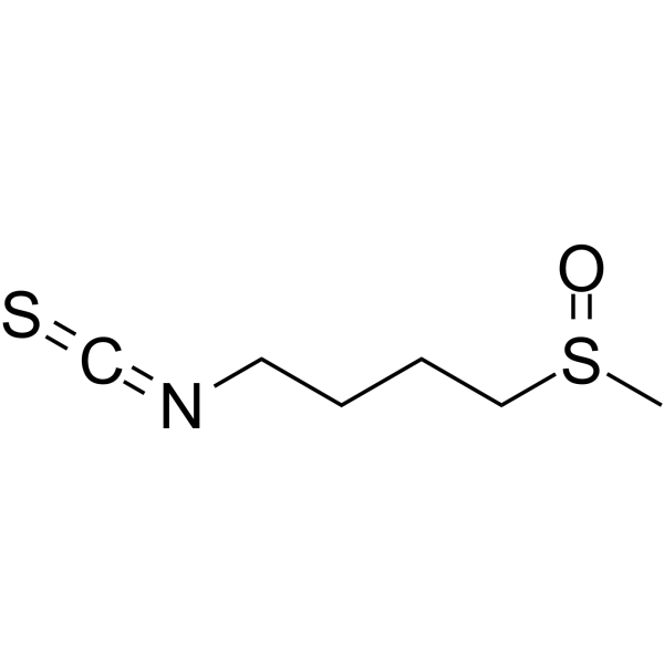
SulforaphaneKeap1/Nrf2/ARE Inducer -
HY-17006
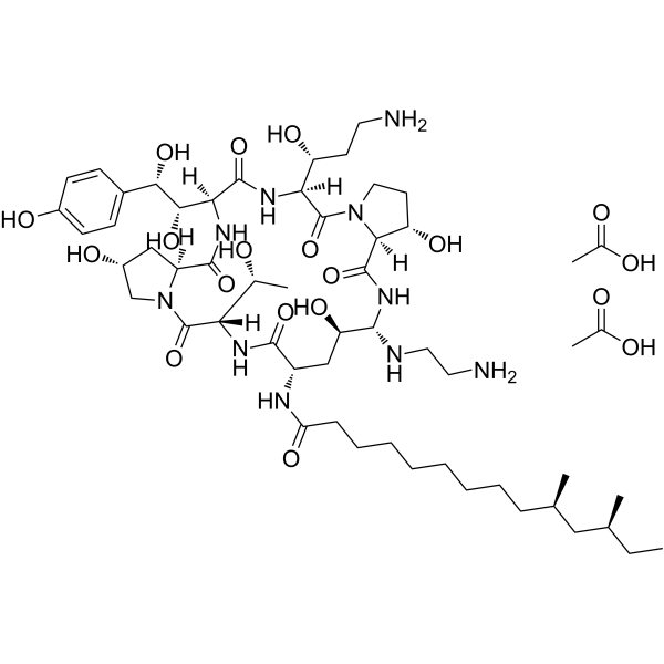
Caspofungin diacetateAntifungal Agent -
HY-B0853
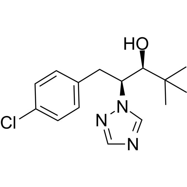
PaclobutrazolFungal Inhibitor -
HY-B0454A
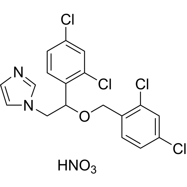
Miconazole nitrateAntifungal Agent -
HY-B0221
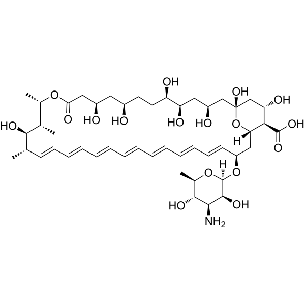
Amphotericin BAntifungal Agent -

HY-B0853R
Paclobutrazol (Standard)Fungal Inhibitor -
HY-14273
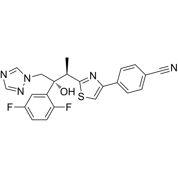
IsavuconazoleTriazole Antifungal -
HY-17006R
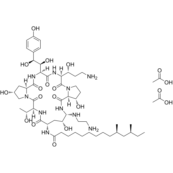
Caspofungin diacetate (Standard)Antifungal Agent -
HY-P1975
Aureobasidin AAntifungal Antibiotic -
HY-B0518A
Naftifine hydrochlorideAntibiotics -

HY-14273R
Isavuconazole (Standard)Triazole Antifungal


Biological Activity
Description
In Vitro
Mancozeb (0.1-500 ppm; 24-48 h) is highly toxic to HepG2 cells, and can affect cell metabolism and induce cell damage[2].
MedChemExpress (MCE) has not independently confirmed the accuracy of these methods. They are for reference only. Further protocols information, click here.
-
Cell Line:HepG2 cells
-
Concentration:0.1, 1, 10, 100 and 500 ppm;
1, 10 and 100 ppm -
Incubation Time:24 h;
48 h -
Result:Was toxic to cells at all concentrations.
Reduced cell viability (to below 50%) at 24 h, resulted in total cell death with a 48-hour exposure to 100ppm.
-
Cell Line:HepG2 cells
-
Concentration:0.1, 1, 10 and 100 ppm
-
Incubation Time:24 h and 48 h
-
Result:Reduced the levels of LDH and cytochrome c.
In Vivo
Mancozeb (100 mg/kg; Gavage; 30 days) can cause ovarian injury and apoptosis in mice[4].
MedChemExpress (MCE) has not independently confirmed the accuracy of these methods. They are for reference only.
-
Animal Model:Female Kunming mice aged 6 weeks old[3]
-
Dosage:0, 50, 100, 150, 200, 250 and 300 mg/kg
-
Administration:Oral administration (p.o.); 30 days
-
Result:Reduced body weight in mice in a concentration-dependent manner.
Significantly reduced the liver organ index of mice at doses of 250 mg/kg and 300 mg/kg.
Reduced the levels of CAT, SOD and ROS in a concentration-dependent manner.
Significantly up-regulated the levels of Keap1 and Nrf2 in the liver.
-
Animal Model:Kunming mice aged 6 weeks old[4]
-
Dosage:100 mg/kg
-
Administration:Oral gavage (i.g.); 30 days
-
Result:Reduced body weight in mice.
Destroyed the ovary structure of mice and decreased the levels of follicle stimulating hormone and luteinizing hormone.
Caused mitochondrial oxidative phosphorylation dysfunction and oxidative stress.
Activated the Bax/Bcl-2 pathway and caspase family, thereby inducing apoptosis.


Chemical Information
-
CAS No. 8018-01-7
-
Appearance Solid
-
Molecular Weight 661.36
-
Formula C8H12Mn2N4S8Zn22-
-
Color Light yellow to yellow
-
SMILES
S=C([S-])NCCNC1=[S][Mn]([Mn+])[S-]1.S=C([S-])NCCNC2=[S][Zn]([Zn+])[S-]2
-
Shipping
Room temperature in continental US; may vary elsewhere.
-
Storage
Powder -20°C 3 years 4°C 2 years In solvent -80°C 2 years -20°C 1 year


Solvent & Solubility
In Vitro:
DMSO : 1 mg/mL (1.51 mM; Need ultrasonic; Hygroscopic DMSO has a significant impact on the solubility of product, please use newly opened DMSO)
H2O : < 0.1 mg/mL (insoluble)
Please refer to the solubility information to select the appropriate solvent. Once prepared, please aliquot and store the solution to prevent product inactivation from repeated freeze-thaw cycles.
Storage method and period of stock solution: -80°C, 2 years; -20°C, 1 year. When stored at -80°C, please use it within 2 years. When stored at -20°C, please use it within 1 year.
Please refer to the solubility information to select the appropriate solvent. Once prepared, please aliquot and store the solution to prevent product inactivation from repeated freeze-thaw cycles.
Storage method and period of stock solution: -80°C, 2 years; -20°C, 1 year. When stored at -80°C, please use it within 2 years. When stored at -20°C, please use it within 1 year.
Concentration (start) × Volume (start) = Concentration (final) × Volume (final)

Protocols
-
Reproductive and Developmental Toxicity Study
Reproductive and developmental toxicity studies detect adverse effects of prenatal or peri/postnatal exposure on maternal condition, pregnancy maintenance, embryo-fetal survival, fetal growth, structural development, and offspring reproductive or developmental endpoints; classic rat protocols generate readouts by comparing treated groups with vehicle, pair-fed, or untreated controls for implantation, resorption, fetal weight, crown-rump length, external morphology, visceral morphology, skeletal ossification, anogenital distance, nipple/areola retention, and postnatal cohort outcomes.
-
Apoptosis
Apoptosis, also called programmed cell death, is generally characterized by distinct morphological characteristics.
-
TUNEL staining for apoptotic DNA fragmentation
TUNEL staining detects DNA strand breaks by using terminal deoxynucleotidyl transferase to add labeled nucleotides to exposed 3′-OH DNA termini, generating either microscopic staining in fixed cells or tissue sections, or fluorescence/cytometric signal in cell suspensions. TUNEL positivity reflects DNA fragmentation but should not be interpreted alone as definitive apoptosis, because TUNEL can also label necrotic, autolytic, mechanically damaged, or DNA-repair-associated DNA breaks.
-
Cell Cytotoxicity Assay
Cytotoxicity assays are usually based on the assessment of cell membrane damage, which can also be indirectly detected by measuring cell viability. Detection methods include MTT assay, CKK-8 assay, LDH assay and ATP assay, etc.
-
Mammalian live/dead viability and cytotoxicity staining
Live/dead viability and cytotoxicity staining assays are based on the simultaneous detection of intracellular esterase activity in metabolically active (viable) cells and membrane integrity loss in non-viable cells. In commonly used dual-staining approaches, membrane-permeant fluorogenic substrates are converted by intracellular esterases into fluorescent products in live cells, while impermeant DNA-binding dyes selectively enter cells with compromised plasma membranes and label nucleic acids in dead or dying cells, enabling discrimination between viable and non-viable populations by fluorescence microscopy or flow cytometry.
-
Annexin V plus membrane-impermeant dye apoptosis staining
Annexin V-based apoptosis assays rely on the detection of phosphatidylserine (PS) externalization from the inner leaflet of the plasma membrane to the outer leaflet, an early biochemical hallmark of apoptosis. Fluorescently labeled Annexin V binds PS in a calcium-dependent manner, enabling identification of early apoptotic cells by flow cytometry or fluorescence microscopy. When combined with a membrane-impermeant DNA-binding dye (e. g. , propidium iodide), this approach allows discrimination between viable (Annexin V−/dye−), early apoptotic (Annexin V+/dye−), and late apoptotic or necrotic (Annexin V+/dye+) cell populations by assessing membrane integrity and PS exposure.
-
Apoptosis Solutions
Apoptosis is a regulated, generally non-lytic cell-death pathway that removes unwanted, damaged, infected, or abnormal cells through coordinated morphological changes, caspase activation, DNA fragmentation, and membrane remodeling. The intrinsic apoptosis pathway is controlled mainly by mitochondrial outer membrane permeabilization, BCL-2 family proteins, cytochrome c release, apoptosome formation, caspase-9 activation, and downstream executioner caspase-3/7 activation. The extrinsic apoptosis pathway is initiated by death receptors such as Fas, TNFR, and TRAIL receptors, which recruit adaptor proteins and activate caspase-8 before engaging executioner caspases or mitochondrial amplification through BID cleavage. Apoptosis is linked to many phenotypes, including cancer cell killing, tissue homeostasis, immune regulation, neurodegeneration, infection response, and treatment-induced cytotoxicity; unresolved questions include how apoptosis interacts with necroptosis, pyroptosis, ferroptos
-
Hepatotoxicity Study
This protocol evaluates hepatotoxicity using complementary in vivo mouse APAP acute liver injury and in vitro hepatocyte-based cytotoxicity readouts. In vivo APAP injury is assessed by serum ALT/AST, liver histology, hepatic glutathione, protein adducts, necrosis, inflammation, and regeneration-related endpoints. In vitro hepatotoxicity is assessed by loss of viability, leakage of ALT/AST/LDH, oxidative-stress markers, mitochondrial function, nuclear morphology, intracellular calcium, and high-content imaging endpoints.


Purity & Documentation
-
Data Sheet (283 KB)
-
SDS (761 KB)
- English - EN (761 KB)
- Français - FR (761 KB)
- Deutsch - DE (761 KB)
- Norwegian - NO (761 KB)
- Español - ES (761 KB)
- Swedish - SV (761 KB)
- Italian - IT (761 KB)
- Korean - KR (761 KB)
- Portuguese - PT (761 KB)
-
Handling Instructions (2659 KB)
References
[1]. Santos R, er al. Thyroid and reproductive hormones in relation to pesticide use in an agricultural population in Southern Brazil. Environ Res. 2019 Jun;173:221-231. [Content Brief]
[2]. WENX Pirozzi AV, et al. Mancozeb, a fungicide routinely used in agriculture, worsens nonalcoholic fatty liver disease in the human HepG2 cell model. Toxicol Lett. 2016 May 13;249:1-4. [Content Brief]
[3]. Gao N, et al. The administration of Glycyrrhiza polysaccharides mitigates liver injury in mice caused by mancozeb via the Keap1-Nrf2/NF-κB pathway. Food Chem Toxicol. 2024 Nov 4:115088. [Content Brief]
[4]. Bao J, et al. Low level of mancozeb exposure affects ovary in mice. Ecotoxicol Environ Saf. 2022 Jul 1;239:113670. [Content Brief]


Complete Stock Solution Preparation Table
Please refer to the solubility information to select the appropriate solvent. Once prepared, please aliquot and store the solution to prevent product inactivation from repeated freeze-thaw cycles.
Storage method and period of stock solution: -80°C, 2 years; -20°C, 1 year. When stored at -80°C, please use it within 2 years. When stored at -20°C, please use it within 1 year.
| Optional Solvent | Concentration Solvent Mass | 1 mg | 5 mg | 10 mg | 25 mg |
|---|---|---|---|---|---|
| DMSO | 1 mM | 1.5120 mL | 7.5602 mL | 15.1204 mL | 37.8009 mL |


Mancozeb Related Classifications
HY-B0854 Related Classifications
 Powered by Bioz