



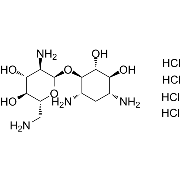
Neamine tetrahydrochloride

Based on 1 Customer Validation
Neamine tetrahydrochloride, a degradation product of Neomycin, is a broad-spectrum aminoglycoside antibiotic. Neamine tetrahydrochloride is an anti-angiogenesis agent targeting angiogenin. Neamine tetrahydrochloride has potent antibacterial, antitumor and neuroprotective activities.
For research use only. We do not sell to patients.
- Purity : 99.92%
- CAS No.: 15446-43-2
- Formula: C12H30Cl4N4O6
- Molecular Weight:468.20
-
Storage:
-20°C, sealed storage, away from moisture
* In solvent : -80°C, 6 months; -20°C, 1 month (sealed storage, away from moisture)

To place orders, for customer services and technical support, please contact: MedChemExpress USA
Tel: 609-228-6898 E-mail: [email protected] [email protected]


-
 Biological Activity
-
Chemical Information
- Protocols
- Purity & Documentation
- References
-
 Help & FAQs


-
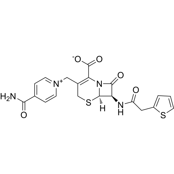
HY-B1252
CefaloniumBactericide -
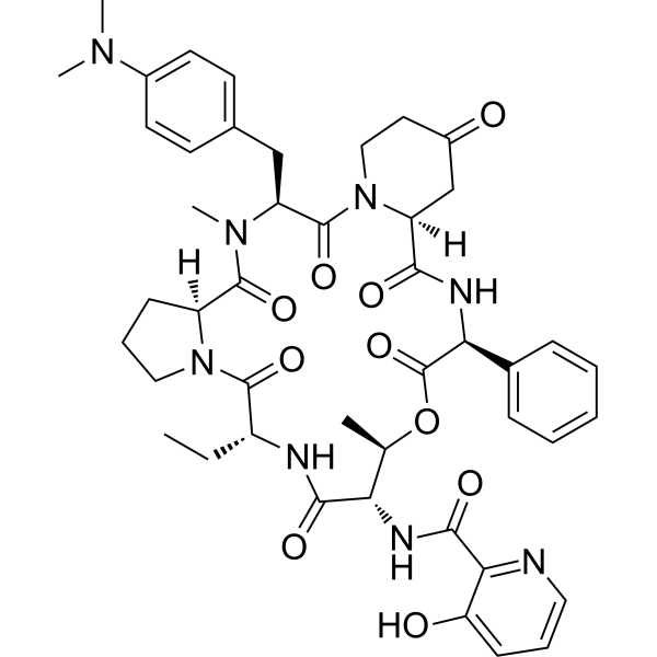
HY-A0279A
Pristinamycin IACycle-Peptidic Macrolactone -
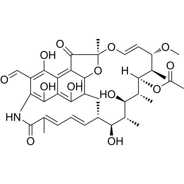
HY-129043
3-Formyl rifamycinRifampicin Product -

HY-125569
Leucomycin A5Antibiotic -
HY-B1802C
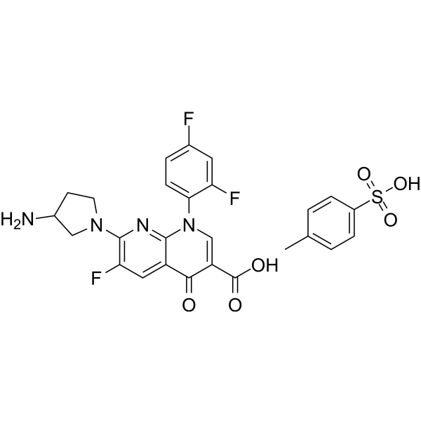
Tosufloxacin tosylateFluoroquinolone Antibiotic -

HY-B1802CR
Tosufloxacin tosylate (Standard)Fluoroquinolone Antibiotic -

HY-B1252R
Cefalonium (Standard)Reference Standard -

HY-A0279AR
Pristinamycin IA (Standard)Cycle-Peptidic Macrolactone -
HY-17466A
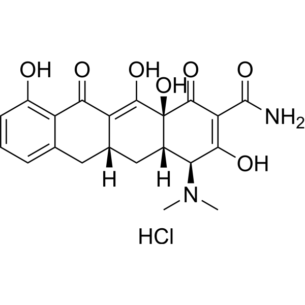
Sancycline hydrochlorideBacterial Inhibitor, Translation Inhibitor -

HY-156074
Erythromycin propionateAntibiotic -
HY-129043R
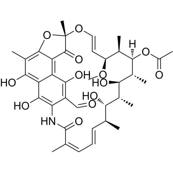
3-Formyl rifamycin (Standard)Standard
All Antibiotic Isoforms
More

Biological Activity
Description
IC50 & Target
|
Aminoglycoside |
In Vitro
Neamine (0.625-25 μM; 48 hours; AsPC-1 cells) treatment inhibits ANG-mediated AsPC-1 cell proliferation in a dose-dependent manner in the range of 0.625-25 μM[1].
Neamine (100 μM) also blocks the nuclear translocation of ANG effectively[1].
MedChemExpress (MCE) has not independently confirmed the accuracy of these methods. They are for reference only. Further protocols information, click here.
-
Cell Line:AsPC-1 cells
-
Concentration:0.625 µM, 1.25 µM, 2.5 µM, 5 µM, 10 µM, 25 µM
-
Incubation Time:48 hours
-
Result:Inhibited angiogenin (ANG)-induced AsPC-1 cells proliferation.
In Vivo
MedChemExpress (MCE) has not independently confirmed the accuracy of these methods. They are for reference only.
-
Animal Model:Male Balb/c nude mice (4 weeks old) injected with AsPC-1 cells[1]
-
Dosage:30 mg/kg, 60 mg/kg
-
Administration:Subcutaneous injection; daily; for 14 days
-
Result:Had anti-tumor effects on AsPC-1 xenograft models.


Chemical Information
-
CAS No. 15446-43-2
-
Appearance Solid
-
Molecular Weight 468.20
-
Formula C12H30Cl4N4O6
-
Color White to off-white
-
SMILES
O[C@H]([C@H]([C@@H](C[C@@H]1N)N)O)[C@@H]1O[C@H]2O[C@@H]([C@H]([C@@H]([C@H]2N)O)O)CN.Cl.Cl.Cl.Cl
-
Shipping
Room temperature in continental US; may vary elsewhere.
-
Storage
-20°C, sealed storage, away from moisture
* In solvent : -80°C, 6 months; -20°C, 1 month (sealed storage, away from moisture)

Protocols
-
Research Protocol for Infectious Diseases
Infectious-disease experiments test how pathogens interact with host barriers, innate immune receptors, inflammatory signaling, pathogen replication, and tissue injury; pattern-recognition receptors such as TLRs, RIG-I-like receptors, NOD-like receptors, and inflammasomes detect microbial molecules and activate NF-κB, interferon, and cytokine responses. The central hypothesis is that infection severity reflects the balance between pathogen burden and host response: protective inflammation restricts pathogen growth, whereas excessive or mislocalized inflammation contributes to tissue damage and disease phenotype. Unresolved questions include which host pathways are protective versus pathogenic, why some infection models fail to translate to human disease, and which combined readouts best predict clinically relevant infection outcomes.
-
Human pluripotent stem cell endothelial-cell differentiation
Human pluripotent stem cell endothelial differentiation is based on stepwise developmental patterning: early activation of WNT/GSK3β inhibition promotes mesodermal or vascular progenitor entry, followed by endothelial specification using VEGF-related signaling, BMP4, FGF2, Notch modulation, or cAMP depending on the published protocol. Endothelial differentiation is read out by acquisition of CD31, CD34, VE-cadherin/CD144, KDR/VEGFR2, vWF, Tie2, NOS3, acetylated LDL uptake, tube/network formation, barrier function, and in vivo vessel-forming capacity where tested.
-
Vascular/Branching Fractal Analysis
Vascular/branching fractal analysis quantifies the geometric complexity of vessel trees or vascular networks from segmented 2D images, commonly by converting vessels into binary and/or skeletonized maps and estimating fractal dimension using box-counting or related approaches. Fractal dimension is interpreted as an image-derived readout of vascular branching complexity, space filling, or density, and has been applied to retinal photographs, fluorescein angiography, OCT angiography, capillary perfusion maps, and in vitro Matrigel angiogenesis networks. The assay readout is generated from vessel-positive pixels after image preprocessing, vessel segmentation, binarization, and optional skeletonization; reported outputs include fractal dimension, vessel density, branchpoint density, endpoint density, vessel length density, tortuosity, and generation-based branching metrics when VESGEN-style analysis is used. The biological interpretation is limited to quantitative vascular patterning and s
-
Endothelial Tube Formation Assay
Endothelial tube formation assay evaluates the ability of endothelial cells to attach, migrate, align, and organize into capillary-like networks when cultured on gelled basement membrane extract or Matrigel; the readout is the morphology and quantity of tube-like networks, which reflects an in vitro endothelial morphogenesis step related to angiogenesis. Basement membrane extract/Matrigel provides laminin-rich extracellular matrix cues that support endothelial differentiation into capillary-like structures, but it can contain biologically active growth factors, so growth-factor-reduced matrix is preferred when testing defined angiogenic stimulators or inhibitors.
-
Endothelial Cell Migration/Angiogenic Sprouting Assay
Endothelial cell migration and angiogenic sprouting assays are in vitro (and partially ex vivo-adapted) functional models that quantify the ability of endothelial cells to undergo coordinated migration, extracellular matrix invasion, and multicellular organization into capillary-like sprouts in response to pro-angiogenic stimuli such as VEGF, bFGF, or conditioned microenvironments. These assays are used to model early angiogenic events including tip-cell formation, directional migration, and lumen-like sprout extension, which collectively reflect angiogenic activation and vascular morphogenesis processes observed in vivo.


Purity & Documentation
-
Data Sheet (274 KB)
-
SDS (252 KB)
- English - EN (252 KB)
- Français - FR (252 KB)
- Deutsch - DE (252 KB)
- Norwegian - NO (252 KB)
- Español - ES (252 KB)
- Swedish - SV (252 KB)
- Italian - IT (252 KB)
- Korean - KR (252 KB)
- Portuguese - PT (252 KB)
-
Handling Instructions (2659 KB)
References
[1]. Ya-Ping Liu, et al. Neamine Inhibits Growth of Pancreatic Cancer Cells in Vitro and in Vivo. J Huazhong Univ Sci Technolog Med Sci. 2016 Feb;36(1):82-87. [Content Brief]
[2]. R Ning, et al. Neamine Induces Neuroprotection After Acute Ischemic Stroke in Type One Diabetic Rats. Neuroscience. 2014 Jan 17;257:76-85. [Content Brief]

Calculators
Concentration (start) × Volume (start) = Concentration (final) × Volume (final)


Neamine tetrahydrochloride Related Classifications
HY-115349 Related Classifications
 Powered by Bioz