



Neamine

Based on 1 Customer Validation
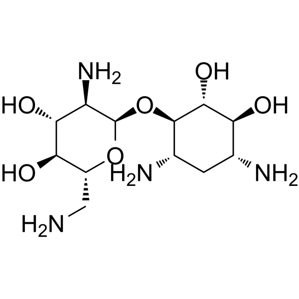
Neamine, a degradation product of Neomycin, is a broad-spectrum aminoglycoside antibiotic. Neamine is an anti-angiogenesis agent targeting angiogenin. Neamine has potent antibacterial, antitumor and neuroprotective activities.
Nur für Forschungszwecke. Wir verkaufen nicht an Patienten.
- Reinheit : 99.96%
- CAS. Nr.: 3947-65-7
- Formel: C12H26N4O6
- Molecular Weight:322.36
-
Speicherung:
-20°C, sealed storage, away from moisture
* In solvent : -80°C, 6 months; -20°C, 1 month (sealed storage, away from moisture)

To place orders, for customer services and technical support, please contact: MedChemExpress USA
Tel: 609-228-6898 E-mail: [email protected] [email protected]


-
 Biologische Aktivität
-
Chemical Information
-
Lösungsmittel & Löslichkeit
- Protokoll
- Reinheit & Dokumentation
- Verweise
-
 Help & FAQs



Alle Antibiotic Isoform-spezifische Produkte anzeigen
More

Biologische Aktivität
Beschreibung
IC50 & Target
|
Aminoglycoside |
In Vitro
Neamine (0.625-25 μM; 48 hours; AsPC-1 cells) treatment inhibits ANG-mediated AsPC-1 cell proliferation in a dose-dependent manner in the range of 0.625-25 μM[1].
Neamine (100 μM) also blocks the nuclear translocation of ANG effectively[1].
MedChemExpress (MCE) has not independently confirmed the accuracy of these methods. They are for reference only.
-
Cell Line:AsPC-1 cells
-
Concentration:0.625 µM, 1.25 µM, 2.5 µM, 5 µM, 10 µM, 25 µM
-
Incubation Time:48 hours
-
Result:Inhibited angiogenin (ANG)-induced AsPC-1 cells proliferation.
In Vivo
MedChemExpress (MCE) has not independently confirmed the accuracy of these methods. They are for reference only.
-
Animal Model:Male Balb/c nude mice (4 weeks old) injected with AsPC-1 cells[1]
-
Dosage:30 mg/kg, 60 mg/kg
-
Administration:Subcutaneous injection; daily; for 14 days
-
Result:Had anti-tumor effects on AsPC-1 xenograft models.


Chemical Information
-
CAS. Nr. 3947-65-7
-
Appearance Solid
-
Molecular Weight 322.36
-
Formel C12H26N4O6
-
Color White to off-white
-
SMILES
O[C@H]([C@H]1O)[C@@H]([C@H](C[C@H]1N)N)O[C@H]([C@@H]([C@H]2O)N)O[C@@H]([C@H]2O)CN
-
Versand
Room temperature in continental US; may vary elsewhere.
-
Speicherung
-20°C, sealed storage, away from moisture
* In solvent : -80°C, 6 months; -20°C, 1 month (sealed storage, away from moisture)


Lösungsmittel & Löslichkeit
In Vitro:
H2O : 100 mg/mL (310.21 mM; Need ultrasonic)
Please refer to the solubility information to select the appropriate solvent. Once prepared, please aliquot and store the solution to prevent product inactivation from repeated freeze-thaw cycles.
Storage method and period of stock solution: -80°C, 6 months; -20°C, 1 month (sealed storage, away from moisture). When stored at -80°C, please use it within 6 months. When stored at -20°C, please use it within 1 month.
* Note: If you choose water as the stock solution, please dilute it to the working solution, then filter and sterilize it with a 0.22 μm filter before use.
Please refer to the solubility information to select the appropriate solvent. Once prepared, please aliquot and store the solution to prevent product inactivation from repeated freeze-thaw cycles.
Storage method and period of stock solution: -80°C, 6 months; -20°C, 1 month (sealed storage, away from moisture). When stored at -80°C, please use it within 6 months. When stored at -20°C, please use it within 1 month.
* Note: If you choose water as the stock solution, please dilute it to the working solution, then filter and sterilize it with a 0.22 μm filter before use.
Konzentration (Stammlösung) × Volumen (Stammlösung) = Konzentration (Ziellösung) × Volumen (Ziellösung)


Reinheit & Dokumentation
-
Data Sheet (277 KB)
-
SDS (392 KB)
- English - EN (392 KB)
- Français - FR (392 KB)
- Deutsch - DE (392 KB)
- Norwegian - NO (392 KB)
- Español - ES (392 KB)
- Swedish - SV (392 KB)
- Italian - IT (392 KB)
- Korean - KR (392 KB)
- Portuguese - PT (392 KB)
-
Handling Instructions (2659 KB)
Verweise
[1]. Ya-Ping Liu, et al. Neamine Inhibits Growth of Pancreatic Cancer Cells in Vitro and in Vivo. J Huazhong Univ Sci Technolog Med Sci. 2016 Feb;36(1):82-87. [Content Brief]
[2]. R Ning, et al. Neamine Induces Neuroprotection After Acute Ischemic Stroke in Type One Diabetic Rats. Neuroscience. 2014 Jan 17;257:76-85. [Content Brief]


Complete Stock Solution Preparation Table
Please refer to the solubility information to select the appropriate solvent. Once prepared, please aliquot and store the solution to prevent product inactivation from repeated freeze-thaw cycles.
Storage method and period of stock solution: -80°C, 6 months; -20°C, 1 month (sealed storage, away from moisture). When stored at -80°C, please use it within 6 months. When stored at -20°C, please use it within 1 month.
| Optional Solvent | Concentration Solvent Mass | 1 mg | 5 mg | 10 mg | 25 mg |
|---|---|---|---|---|---|
| H2O | 1 mM | 3.1021 mL | 15.5106 mL | 31.0212 mL | 77.5530 mL |
| 5 mM | 0.6204 mL | 3.1021 mL | 6.2042 mL | 15.5106 mL | |
| 10 mM | 0.3102 mL | 1.5511 mL | 3.1021 mL | 7.7553 mL | |
| 15 mM | 0.2068 mL | 1.0340 mL | 2.0681 mL | 5.1702 mL | |
| 20 mM | 0.1551 mL | 0.7755 mL | 1.5511 mL | 3.8777 mL | |
| 25 mM | 0.1241 mL | 0.6204 mL | 1.2408 mL | 3.1021 mL | |
| 30 mM | 0.1034 mL | 0.5170 mL | 1.0340 mL | 2.5851 mL | |
| 40 mM | 0.0776 mL | 0.3878 mL | 0.7755 mL | 1.9388 mL | |
| 50 mM | 0.0620 mL | 0.3102 mL | 0.6204 mL | 1.5511 mL | |
| 60 mM | 0.0517 mL | 0.2585 mL | 0.5170 mL | 1.2926 mL | |
| 80 mM | 0.0388 mL | 0.1939 mL | 0.3878 mL | 0.9694 mL | |
| 100 mM | 0.0310 mL | 0.1551 mL | 0.3102 mL | 0.7755 mL |
* Note: If you choose water as the stock solution, please dilute it to the working solution, then filter and sterilize it with a 0.22 μm filter before use.


Neamine Related Classifications
HY-N7449 Related Classifications
 Powered by Bioz