



Neospiramycin I

Based on 1 Customer Validation
Neospiramycin I is a macrolide antibiotic and a derivative of Spiramycin I (HY-N7141). Neospiramycin I is effective against the macrolide-sensitive KB210 strain of S. aureus, but ineffective against the macrolide-resistant KB224 strain, with minimum inhibitory concentrations (MIC) of 3.12 and greater than 100 µg/mL, respectively; it is also effective against B. cereus, B. subtilis, M. luteus, E. coli, and K. pneumoniae, with respective MIC values of 1.56, 3.12, 3.12, 0.2, 50, and 12.5 µg/mL. Neospiramycin I binds to the ribosomes of E. coli, with an inhibitory concentration 50 (IC50) of 1.2 µM. It protects mice from death in a type III S. pneumoniae infection model, with an effective dose 50 (ED50) of 399.8 mg/kg.
For research use only. We do not sell to patients.
- Purity : 96.27%
- CAS No.: 70253-62-2
- Formula: C36H62N2O11
- Molecular Weight:698.88
-
Storage:Powder -20°C, 3 years ; In solvent -80°C, 6 months , -20°C, 1 month


To place orders, for customer services and technical support, please contact: MedChemExpress USA
Tel: 609-228-6898 E-mail: [email protected] [email protected]


-
 Biological Activity
-
Chemical Information
- Protocols
- Purity & Documentation
- References
-
 Help & FAQs


-
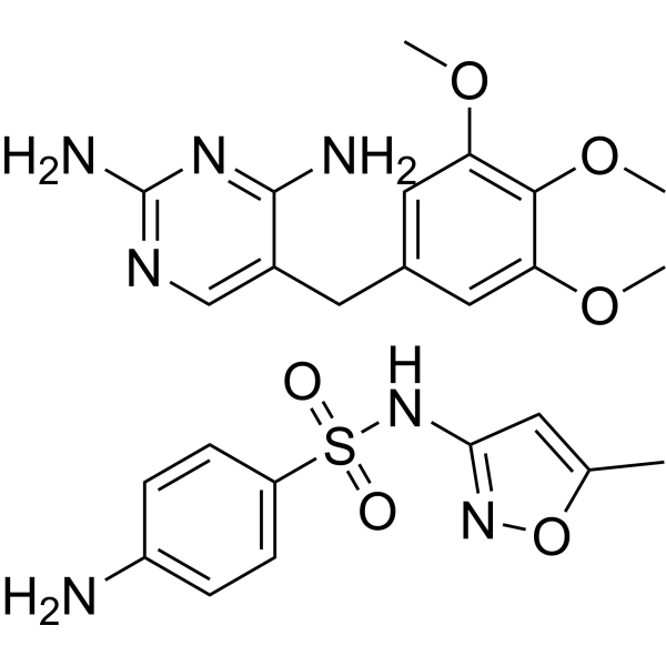
HY-141619A
Cotrimoxazole (Trimethoprim/sulfamethoxazole 1:19)Antibacterial Agent -
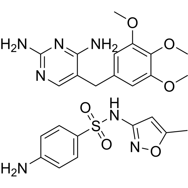
HY-141619
Cotrimoxazole (Trimethoprim/sulfamethoxazole 1:5)Antibacterial Agent -
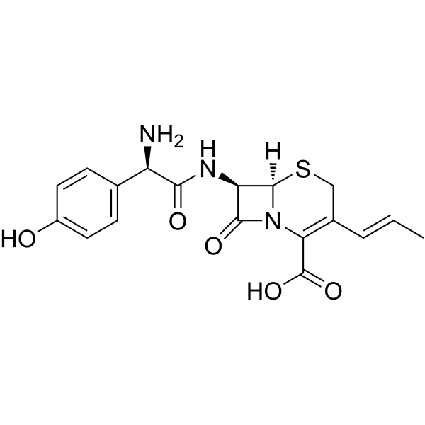
HY-B0458A
CefprozilAntibiotic -
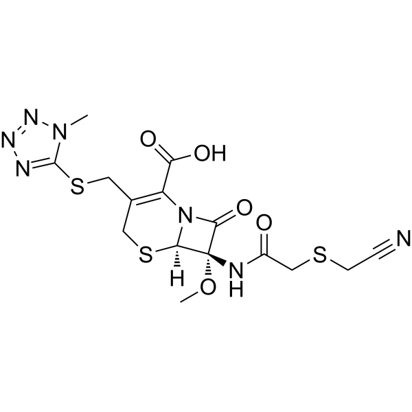
HY-B1595
CefmetazoleCephamycin Antibiotic -
HY-111402
PyridomycinInhA Inhibitor/Antibiotic -

HY-P1708
Enopeptin AAntibiotic
All Antibiotic Isoforms
More

Biological Activity
Description


Chemical Information
-
CAS No. 70253-62-2
-
Appearance Solid
-
Molecular Weight 698.88
-
Formula C36H62N2O11
-
Color Off-white to light yellow
-
SMILES
O=CC[C@@H](C[C@H]([C@H](/C=C/C=C/C[C@H](OC(C[C@H]([C@@H]1OC)O)=O)C)O[C@@H]2O[C@@H]([C@H](CC2)N(C)C)C)C)[C@@H]1O[C@H]3[C@@H]([C@H]([C@@H]([C@H](O3)C)O)N(C)C)O
-
Shipping
Room temperature in continental US; may vary elsewhere.
-
Storage
Powder -20°C 3 years In solvent -80°C 6 months -20°C 1 month

Protocols
-
Research Protocol for Infectious Diseases
Infectious-disease experiments test how pathogens interact with host barriers, innate immune receptors, inflammatory signaling, pathogen replication, and tissue injury; pattern-recognition receptors such as TLRs, RIG-I-like receptors, NOD-like receptors, and inflammasomes detect microbial molecules and activate NF-κB, interferon, and cytokine responses. The central hypothesis is that infection severity reflects the balance between pathogen burden and host response: protective inflammation restricts pathogen growth, whereas excessive or mislocalized inflammation contributes to tissue damage and disease phenotype. Unresolved questions include which host pathways are protective versus pathogenic, why some infection models fail to translate to human disease, and which combined readouts best predict clinically relevant infection outcomes.
-
Cell Cytotoxicity Assay
Cytotoxicity assays are usually based on the assessment of cell membrane damage, which can also be indirectly detected by measuring cell viability. Detection methods include MTT assay, CKK-8 assay, LDH assay and ATP assay, etc.


Purity & Documentation
-
Data Sheet (273 KB)
-
SDS (252 KB)
- English - EN (252 KB)
- Français - FR (252 KB)
- Deutsch - DE (252 KB)
- Norwegian - NO (252 KB)
- Español - ES (252 KB)
- Swedish - SV (252 KB)
- Italian - IT (252 KB)
- Korean - KR (252 KB)
- Portuguese - PT (252 KB)
-
Handling Instructions (2659 KB)
References
[1]. Sano H, et al. Chemical modification of spiramycins. II. Synthesis and antimicrobial activity of 4'-deoxy derivatives of neospiramycin I and their 12-(Z)-isomers. J Antibiot (Tokyo). 1984 Jul;37(7):738-49. [Content Brief]
[2]. Sano H, et al. Chemical modification of spiramycins. I. Synthesis of the acetal derivatives of neospiramycin I. J Antibiot (Tokyo). 1983 Oct;36(10):1336-44. [Content Brief]

Calculators
Concentration (start) × Volume (start) = Concentration (final) × Volume (final)


Neospiramycin I Related Classifications
HY-W587414 Related Classifications
 Powered by Bioz