



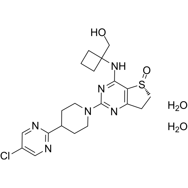
Nerandomilast dihydrate
Based on 2 publication(s) in Google Scholar
Nerandomilast (BI 1015550) dihydrate is an orally active inhibitor of PDE4B with an IC50 value of 7.2 nM. Nerandomilast (dihydrate) has good safety and potential applications in inflammation, allergic diseases, pulmonary fibrosis, and chronic obstructive pulmonary disease (COPD).
For research use only. We do not sell to patients.
- Purity : 99.93%
- Formula: C20H29ClN6O4S
- Molecular Weight:485.00
-
Storage:
4°C, sealed storage, away from moisture
* In solvent : -80°C, 6 months; -20°C, 1 month (sealed storage, away from moisture)

To place orders, for customer services and technical support, please contact: MedChemExpress USA
Tel: 609-228-6898 E-mail: [email protected] [email protected]


-
 Biological Activity
-
Chemical Information
-
Solvent & Solubility
- Protocols
- Purity & Documentation
- References
-
 Help & FAQs


Publications Citing Use of MedChemExpress (MCE) Nerandomilast dihydrate
More

Biological Activity
Description
In Vitro
Nerandomilast (dihydrate) inhibits Lipopolysaccharides (HY-D1056) induced TNF-α release and Phytohemagglutinin P (HY-N7038A) induced IL-2 release in human PBMCs with IC50 values of 35 nM and 9 nM, respectively[2].
Nerandomilast (dihydrate) inhibits TNF-α release in rat whole blood with an IC50 value of 91 nM[2].
MedChemExpress (MCE) has not independently confirmed the accuracy of these methods. They are for reference only.
In Vivo
Nerandomilast (dihydrate) prevents inflammation in rat lung tissue with an ED50 value of 0.1 mg/kg[1].
Nerandomilast (dihydrate) (0.01, 0.1 and 1.0 mg/kg; p.o.; single dose) reduces the Lipopolysaccharides (HY-D1056) induced TNF-α release with dose-dependent manner in mice plasma[2].
Nerandomilast (dihydrate) (0.1, 0.3 and 1.0 mg/kg; p.o.; single dose) inhibits lipopolysaccharides induced neutrophil entry into bronchoalveolar lavage fluid of male Suncus Murinus and Wistar rats[2].
Nerandomilast (dihydrate) (2.5 mg/kg and 12.5 mg/kg; p.o.; twice daily for 6 d) effectively improves the damage of Bleomycin (HY-108345) to mice[2].
MedChemExpress (MCE) has not independently confirmed the accuracy of these methods. They are for reference only.
-
Animal Model:Rats[1].
-
Dosage:0, 0.3, 1.0 and 3.0 mg/kg.
-
Administration:Oral gavage; single dose.
-
Result:Had minimal toxic and side effects on the intestines and stomach of rats, demonstrating biosafety.
-
Animal Model:Male Suncus Murinus and Wistar rats; mice[2].
-
Dosage:0.01, 0.1, 0.3, 1.0, 2.5 or 12.5 mg/kg.
-
Administration:Oral gavage; single dose or twice daily for 6 d.
-
Result:Effectively improved inflammation in lung tissue and reduced the pro-inflammatory factor TNF-α release.
Clinical Trial
| NCT Number | Sponsor | Condition | Start Date |
Phase

|
|---|---|---|---|---|
| NCT01329991 | 2011-05 | PHASE1 |


Chemical Information
-
Appearance Solid
-
Molecular Weight 485.00
-
Formula C20H29ClN6O4S
-
Color White to off-white
-
SMILES
O=[S@]1C2=C(CC1)N=C(N3CCC(CC3)C4=NC=C(C=N4)Cl)N=C2NC5(CO)CCC5.O.O
-
Synonyms
BI 1015550 dihydrate
-
Shipping
Room temperature in continental US; may vary elsewhere.
-
Storage
4°C, sealed storage, away from moisture
* In solvent : -80°C, 6 months; -20°C, 1 month (sealed storage, away from moisture)


Publications (2)
-
Journal Impact Factor
-
Most Recent
-
-
PLoS Pathog
PDE4B deficiency aids macrophage differentiation and contributes to Cryptococcus neoformans brain infection. [Abstract]2026 Mar 10;22(3):e1014040. PMID: 41805786


Solvent & Solubility
In Vitro:
DMSO : 6.25 mg/mL (12.89 mM; ultrasonic and warming and heat to 60°C; Hygroscopic DMSO has a significant impact on the solubility of product, please use newly opened DMSO)
Please refer to the solubility information to select the appropriate solvent. Once prepared, please aliquot and store the solution to prevent product inactivation from repeated freeze-thaw cycles.
Storage method and period of stock solution: -80°C, 6 months; -20°C, 1 month (sealed storage, away from moisture). When stored at -80°C, please use it within 6 months. When stored at -20°C, please use it within 1 month.
Please refer to the solubility information to select the appropriate solvent. Once prepared, please aliquot and store the solution to prevent product inactivation from repeated freeze-thaw cycles.
Storage method and period of stock solution: -80°C, 6 months; -20°C, 1 month (sealed storage, away from moisture). When stored at -80°C, please use it within 6 months. When stored at -20°C, please use it within 1 month.
Concentration (start) × Volume (start) = Concentration (final) × Volume (final)


Purity & Documentation
-
Data Sheet (277 KB)
-
SDS (252 KB)
- English - EN (252 KB)
- Français - FR (252 KB)
- Deutsch - DE (252 KB)
- Norwegian - NO (252 KB)
- Español - ES (252 KB)
- Swedish - SV (252 KB)
- Italian - IT (252 KB)
- Korean - KR (252 KB)
- Portuguese - PT (252 KB)
-
Handling Instructions (2659 KB)
References


Complete Stock Solution Preparation Table
Please refer to the solubility information to select the appropriate solvent. Once prepared, please aliquot and store the solution to prevent product inactivation from repeated freeze-thaw cycles.
Storage method and period of stock solution: -80°C, 6 months; -20°C, 1 month (sealed storage, away from moisture). When stored at -80°C, please use it within 6 months. When stored at -20°C, please use it within 1 month.
| Optional Solvent | Concentration Solvent Mass | 1 mg | 5 mg | 10 mg | 25 mg |
|---|---|---|---|---|---|
| DMSO | 1 mM | 2.0619 mL | 10.3093 mL | 20.6186 mL | 51.5464 mL |
| 5 mM | 0.4124 mL | 2.0619 mL | 4.1237 mL | 10.3093 mL | |
| 10 mM | 0.2062 mL | 1.0309 mL | 2.0619 mL | 5.1546 mL |


Nerandomilast dihydrate Related Classifications
HY-153192A Related Classifications
 Powered by Bioz