



Phytosphingosine
Based on 4 publication(s) in Google Scholar
Phytosphingosine is a phospholipid with anti-inflammatory, antibacterial, and anti-cancer activities, which can induce apoptosis. Phytosphingosine is an immune regulator and can be used in the study of inflammatory skin diseases. Phytosphingosine is also an activator of GPR120 with an IC50 value of 33.4 μM and can be used in the study of type II diabetes.
For research use only. We do not sell to patients.
- Purity : 99.85%
- CAS No.: 554-62-1
- Formula: C18H39NO3
- Molecular Weight:317.51
-
Storage:Powder -20°C, 3 years , 4°C, 2 years ; In solvent -80°C, 6 months , -20°C, 1 month

To place orders, for customer services and technical support, please contact: MedChemExpress USA
Tel: 609-228-6898 E-mail: [email protected] [email protected]


-
 Biological Activity
-
Chemical Information
-
Solvent & Solubility
- Protocols
- Purity & Documentation
- References
-
 Help & FAQs


-
HY-101047
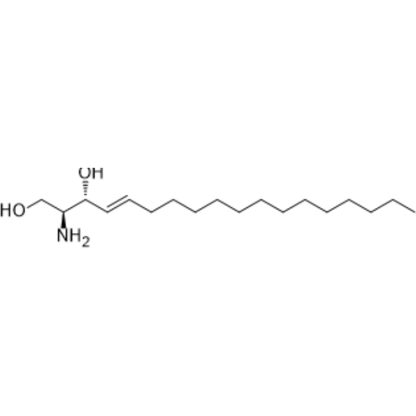
D-erythro-SphingosinePKC Inhibitor -
HY-W019838
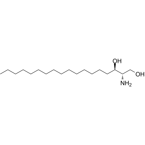
D-Erythro-dihydrosphingosine -
HY-109590
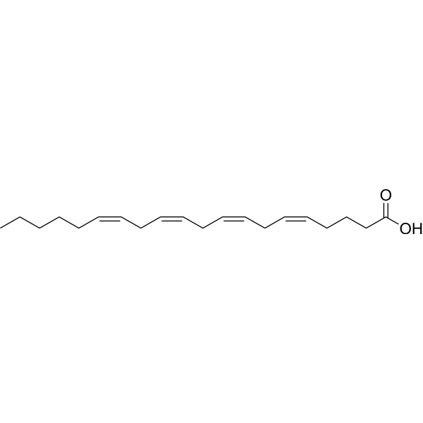
Arachidonic acidPolyunsaturated Fatty Acid -
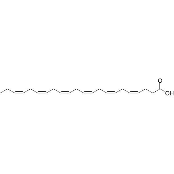
HY-B2167
Docosahexaenoic acidomega-3 Fatty Acid -
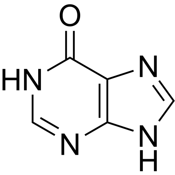
HY-N0091
HypoxanthineEndogenous Metabolite -
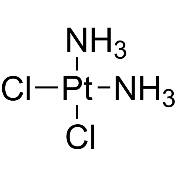
HY-17394
CisplatinDNA Alkylator -
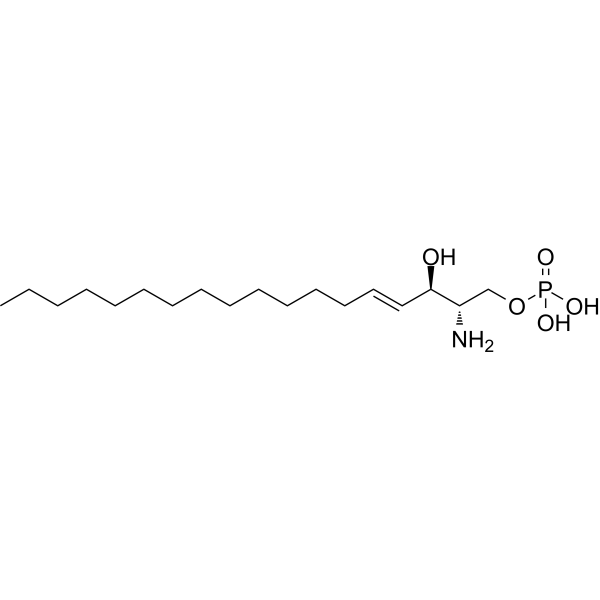
HY-108496
Sphingosine-1-phosphateS1PR Agonist -
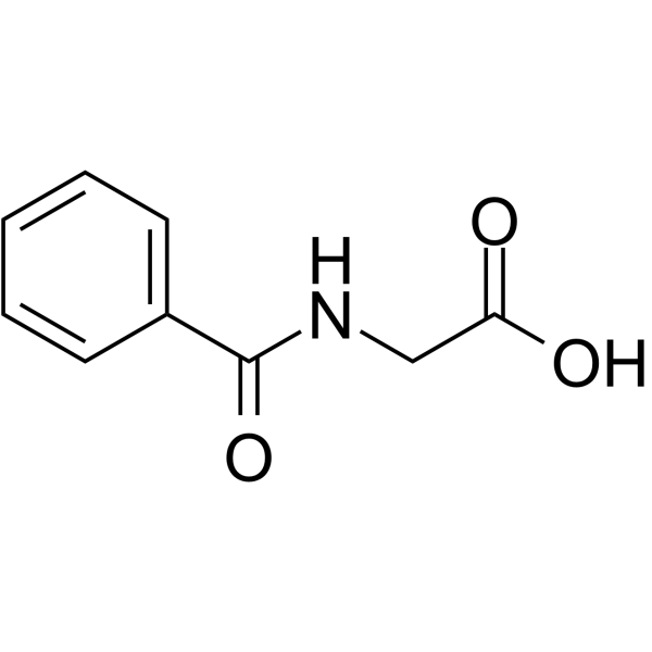
HY-W016562
Hippuric acidMetabolite -
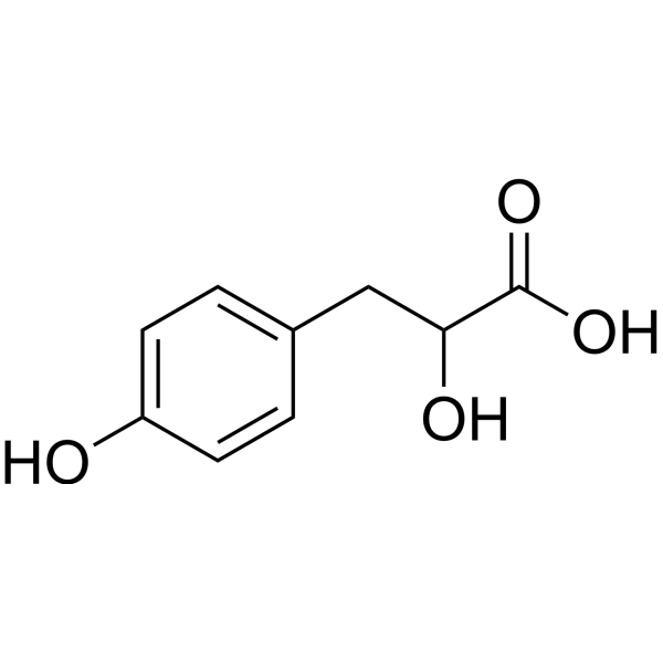
HY-113219
Hydroxyphenyllactic acidEndogenous Metabolite -
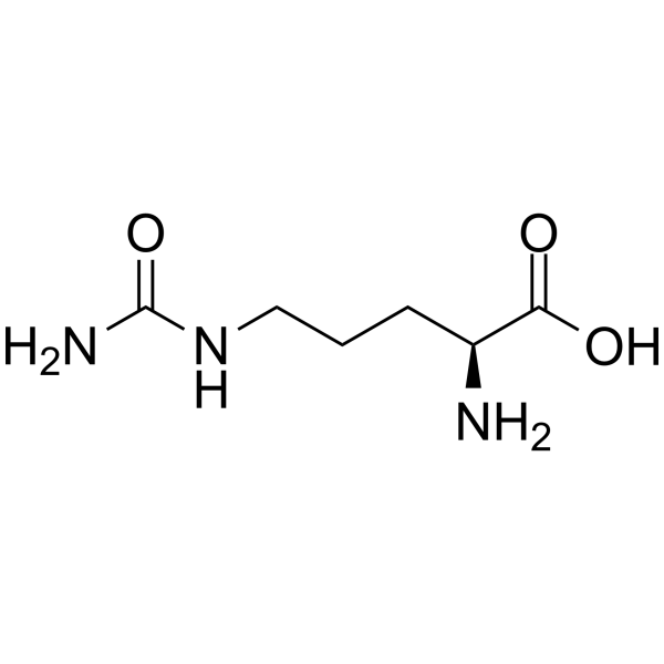
HY-N0391
L-CitrullineEndogenous Metabolite -
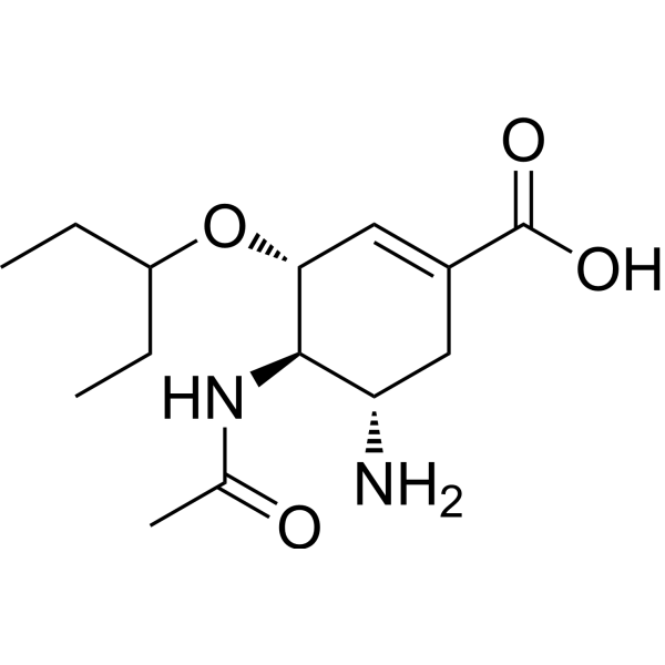
HY-13318
Oseltamivir acidOseltamivir phosphate Metabolite -
HY-100881
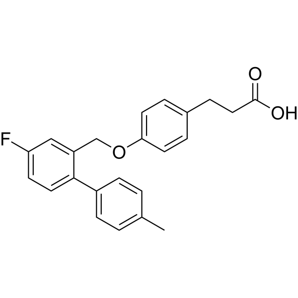
TUG-891FFA4/GPR120 Agonist
Publications Citing Use of MedChemExpress (MCE) Phytosphingosine
More Customer Validation & Images

-
ELISA
-
Histological Imaging/Staining
-
Cell Imaging/Staining
-
WB
-
IF
All Endogenous Metabolite Isoforms
More

Biological Activity
Description
IC50 & Target
|
Human Endogenous Metabolite |
Cellular Effect
|
Cell Line

|
Type | Value | Description | References |
|---|---|---|---|---|
| Jurkat | IC50 |
3.75 μM
Compound: Phytosphingosine
|
Inhibition of wild type human Jurkat cells after 18 hrs by MTT assay
Inhibition of wild type human Jurkat cells after 18 hrs by MTT assay
|
[PMID: 17400555] |
| Jurkat | IC50 |
4.12 μM
Compound: Phytosphingosine
|
Inhibition of human 2-amino-4-(4-heptyloxyphenyl)-2-methylbutanol-resistant human Jurkat SBR3 mutant cells proliferation after 18 hrs by MTT assay
Inhibition of human 2-amino-4-(4-heptyloxyphenyl)-2-methylbutanol-resistant human Jurkat SBR3 mutant cells proliferation after 18 hrs by MTT assay
|
[PMID: 17400555] |
| Jurkat | IC50 |
5.29 μM
Compound: Phytosphingosine
|
Inhibition of 2-amino-4-(4-heptyloxyphenyl)-2-methylbutanol-resistant human Jurkat SBR2 mutant cells proliferation after 18 hrs by MTT assay
Inhibition of 2-amino-4-(4-heptyloxyphenyl)-2-methylbutanol-resistant human Jurkat SBR2 mutant cells proliferation after 18 hrs by MTT assay
|
[PMID: 17400555] |
| Jurkat | IC50 |
6.57 μM
Compound: Phytosphingosine
|
Inhibition of 2-amino-4-(4-heptyloxyphenyl)-2-methylbutanol-resistant human Jurkat SBR1 mutant cells proliferation after 18 hrs by MTT assay
Inhibition of 2-amino-4-(4-heptyloxyphenyl)-2-methylbutanol-resistant human Jurkat SBR1 mutant cells proliferation after 18 hrs by MTT assay
|
[PMID: 17400555] |
In Vitro
Phytosphingosine (200-400 μg/mL; 60-240 min) can inhibit the growth of Gram-positive and Gram-negative bacteria, yeast, and molds, and significantly inhibit the release of IL-1a, exhibiting anti-inflammatory activity[1].
Phytosphingosine (1-5 μg/mL; 24-48 h) inhibits lung adenoma cell proliferation by inducing G2/M phase arrest, apoptosis and mitochondria-dependent pathway cell death in A549 and LLC cells, with IC50 values of 4.3 μg/mL and 4.5μg/mL, respectively[2].
Phytosphingosine (80 μM; 1-10 days) has antibacterial activity and can inhibit the growth ofS. sclerotiorum, S. indica and V. longisporum[3].
MedChemExpress (MCE) has not independently confirmed the accuracy of these methods. They are for reference only. Further protocols information, click here.
-
Cell Line:A549, LLC, BEAS-2B
-
Concentration:1 μg/mL, 2 μg/mL, 3 μg/mL, 4 μg/mL, 5 μg/mL
-
Incubation Time:24 h, 48 h
-
Result:Significantly inhibited cell proliferation in a concentration- and time-dependent manner.
-
Cell Line:A549
-
Concentration:1 μg/mL, 3 μg/mL, 5 μg/mL
-
Incubation Time:24 h
-
Result:Significantly increased the proportion of G2/M phase cells.
-
Cell Line:A549
-
Concentration:1 μg/mL, 3 μg/mL, 5 μg/mL
-
Incubation Time:24 h
-
Result:Up-regulated the pro-apoptotic factor Bax protein level and down-regulated the anti-apoptotic factor Bcl-2 protein level.
Promoted the release of cytochrome c to up-regulate caspase 9 and caspase 3 levels, leading to PARP cleavage.
In Vivo
MedChemExpress (MCE) has not independently confirmed the accuracy of these methods. They are for reference only.


Chemical Information
-
CAS No. 554-62-1
-
Appearance Solid
-
Molecular Weight 317.51
-
Formula C18H39NO3
-
Color White to off-white
-
SMILES
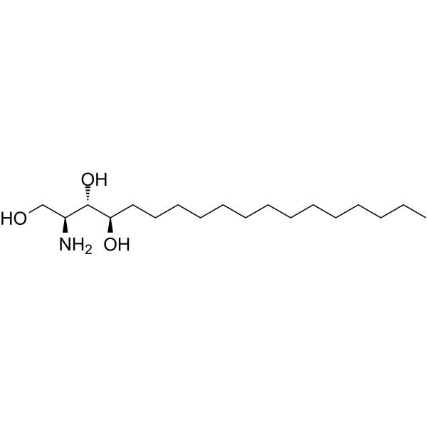
CCCCCCCCCCCCCC[C@@H](O)[C@@H](O)[C@@H](N)CO
-
Synonyms
4-Hydroxysphinganine
-
Structure Classification
-
Initial Source
-
Shipping
Room temperature in continental US; may vary elsewhere.
-
Storage
Powder -20°C 3 years 4°C 2 years In solvent -80°C 6 months -20°C 1 month


Publications (4)
-
Journal Impact Factor
-
Most Recent
-
MedComm (2020)
Phytosphingosine Alleviates Cigarette Smoke-Induced Bronchial Epithelial Cell Senescence in Chronic Obstructive Pulmonary Disease by Targeting the Free Fatty Acid Receptor 4. [Abstract]2025 Aug 29;6(9):e70345. PMID: 40895192
Phytosphingosine purchased from MedChemExpress. Usage Cited in: MedComm (2020). 2025 Aug 29;6(9):e70345. [Abstract]
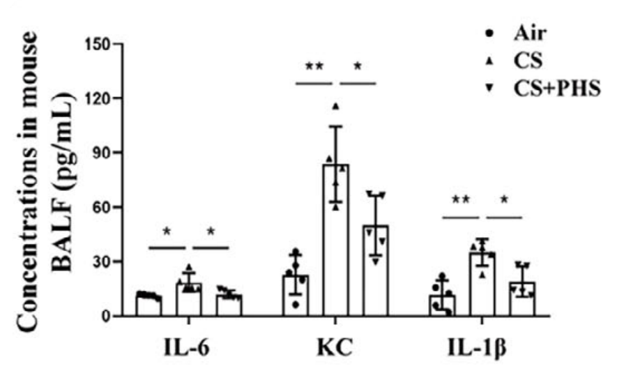
Phytosphingosine (PHS) (25 mg/kg; p.o.; once daily for 3 weeks) significantly decreased the levels of IL-6, KC, and IL-1β in CS-exposed mice.
Phytosphingosine purchased from MedChemExpress. Usage Cited in: MedComm (2020). 2025 Aug 29;6(9):e70345. [Abstract]
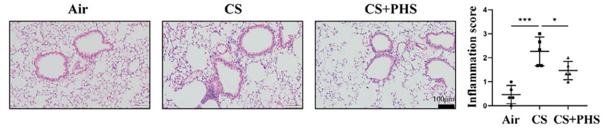
Phytosphingosine (PHS) (25 mg/kg; p.o.; once daily for 3 weeks) reduced infiltration of inflammatory cells around the airways in CS-exposed mice and inflammation scores.
Phytosphingosine purchased from MedChemExpress. Usage Cited in: MedComm (2020). 2025 Aug 29;6(9):e70345. [Abstract]
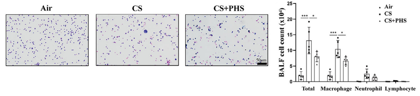
Phytosphingosine (PHS) (25 mg/kg; p.o.; once daily for 3 weeks) significantly decreased total and various inflammatory cell counts, particularly macrophages, in bronchoalveolar lavage fluid (BALF) of CS-exposed mice.
Phytosphingosine purchased from MedChemExpress. Usage Cited in: MedComm (2020). 2025 Aug 29;6(9):e70345. [Abstract]
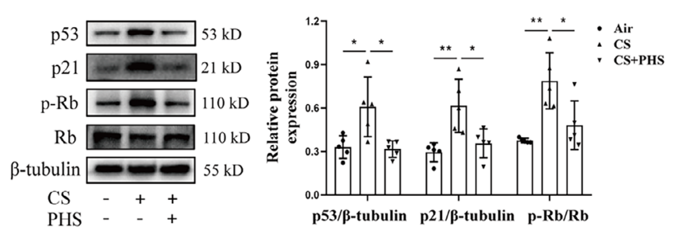
Phytosphingosine (PHS) (25 mg/kg; p.o.; once daily for 3 weeks) significantly downregulated p53, p21, and p-Rb in CS-exposed mice.
Phytosphingosine purchased from MedChemExpress. Usage Cited in: MedComm (2020). 2025 Aug 29;6(9):e70345. [Abstract]
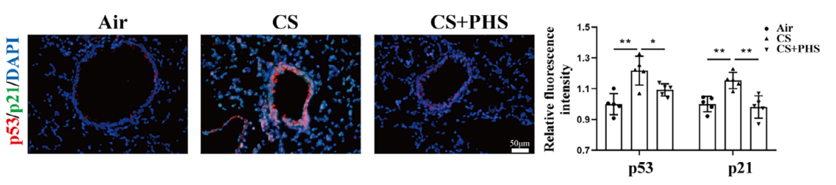
Co-immunofluorescence targeting p53 and p21 revealed that PHS treatment reduced the expression of senescent proteins in the airway epithelium of CS-exposed mice.
-
Chem Biol Interact
Phytosphingosine suppresses gastric cancer through SFRP4/β-catenin axis-mediated Wnt signaling pathway inhibition. [Abstract]2025 Sep 20:111749. PMID: 40983245 -
Molecules
Untargeted Metabolomics Using UHPLC-HRMS Reveals Metabolic Changes of Fresh-Cut Potato during Browning Process. [Abstract]2023 Apr 11;28(8):3375. PMID: 37110608 -


Solvent & Solubility
In Vitro:
DMSO : 5 mg/mL (15.75 mM; ultrasonic and warming and heat to 60°C; Hygroscopic DMSO has a significant impact on the solubility of product, please use newly opened DMSO)
Please refer to the solubility information to select the appropriate solvent. Once prepared, please aliquot and store the solution to prevent product inactivation from repeated freeze-thaw cycles.
Storage method and period of stock solution: -80°C, 6 months; -20°C, 1 month. When stored at -80°C, please use it within 6 months. When stored at -20°C, please use it within 1 month.
Please refer to the solubility information to select the appropriate solvent. Once prepared, please aliquot and store the solution to prevent product inactivation from repeated freeze-thaw cycles.
Storage method and period of stock solution: -80°C, 6 months; -20°C, 1 month. When stored at -80°C, please use it within 6 months. When stored at -20°C, please use it within 1 month.
Concentration (start) × Volume (start) = Concentration (final) × Volume (final)
In Vivo:
Select the appropriate dissolution method based on your experimental animal and administration route.
- For the following dissolution methods, please ensure to first prepare a clear stock solution using an In Vitro approach and then sequentially add co-solvents:
- To ensure reliable experimental results, the clarified stock solution can be appropriately stored based on storage conditions. As for the working solution for In Vivo experiments, it is recommended to prepare freshly and use it on the same day.
- The percentages shown for the solvents indicate their volumetric ratio in the final prepared solution. If precipitation or phase separation occurs during preparation, heat and/or sonication can be used to aid dissolution.
Add each solvent one by one: 10% DMSO 40% PEG300 5% Tween-80 45% Saline
Solubility: ≥ 2.5 mg/mL (7.87 mM); Clear solution
This protocol yields a clear solution of ≥ 2.5 mg/mL (saturation unknown).
Taking 1 mL working solution as an example, add 100 μL DMSO stock solution (25.0 mg/mL) to 400 μL PEG300, and mix evenly; then add 50 μL Tween-80 and mix evenly; then add 450 μL Saline to adjust the volume to 1 mL.
Preparation of Saline: Dissolve 0.9 g sodium chloride in ddH₂O and dilute to 100 mL to obtain a clear Saline solution.
Add each solvent one by one: 10% DMSO 90% (20% SBE-β-CD in Saline)
Solubility: ≥ 2.5 mg/mL (7.87 mM); Clear solution
This protocol yields a clear solution of ≥ 2.5 mg/mL (saturation unknown).
Taking 1 mL working solution as an example, add 100 μL DMSO stock solution (25.0 mg/mL) to 900 μL 20% SBE-β-CD in Saline, and mix evenly.
Preparation of 20% SBE-β-CD in Saline (4°C, storage for one week): 2 g SBE-β-CD powder is dissolved in 10 mL Saline, completely dissolve until clear.
For the following dissolution methods, please prepare the working solution directly:
It is recommended to prepare fresh solutions and use them promptly within a short period of time.
The percentages shown for the solvents indicate their volumetric ratio in the final prepared solution. If precipitation or phase separation occurs during preparation, heat and/or sonication can be used to aid dissolution.
Add each solvent one by one: 50% PEG300 50% Saline
Solubility: 3.33 mg/mL (10.49 mM); Suspended solution; Need ultrasonic
In Vivo Dissolution Calculator
Please enter the basic information of animal experiments:
-
-
-
-
Recommended: Prepare an additional quantity of animals to account for potential losses during experiments.
Please enter your animal formula composition:
-
%DMSO +
Recommended: Keep the proportion of DMSO in working solution below 2% if your animal is weak.
-
%+
-
+%Tween-80 + +
-
%Saline +
The co-solvents required include: DMSO, . All of co-solvents are available by MedChemExpress (MCE). , Tween 80. All of co-solvents are available by MedChemExpress (MCE).
Working solution concentration: 0.22 mg/mL
Method for preparing stock solution: mg drug dissolved in μL DMSO. Stock solution concentration: mg/mL.
1. Take μL DMSO stock solution;
2. Add μL .
μL , mix evenly;
3. Then add μL Tween 80, mix evenly;
4. Then add μL
Please ensure that the stock solution in the first step is dissolved to a clear state, and add co-solvents in sequence. You can use ultrasonic heating (ultrasonic cleaner, recommended frequency 20-40 kHz), vortexing, etc. to assist dissolution.

Protocols
-
Research Protocol for Infectious Diseases
Infectious-disease experiments test how pathogens interact with host barriers, innate immune receptors, inflammatory signaling, pathogen replication, and tissue injury; pattern-recognition receptors such as TLRs, RIG-I-like receptors, NOD-like receptors, and inflammasomes detect microbial molecules and activate NF-κB, interferon, and cytokine responses. The central hypothesis is that infection severity reflects the balance between pathogen burden and host response: protective inflammation restricts pathogen growth, whereas excessive or mislocalized inflammation contributes to tissue damage and disease phenotype. Unresolved questions include which host pathways are protective versus pathogenic, why some infection models fail to translate to human disease, and which combined readouts best predict clinically relevant infection outcomes.
-
Apoptosis
Apoptosis, also called programmed cell death, is generally characterized by distinct morphological characteristics.
-
TUNEL staining for apoptotic DNA fragmentation
TUNEL staining detects DNA strand breaks by using terminal deoxynucleotidyl transferase to add labeled nucleotides to exposed 3′-OH DNA termini, generating either microscopic staining in fixed cells or tissue sections, or fluorescence/cytometric signal in cell suspensions. TUNEL positivity reflects DNA fragmentation but should not be interpreted alone as definitive apoptosis, because TUNEL can also label necrotic, autolytic, mechanically damaged, or DNA-repair-associated DNA breaks.
-
Cell Cytotoxicity Assay
Cytotoxicity assays are usually based on the assessment of cell membrane damage, which can also be indirectly detected by measuring cell viability. Detection methods include MTT assay, CKK-8 assay, LDH assay and ATP assay, etc.
-
Annexin V plus membrane-impermeant dye apoptosis staining
Annexin V-based apoptosis assays rely on the detection of phosphatidylserine (PS) externalization from the inner leaflet of the plasma membrane to the outer leaflet, an early biochemical hallmark of apoptosis. Fluorescently labeled Annexin V binds PS in a calcium-dependent manner, enabling identification of early apoptotic cells by flow cytometry or fluorescence microscopy. When combined with a membrane-impermeant DNA-binding dye (e. g. , propidium iodide), this approach allows discrimination between viable (Annexin V−/dye−), early apoptotic (Annexin V+/dye−), and late apoptotic or necrotic (Annexin V+/dye+) cell populations by assessing membrane integrity and PS exposure.
-
Apoptosis Solutions
Apoptosis is a regulated, generally non-lytic cell-death pathway that removes unwanted, damaged, infected, or abnormal cells through coordinated morphological changes, caspase activation, DNA fragmentation, and membrane remodeling. The intrinsic apoptosis pathway is controlled mainly by mitochondrial outer membrane permeabilization, BCL-2 family proteins, cytochrome c release, apoptosome formation, caspase-9 activation, and downstream executioner caspase-3/7 activation. The extrinsic apoptosis pathway is initiated by death receptors such as Fas, TNFR, and TRAIL receptors, which recruit adaptor proteins and activate caspase-8 before engaging executioner caspases or mitochondrial amplification through BID cleavage. Apoptosis is linked to many phenotypes, including cancer cell killing, tissue homeostasis, immune regulation, neurodegeneration, infection response, and treatment-induced cytotoxicity; unresolved questions include how apoptosis interacts with necroptosis, pyroptosis, ferroptos
-
Research Protocol for Inflammation-related Diseases
The NLRP3 inflammasome is a cytosolic innate immune signaling platform that integrates priming signals and danger-signal activation to promote caspase-1 activation, maturation of IL-1β and IL-18, and gasdermin D-mediated pyroptotic cell death. The core experimental logic is to determine whether inflammatory disease phenotypes are driven by increased NLRP3 expression, ASC-containing inflammasome assembly, caspase-1 cleavage, GSDMD cleavage, and extracellular release of IL-1β/IL-18 rather than by nonspecific cell injury alone. The pathway is strongly linked to inflammation-related disease phenotypes because monosodium urate crystals activate NALP3/NLRP3 inflammasome signaling in gout-like crystal inflammation, cholesterol crystals activate NLRP3 inflammasomes in atherogenesis models, and DSS-induced intestinal inflammation has been reported to involve NLRP3 inflammasome activity. However, experimental colitis studies also show context-dependent protective effects of NLRP3 inflammasome co
-
Research Protocol for Metabolic Diseases
AMP-activated protein kinase, AMPK, is a conserved cellular energy sensor that responds to reduced cellular energy status and coordinates metabolism by increasing ATP-generating catabolic pathways while suppressing ATP-consuming anabolic processes. In metabolic disease research, the AMPK pathway is experimentally relevant because it regulates hepatic lipid synthesis, fatty acid oxidation, glucose production, skeletal-muscle glucose disposal, mTORC1-linked biosynthesis, autophagy, mitochondrial homeostasis, and whole-body energy balance. The central pathway logic is that energy stress, metformin, exercise-like stimulation, or direct AMPK activators increase AMPKα Thr172 phosphorylation and downstream substrate phosphorylation, including ACC and RAPTOR. Phosphorylation of ACC suppresses lipogenesis and supports fatty acid oxidation, whereas phosphorylation of RAPTOR suppresses mTORC1 signaling and links cellular energy status to growth and protein synthesis control. The pathway is linked


Purity & Documentation
-
Data Sheet (274 KB)
-
SDS (620 KB)
- English - EN (620 KB)
- Français - FR (620 KB)
- Deutsch - DE (620 KB)
- Norwegian - NO (620 KB)
- Español - ES (620 KB)
- Swedish - SV (620 KB)
- Italian - IT (620 KB)
- Korean - KR (620 KB)
- Portuguese - PT (620 KB)
-
Handling Instructions (2659 KB)
References
[1]. 1. Pavicic T, Wollenweber U, Farwick M, et al. Anti‐microbial and‐inflammatory activity and efficacy of phytosphingosine: an in vitro and in vivo study addressing acne vulgaris[J]. International journal of cosmetic science, 2007, 29(3): 181-190. [Content Brief]
[2]. Cai Q, et al. Phytosphingosine inhibits the growth of lung adenocarcinoma cells by inducing G2/M-phase arrest, apoptosis, and mitochondria-dependent pathway cell death in vitro and in vivo. Chem Biol Interact. 2024 Jan 5;387:110795. [Content Brief]
[3]. Glenz R, et al. The major plant sphingolipid long chain base phytosphingosine inhibits growth of bacterial and fungal plant pathogens. Sci Rep. 2022 Jan 20;12(1):1081. [Content Brief]
[4]. Nagasawa T, et al. Phytosphingosine is a novel activator of GPR120. J Biochem. 2018 Jul 1;164(1):27-32. [Content Brief]


Complete Stock Solution Preparation Table
Please refer to the solubility information to select the appropriate solvent. Once prepared, please aliquot and store the solution to prevent product inactivation from repeated freeze-thaw cycles.
Storage method and period of stock solution: -80°C, 6 months; -20°C, 1 month. When stored at -80°C, please use it within 6 months. When stored at -20°C, please use it within 1 month.
| Optional Solvent | Concentration Solvent Mass | 1 mg | 5 mg | 10 mg | 25 mg |
|---|---|---|---|---|---|
| DMSO | 1 mM | 3.1495 mL | 15.7475 mL | 31.4951 mL | 78.7377 mL |
| 5 mM | 0.6299 mL | 3.1495 mL | 6.2990 mL | 15.7475 mL | |
| 10 mM | 0.3150 mL | 1.5748 mL | 3.1495 mL | 7.8738 mL | |
| 15 mM | 0.2100 mL | 1.0498 mL | 2.0997 mL | 5.2492 mL |


Phytosphingosine Related Classifications
HY-W011303 Related Classifications
 Powered by Bioz