



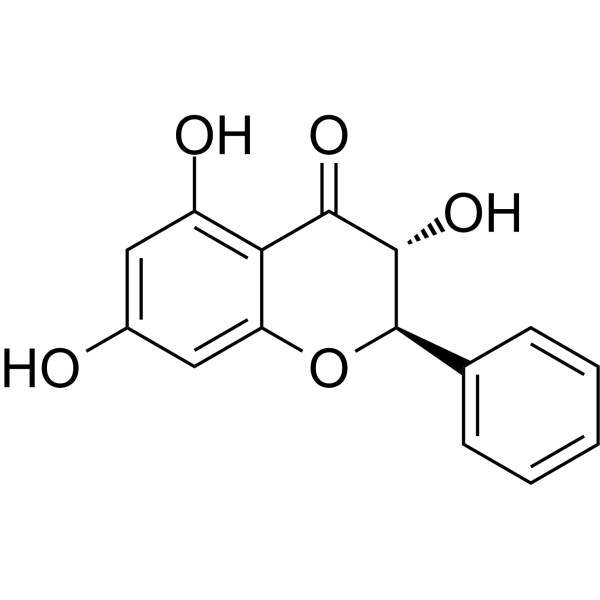
Pinobanksin

Based on 1 Customer Validation
Pinobanksin is an antioxidant and anti-ferroptosis agent found in sunflower honey and propolis, with oral activity. Pinobanksin enhances Nrf2-mediated antioxidant defense, inhibits NF-κB inflammation, and suppresses apoptosis and ferroptosis in in vivo models and normal cells. Pinobanksin exhibits apoptosis-inducing effects on B-cell lymphoma cells. Pinobanksin can be used in research related to perfluorooctane sulfonate-induced renal/cardiac toxicity, acute colitis, and B-cell lymphoma.
Nos produits utilisent uniquement pour la recherche. Nous ne vendons pas aux patients.
- Pureté : 99.30%
- CAS No.: 548-82-3
- Formule: C15H12O5
- Masse moléculaire:272.25
-
Stockage:
4°C, protect from light
* In solvent : -80°C, 6 months; -20°C, 1 month (protect from light)

To place orders, for customer services and technical support, please contact: MedChemExpress USA
Tel: 609-228-6898 E-mail: [email protected] [email protected]


-
 Activité biologique
-
Chemical Information
-
Solvant et solubilité
- Protocole
- Pureté et documentation
- Références
-
 Help & FAQs




Activité biologique
Description
Cellular Effect
|
Cell Line

|
Type | Value | Description | References |
|---|---|---|---|---|
| B16 | IC50 |
>100 μM
Compound: 7
|
Inhibition of melanogenesis in theophylline-stimulated mouse B16-4A5 cells after 72 hrs
Inhibition of melanogenesis in theophylline-stimulated mouse B16-4A5 cells after 72 hrs
|
[PMID: 19615910] |
| B16-4A5 | IC50 |
>100 μM
Compound: 7
|
Inhibition of theophylline-stimulated mouse B16-4A5 cell proliferation assessed as cell viability after 68 hrs by WST8 assay
Inhibition of theophylline-stimulated mouse B16-4A5 cell proliferation assessed as cell viability after 68 hrs by WST8 assay
|
[PMID: 19615910] |
In Vitro
Pinobanksin (0.001-1000 μM; 3-15 days) maintains the total motility, progressive motility, and path velocity of Wenchang boar sperm[2].
Pinobanksin (1 μM; 6-9 days) protects the motility and quality maintenance of Wenchang boar spermatozoa stored at 4°C for 6 and 9 days, and this protective effect is abolished by Dorsomorphin (HY-13418A) inhibition of AMPK, confirming that AMPK is a key downstream mediator of its protective effect on sperm function[2].
Pinobanksin (500 μM; 2 h) does not chemically react with or chelate DSS under cell-free conditions[4].
Pinobanksin (1 μM; 6-9 days) maintains cell membrane integrity, acrosome integrity, DNA integrity, ATP content, mitochondrial membrane potential, and intracellular Ca2+ levels in Wenchang boar sperm[2].
Pinobanksin (1 μM; 6-9 days) enhances the antioxidant defense capacity and reduces oxidative damage in Wenchang boar spermatozoa stored at 4°C for 6 and 9 days by modulating glutathione homeostasis, enhancing antioxidant enzyme activity, and upregulating Nrf2 target proteins[2].
Pinobanksin (1 μM; 6-9 days) inhibits apoptosis of Wenchang boar spermatozoa during cryopreservation by upregulating anti-apoptotic proteins and suppressing pro-apoptotic mediators[2].
Pinobanksin (1 μM) activates the CaMKKβ/AMPK/Nrf2 signaling pathway in Wenchang boar sperm during low-temperature preservation[2].
Pinobanksin (1 μM; 6-9 days) maintains motility, improves quality, and exerts anti-apoptotic effects in Wenchang boar sperm stored at 4°C, and these effects are abolished by STO-609 (HY-19805) inhibition of CaMKKβ, indicating that CaMKKβ-mediated sperm function protection is crucial[2].
Pinobanksin (10-50 μM; 48 h) inhibits ferroptosis in IEC-6 intestinal epithelial cells treated with RSL3 (HY-100218A) or Erastin (HY-15763)[4].
Pinobanksin (50 μM) upregulates GPX4 protein expression, GSH content, cystine uptake, and glutamate release in RSL3-treated IEC-6 cells in an SLC7A11-dependent manner[4].
Pinobanksin (10-50 μM) reduces lipid reactive oxygen species levels in RSL3-induced ferroptotic IEC-6 intestinal epithelial cells[4].
Pinobanksin (50 μM) reduces cellular iron accumulation and inhibits lipid peroxidation in RSL3-treated IEC-6 intestinal epithelial cells through an SLC7A11-dependent mechanism[4].
Pinobanksin (48 h) inhibits the proliferation of mouse B-cell lymphoma M12.C3.F6 cells with an IC50 of 52.1 μM[6].
Pinobanksin (50 μM; 12 h) induces apoptosis in mouse B-cell lymphoma M12.C3.F6 cells[6].
MedChemExpress (MCE) has not independently confirmed the accuracy of these methods. They are for reference only. Further protocols information, click here.
-
Cell Line:IEC-6 rat intestinal epithelial cells
-
Concentration:10, 50 μM (this compound); 0.1, 0.2, 0.5, 1, 10 μM (RSL3 (HY-100218A) or Erastin (HY-15763), co-treated)
-
Incubation Time:48 h
-
Result:Exerted suppressive effects on ferroptotic cell death induced by RSL3 and erastin.
Increased cell viability in a dose-dependent manner under ferroptotic conditions.
-
Cell Line:murine B-cell lymphoma M12.C3.F6 cells
-
Concentration:50 μM
-
Incubation Time:12 h
-
Result:Induced a pro-apoptotic effect in M12.C3.F6 cells, showing a statistically significant increase in the percentage of apoptotic cells compared to the DMSO vehicle control.
In Vivo
Pinobanksin (20 mg/kg; daily; for 30 consecutive days) ameliorates PFOS-induced cardiotoxicity in Sprague Dawley rats by activating the Nrf-2/Keap-1 pathway, restoring antioxidant defense, reducing cardiac injury biomarkers, inhibiting inflammation and apoptosis, and preserving cardiac tissue architecture[3].
Pinobanksin (25-80 mg/kg; p.o.; once daily; for 9 consecutive days) alleviates DSS-induced acute colitis in mice and inhibits colonic epithelial ferroptosis, with the high dose strongly activating SLC7A11 and increasing SLC7A11 mRNA expression[4].
Pinobanksin (80 mg/kg, p.o., once daily for 9 days) alleviates colitis and intestinal epithelial ferroptosis, whereas the SLC7A11 antagonist HG106 (HY-W451275) largely reverses these effects, indicating that its anti-colitis/anti-ferroptosis effects in this model mainly depend on the SLC7A11-glutathione-GPX4 axis[4].
MedChemExpress (MCE) has not independently confirmed the accuracy of these methods. They are for reference only.
-
Animal Model:Male albino rats (190 g; perfluorooctane sulfonate-induced renal injury)[1]
-
Dosage:20 mg/kg
-
Administration:oral gavage; daily; 30 days
-
Result:Reduced Keap-1 mRNA expression and increased Nrf-2, CAT, SOD, GPx, GSR, GST, and HO-1 mRNA expression relative to the PFOS-only group.
Increased CAT activity to 10.37 μmol/mg protein, SOD activity to 10.38 μmol/mg protein, GPx activity to 23.78 μmol/mg protein, GSH content to 15.50 μmol/mg tissue, GSR activity to 7.81 nmol NADPH oxidized/min/mg tissue, GST activity to 31.35 μmol/mg protein, and HO-1 activity to 242.57 μmol/mg protein.
Decreased ROS levels to 2.04 μmol/mg tissue and MDA levels to 1.15 nmol/mg protein.
Reduced urea to 24.40 mg/dl, creatinine to 1.59 mg/dl, KIM-1 to 0.93 mg/mL, and NGAL to 1.31 ng/day, and increased creatinine clearance to 1.71 mL/min.
Reduced NF-κB to 35.89 ng/g tissue, TNF-α to 14.72 ng/g tissue, IL-1β to 23.45 ng/g tissue, IL-6 to 14.78 ng/g tissue, and COX-2 to 31.53 ng/g tissue.
Decreased Bax and Caspase-3 mRNA expression and increased Bcl-2 mRNA expression.
Restored normal renal architecture, reversing PFOS-induced glomerular distortion, tubular damage, inflammation, and hemorrhage.
Showed all measured parameters (antioxidant enzymes, renal function markers, inflammatory cytokines, apoptotic markers, and renal histology) comparable to the control group when administered alone.
-
Animal Model:Sprague Dawley rats (albino, Rattus norvegicus, weight 180-200 g)[3]
-
Dosage:20 mg/kg (monotherapy and co-treatment with PFOS)
-
Administration:daily; 30 days
-
Result:Increased Nrf-2 transcriptional expression and decreased Keap-1 transcriptional expression relative to the PFOS-only group.
Yielded cardiac CAT activity of 10.44 U/mg protein, SOD activity of 7.67 U/mg protein, GSR activity of 5.73 nM NADPH oxidized/min/mg tissue, GPx activity of 16.25 U/mg protein, GSH content of 21.75 μM/g tissue, GST activity of 26.68 nM/min/mg protein, and HO-1 activity of 267.92 pmoles bilirubin/mg protein/h in co-treatment with PFOS.
Yielded cardiac MDA level of 1.36 nmol/g and ROS level of 2.24 nmol/g in co-treatment with PFOS.
Yielded plasma LDH of 312.63 mg/dl, CPK of 35.35 mcg/L, CK-MB of 5.58 ng/mL, and troponin I of 33.03 pg/mL in co-treatment with PFOS.
Yielded cardiac NF-κB of 32.46 ng/g tissue, TNF-α of 23.08 ng/g tissue, IL-1β of 45.56 ng/g tissue, IL-6 of 13.84 ng/g tissue, and COX-2 of 20.62 ng/g tissue in co-treatment with PFOS.
Upregulated Bcl-2 expression and downregulated Bax and caspase-3 expression relative to the PFOS-only group.
Showed minimal cardiomyocyte size abnormality, minimal inflammation, mild intercalated disc disruption, minimal myocardial edema, and mild vascular congestion in co-treatment group, compared to severe damage in the PFOS-only group.
Showed normal cardiac histology (score 0 for all parameters) as monotherapy, identical to control.
Did not significantly alter Nrf-2/Keap-1 transcript levels, redox parameters, cardiac injury markers, or apoptotic marker expression as monotherapy compared to control.
-
Animal Model:C57BL/6 mice (6-8 weeks old; 18-22 g; gender not specified)[4]
-
Dosage:25 mg/kg; 80 mg/kg
-
Administration:i.g.; daily; 9 days
-
Result:Increased body weight and food intake in DSS-treated mice.
Alleviated diarrhea and rectal bleeding in DSS-treated mice.
Decreased disease activity index (DAI) scores in DSS-treated mice.
Restored intestinal villus architecture in DSS-treated mice.
Reduced inflammatory cell infiltration in DSS-treated mice.
Increased colon length in DSS-treated mice.
Decreased colonic myeloperoxidase (MPO) activity in DSS-treated mice.
Decreased proinflammatory cytokine production in DSS-treated mice.
Restored epithelial barrier function, as evidenced by reduced serum fluorescein isothiocyanate-dextran (FD-4) levels.
Inhibited epithelial ferroptosis in colitic mice, characterized by restoration of mitochondrial morphology, decreased 4-hydroxynonenal (4-HNE) expression, and reduced malondialdehyde (MDA) and iron contents in colonic tissue.
Significantly increased colonic glutathione peroxidase 4 (GPX4) protein expression in colitic mice.
Exerted no significant effects on ferroptosis suppressor protein 1 (FSP1) or transferrin receptor (TFR) expression in colitic mice.
Induced a 3.4-fold increase in solute carrier family 7 member 11 (SLC7A11) mRNA expression in colonic tissues.
Significantly increased SLC7A11 protein levels in colonic tissues.
Reversed the DSS-induced decrease in colonic glutathione (GSH) and cystine contents and the GSH/GSSG ratio.
-
Animal Model:C57BL/6 mice (6-8 weeks old; 18-22 g; gender not specified; SLC7A11 inhibition via HG106 pretreatment)[4]
-
Dosage:80 mg/kg
-
Administration:i.g.; daily; 9 days (starting on day 4 after 3 days of HG106 pretreatment)
-
Result:Exhibited largely reversed protective effects in mice pretreated with the SLC7A11 antagonist HG106 compared with pinobanksin-treated colitic mice without HG106.
Showed significantly increased DAI scores in the HG106 + DSS + pinobanksin group compared with pinobanksin-treated colitic mice without HG106.
Showed significantly increased colonic MPO contents in the HG106 + DSS + pinobanksin group compared with pinobanksin-treated colitic mice without HG106.
Showed significantly decreased colonic GPX4 expression in the HG106 + DSS + pinobanksin group compared with pinobanksin-treated colitic mice without HG106.
Showed significantly increased MDA contents in the HG106 + DSS + pinobanksin group compared with pinobanksin-treated colitic mice without HG106.
Resulted in reverted mitochondrial morphology in colonic epithelial cells to a ferroptotic state in the HG106 + DSS + pinobanksin group.
Resulted in damaged intestinal villi with increased inflammatory cell infiltration in the HG106 + DSS + pinobanksin group.
Resulted in reduced colon length in the HG106 + DSS + pinobanksin group.
Showed no significant difference in DAI scores between the DSS control group and the HG106 + DSS + pinobanksin group.
Showed no significant difference in MPO contents between the DSS control group and the HG106 + DSS + pinobanksin group.
Showed no significant difference in GPX4 expression between the DSS control group and the HG106 + DSS + pinobanksin group.
Showed no significant difference in MDA contents between the DSS control group and the HG106 + DSS + pinobanksin group.


Chemical Information
-
CAS No. 548-82-3
-
Appearance Solid
-
Masse moléculaire 272.25
-
Formule C15H12O5
-
Color White to off-white
-
SMILES
O=C1[C@H](O)[C@@H](C2=CC=CC=C2)OC3=CC(O)=CC(O)=C13
-
Synonyms
3,5,7-Trihydroxyflavanone
-
Structure Classification
-
Initial Source
-
Livraison
Room temperature in continental US; may vary elsewhere.
-
Stockage
4°C, protect from light
* In solvent : -80°C, 6 months; -20°C, 1 month (protect from light)


Solvant et solubilité
In Vitro:
DMSO : 100 mg/mL (367.31 mM; Need ultrasonic; Hygroscopic DMSO has a significant impact on the solubility of product, please use newly opened DMSO)
Please refer to the solubility information to select the appropriate solvent. Once prepared, please aliquot and store the solution to prevent product inactivation from repeated freeze-thaw cycles.
Storage method and period of stock solution: -80°C, 6 months; -20°C, 1 month (protect from light). When stored at -80°C, please use it within 6 months. When stored at -20°C, please use it within 1 month.
Please refer to the solubility information to select the appropriate solvent. Once prepared, please aliquot and store the solution to prevent product inactivation from repeated freeze-thaw cycles.
Storage method and period of stock solution: -80°C, 6 months; -20°C, 1 month (protect from light). When stored at -80°C, please use it within 6 months. When stored at -20°C, please use it within 1 month.
Concentration (start) × Volume (start) = Concentration (final) × Volume (final)
In Vivo:
Select the appropriate dissolution method based on your experimental animal and administration route.
- For the following dissolution methods, please ensure to first prepare a clear stock solution using an In Vitro approach and then sequentially add co-solvents:
- To ensure reliable experimental results, the clarified stock solution can be appropriately stored based on storage conditions. As for the working solution for In Vivo experiments, it is recommended to prepare freshly and use it on the same day.
- The percentages shown for the solvents indicate their volumetric ratio in the final prepared solution. If precipitation or phase separation occurs during preparation, heat and/or sonication can be used to aid dissolution.
Add each solvent one by one: 10% DMSO 40% PEG300 5% Tween-80 45% Saline
Solubility: ≥ 2.5 mg/mL (9.18 mM); Clear solution
This protocol yields a clear solution of ≥ 2.5 mg/mL (saturation unknown).
Taking 1 mL working solution as an example, add 100 μL DMSO stock solution (25.0 mg/mL) to 400 μL PEG300, and mix evenly; then add 50 μL Tween-80 and mix evenly; then add 450 μL Saline to adjust the volume to 1 mL.
Preparation of Saline: Dissolve 0.9 g sodium chloride in ddH₂O and dilute to 100 mL to obtain a clear Saline solution.
Add each solvent one by one: 10% DMSO 90% (20% SBE-β-CD in Saline)
Solubility: ≥ 2.5 mg/mL (9.18 mM); Clear solution
This protocol yields a clear solution of ≥ 2.5 mg/mL (saturation unknown).
Taking 1 mL working solution as an example, add 100 μL DMSO stock solution (25.0 mg/mL) to 900 μL 20% SBE-β-CD in Saline, and mix evenly.
Preparation of 20% SBE-β-CD in Saline (4°C, storage for one week): 2 g SBE-β-CD powder is dissolved in 10 mL Saline, completely dissolve until clear.
In Vivo Dissolution Calculator
Please enter the basic information of animal experiments:
-
-
-
-
Recommended: Prepare an additional quantity of animals to account for potential losses during experiments.
Please enter your animal formula composition:
-
%DMSO +
Recommended: Keep the proportion of DMSO in working solution below 2% if your animal is weak.
-
%+
-
+%Tween-80 + +
-
%Saline +
The co-solvents required include: DMSO, . All of co-solvents are available by MedChemExpress (MCE). , Tween 80. All of co-solvents are available by MedChemExpress (MCE).
Working solution concentration: 0.22 mg/mL
Method for preparing stock solution: mg drug dissolved in μL DMSO. Stock solution concentration: mg/mL. * In solvent : -80°C, 6 months; -20°C, 1 month (protect from light)
1. Take μL DMSO stock solution;
2. Add μL .
μL , mix evenly;
3. Then add μL Tween 80, mix evenly;
4. Then add μL
Please ensure that the stock solution in the first step is dissolved to a clear state, and add co-solvents in sequence. You can use ultrasonic heating (ultrasonic cleaner, recommended frequency 20-40 kHz), vortexing, etc. to assist dissolution.

Protocole
-
Detection of Apoptosis
Apoptosis, also called programmed cell death, is generally characterized by distinct morphological characteristics.
-
TUNEL staining for apoptotic DNA fragmentation
TUNEL staining detects DNA strand breaks by using terminal deoxynucleotidyl transferase to add labeled nucleotides to exposed 3′-OH DNA termini, generating either microscopic staining in fixed cells or tissue sections, or fluorescence/cytometric signal in cell suspensions. TUNEL positivity reflects DNA fragmentation but should not be interpreted alone as definitive apoptosis, because TUNEL can also label necrotic, autolytic, mechanically damaged, or DNA-repair-associated DNA breaks.
-
Annexin V plus membrane-impermeant dye apoptosis staining
Annexin V-based apoptosis assays rely on the detection of phosphatidylserine (PS) externalization from the inner leaflet of the plasma membrane to the outer leaflet, an early biochemical hallmark of apoptosis. Fluorescently labeled Annexin V binds PS in a calcium-dependent manner, enabling identification of early apoptotic cells by flow cytometry or fluorescence microscopy. When combined with a membrane-impermeant DNA-binding dye (e. g. , propidium iodide), this approach allows discrimination between viable (Annexin V−/dye−), early apoptotic (Annexin V+/dye−), and late apoptotic or necrotic (Annexin V+/dye+) cell populations by assessing membrane integrity and PS exposure.
-
DSS-Induced Colitis
Dextran sulfate sodium (DSS)-induced colitis is generated by administering DSS in mouse drinking water, producing epithelial injury, barrier disruption, weight loss, diarrhea, fecal blood, colon shortening, histologic mucosal damage, and inflammatory mediator changes; the model is mainly used to study acute or chronic intestinal inflammation resembling selected features of ulcerative colitis. DSS injury is interpreted through clinical and tissue readouts rather than a single molecular endpoint: daily body weight, stool consistency, and bleeding are combined into a disease activity index, while colon length, histology, cytokines, myeloperoxidase activity, intestinal permeability, and tight-junction markers provide complementary measures of inflammation and barrier damage.
-
TNBS-Induced Colitis
TNBS-induced colitis is produced by intrarectal delivery of 2,4,6-trinitrobenzene sulfonic acid in ethanol, where ethanol disrupts the mucosal barrier and TNBS haptenates colonic proteins, generating immune-mediated colonic inflammation with weight loss, diarrhea, ulceration, transmural injury, inflammatory-cell infiltration, and cytokine responses. The model is used as an experimental intestinal inflammation model with Crohn’s disease–like features, especially when Th1-type responses, IL-12–dependent inflammation, chronic relapsing inflammation, or fibrosis-related endpoints are studied.
-
Apoptosis Solutions
Apoptosis is a regulated, generally non-lytic cell-death pathway that removes unwanted, damaged, infected, or abnormal cells through coordinated morphological changes, caspase activation, DNA fragmentation, and membrane remodeling. The intrinsic apoptosis pathway is controlled mainly by mitochondrial outer membrane permeabilization, BCL-2 family proteins, cytochrome c release, apoptosome formation, caspase-9 activation, and downstream executioner caspase-3/7 activation. The extrinsic apoptosis pathway is initiated by death receptors such as Fas, TNFR, and TRAIL receptors, which recruit adaptor proteins and activate caspase-8 before engaging executioner caspases or mitochondrial amplification through BID cleavage. Apoptosis is linked to many phenotypes, including cancer cell killing, tissue homeostasis, immune regulation, neurodegeneration, infection response, and treatment-induced cytotoxicity; unresolved questions include how apoptosis interacts with necroptosis, pyroptosis, ferroptos
-
Ferroptosis Solutions
Ferroptosis is an iron-dependent, non-apoptotic form of regulated cell death characterized by lethal lipid peroxidation and sensitivity to suppression by iron chelators or lipophilic radical-trapping antioxidants. The core pathway links cystine uptake through system Xc−, glutathione availability, GPX4-dependent detoxification of phospholipid hydroperoxides, iron-dependent oxidative reactions, and polyunsaturated-phospholipid metabolism into a cell-death program that is biochemically and morphologically distinct from apoptosis, necrosis, and autophagy. The ferroptosis pathway is experimentally linked to phenotype through chemical and genetic perturbation. Erastin induces ferroptosis by inhibiting cystine uptake through system Xc− and weakening antioxidant defenses, while GPX4 inhibition or depletion causes lipid peroxide accumulation and ferroptotic cancer-cell death. ACSL4 and oxidizable arachidonoyl- or adrenoyl-containing phosphatidylethanolamines shape ferroptosis sensitivity by con
-
Research Protocol for Inflammation-related Diseases
The NLRP3 inflammasome is a cytosolic innate immune signaling platform that integrates priming signals and danger-signal activation to promote caspase-1 activation, maturation of IL-1β and IL-18, and gasdermin D-mediated pyroptotic cell death. The core experimental logic is to determine whether inflammatory disease phenotypes are driven by increased NLRP3 expression, ASC-containing inflammasome assembly, caspase-1 cleavage, GSDMD cleavage, and extracellular release of IL-1β/IL-18 rather than by nonspecific cell injury alone. The pathway is strongly linked to inflammation-related disease phenotypes because monosodium urate crystals activate NALP3/NLRP3 inflammasome signaling in gout-like crystal inflammation, cholesterol crystals activate NLRP3 inflammasomes in atherogenesis models, and DSS-induced intestinal inflammation has been reported to involve NLRP3 inflammasome activity. However, experimental colitis studies also show context-dependent protective effects of NLRP3 inflammasome co


Pureté et documentation
-
Fiche technique (303 KB)
-
SDS (252 KB)
- English - EN (252 KB)
- Français - FR (252 KB)
- Deutsch - DE (252 KB)
- Norwegian - NO (252 KB)
- Español - ES (252 KB)
- Swedish - SV (252 KB)
- Italian - IT (252 KB)
- Korean - KR (252 KB)
- Portuguese - PT (252 KB)
-
Instruction de manipulation (2659 KB)
Références
[2]. Yang D, et al. Pinobanksin alleviates 4℃ semen preservation-induced oxidative damage and apoptosis in Wenchang Pig spermatozoa via the CaMKKβ/AMPK/Nrf2 pathway. Veterinary journal (London, England : 1997). 2026 Apr;316:106599. [Content Brief]
[6]. Alday E, et al. Apoptotic induction by pinobanksin and some of its ester derivatives from Sonoran propolis in a B-cell lymphoma cell line. Chemico-biological interactions. 2015 Dec 05;242:35-44. [Content Brief]


Complete Stock Solution Preparation Table
Please refer to the solubility information to select the appropriate solvent. Once prepared, please aliquot and store the solution to prevent product inactivation from repeated freeze-thaw cycles.
Storage method and period of stock solution: -80°C, 6 months; -20°C, 1 month (protect from light). When stored at -80°C, please use it within 6 months. When stored at -20°C, please use it within 1 month.
| Optional Solvent | Concentration Solvent Mass | 1 mg | 5 mg | 10 mg | 25 mg |
|---|---|---|---|---|---|
| DMSO | 1 mM | 3.6731 mL | 18.3655 mL | 36.7309 mL | 91.8274 mL |
| 5 mM | 0.7346 mL | 3.6731 mL | 7.3462 mL | 18.3655 mL | |
| 10 mM | 0.3673 mL | 1.8365 mL | 3.6731 mL | 9.1827 mL | |
| 15 mM | 0.2449 mL | 1.2244 mL | 2.4487 mL | 6.1218 mL | |
| 20 mM | 0.1837 mL | 0.9183 mL | 1.8365 mL | 4.5914 mL | |
| 25 mM | 0.1469 mL | 0.7346 mL | 1.4692 mL | 3.6731 mL | |
| 30 mM | 0.1224 mL | 0.6122 mL | 1.2244 mL | 3.0609 mL | |
| 40 mM | 0.0918 mL | 0.4591 mL | 0.9183 mL | 2.2957 mL | |
| 50 mM | 0.0735 mL | 0.3673 mL | 0.7346 mL | 1.8365 mL | |
| 60 mM | 0.0612 mL | 0.3061 mL | 0.6122 mL | 1.5305 mL | |
| 80 mM | 0.0459 mL | 0.2296 mL | 0.4591 mL | 1.1478 mL | |
| 100 mM | 0.0367 mL | 0.1837 mL | 0.3673 mL | 0.9183 mL |


Pinobanksin Related Classifications
HY-N3062 Related Classifications
 Powered by Bioz
Keywords
- Pinobanksin
- 548-82-3
- 3,5,7-Trihydroxyflavanone
- Apoptosis
- Ferroptosis
- NF-κB
- Keap1-Nrf2
- Wenchang pig spermatozoa
- propolis
- murine B-cell lymphoma M12.C3.F6 cells
- CaMKKβ/AMPK/Nrf2 signaling pathway
- SLC7A11
- ferroptosis
- IEC-6 intestinal epithelial cells
- dihydroflavonol
- sunflower honey
- flavonoid aglycone
- Inhibitor
- inhibitor
- inhibit