



Pseudolaric Acid B
Based on 2 publication(s) in Google Scholar
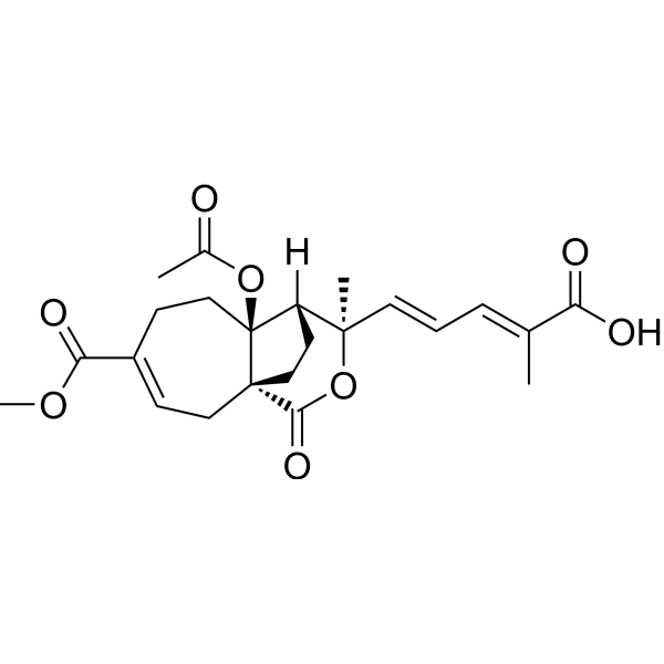
Pseudolaric Acid B is an orally active diterpene acid. Pseudolaric Acid B has anti-fungal, anti-fertility, anti-angiogenesis and anticancer activity, and can induce tumor cell apoptosis and autophagy. In addition, Pseudolaric Acid B can inhibit the secretion of hepatitis B virus (HBV) and has immunosuppressive effect, selectively inhibiting the proliferation of T lymphocytes and the production of IL-2.
For research use only. We do not sell to patients.
- Purity : 98.67%
- CAS No.: 82508-31-4
- Formula: C23H28O8
- Molecular Weight:432.46
-
Storage:
4°C, protect from light
* In solvent : -80°C, 6 months; -20°C, 1 month (protect from light)

To place orders, for customer services and technical support, please contact: MedChemExpress USA
Tel: 609-228-6898 E-mail: [email protected] [email protected]


-
 Biological Activity
-
Chemical Information
-
Solvent & Solubility
- Protocols
- Purity & Documentation
- References
-
 Help & FAQs


-
HY-N0176
DihydroartemisininAntimalarial Agent -

HY-N6939R
Pseudolaric Acid B (Standard)Anticancer Agent -
HY-N0111
Coenzyme Q10Antioxidant -
HY-N0523
Gallic acidBioactive Polyhydroxyphenolic Compound -
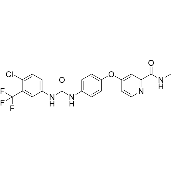
HY-10201
SorafenibMultikinase Inhibitor -

HY-N0523R
Gallic acid (Standard)Bioactive Polyhydroxyphenolic Compound -

HY-N0111R
Coenzyme Q10 (Standard)Antioxidant -

HY-N0176R
Dihydroartemisinin (Standard)Reference Standard -
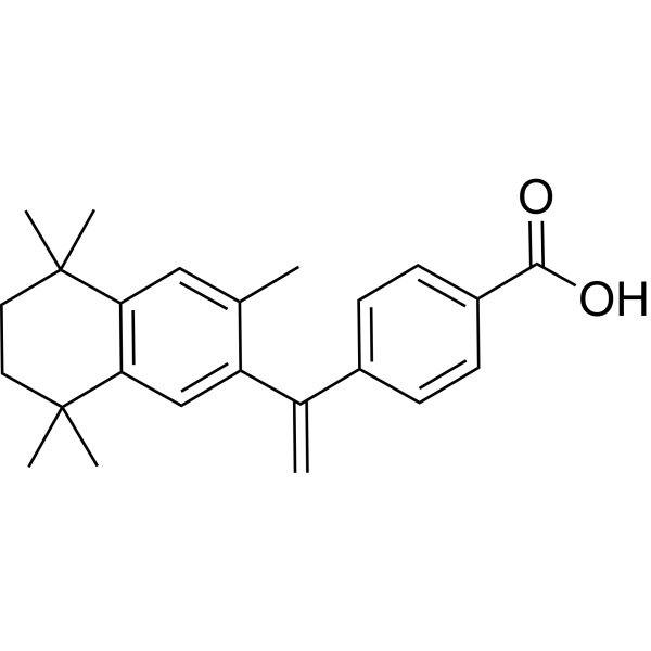
HY-14171
BexaroteneRXR Agonist -
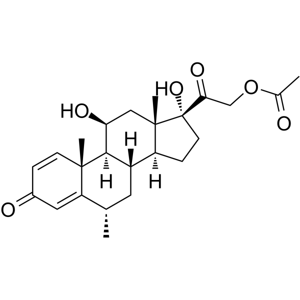
HY-13681
Methylprednisolone acetateCorticosteroid Hormone -
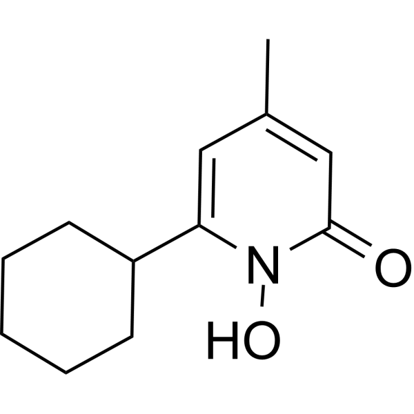
HY-B0450
CiclopiroxAntifungal Agent -
HY-W016412
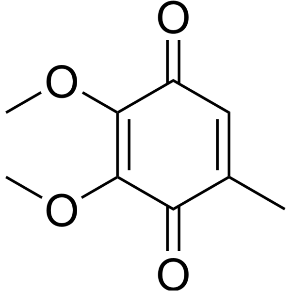
Coenzyme Q0Apoptosis Inducer
Publications Citing Use of MedChemExpress (MCE) Pseudolaric Acid B
More

Biological Activity
Description
IC50 & Target
[3]|
HIF-1α |
Cellular Effect
|
Cell Line

|
Type | Value | Description | References |
|---|---|---|---|---|
| A549 | IC50 |
0.3 μM
Compound: PAB
|
Cytotoxicity against human A549 cells by SRB method
Cytotoxicity against human A549 cells by SRB method
|
[PMID: 17253850] |
| A549 | IC50 |
0.3 μM
Compound: pseudolaric acid B
|
Cytotoxicity against human A549 cells by SRB method
Cytotoxicity against human A549 cells by SRB method
|
[PMID: 17323995] |
| A549 | IC50 |
0.3 μM
Compound: pseudolaricacid B
|
Cytotoxicity against human A549 cells by MTT assay
Cytotoxicity against human A549 cells by MTT assay
|
[PMID: 18651771] |
| A549 | IC50 |
0.86 μg/mL
Compound: PAB
|
Cytotoxicity against human A549 cells after 24 hrs
Cytotoxicity against human A549 cells after 24 hrs
|
[PMID: 15387644] |
| A549 | IC50 |
1.6 μM
Compound: Pseudolaric acid B
|
Cytotoxicity against human A549 cells after 72 hrs by SRB assay
Cytotoxicity against human A549 cells after 72 hrs by SRB assay
|
[PMID: 20038159] |
| Bel-7402 | IC50 |
>100 μM
Compound: 7
|
Cytotoxicity against human Bel7402 cells after 48 hrs by MTT assay
Cytotoxicity against human Bel7402 cells after 48 hrs by MTT assay
|
[PMID: 17291040] |
| Bel-7402 | IC50 |
0.704 μM
Compound: 1, pseudolaric acid B, PAB
|
Antiproliferative activity against human BEL-7402 cells by SRB assay
Antiproliferative activity against human BEL-7402 cells by SRB assay
|
[PMID: 18078313] |
| Bel-7402 | IC50 |
1.3 μM
Compound: Pseudolaric acid B
|
Cytotoxicity against human Bel7402 cells after 72 hrs by SRB assay
Cytotoxicity against human Bel7402 cells after 72 hrs by SRB assay
|
[PMID: 20038159] |
| BGC-823 | IC50 |
>100 μM
Compound: 7
|
Cytotoxicity against human BGC823 cells after 48 hrs by MTT assay
Cytotoxicity against human BGC823 cells after 48 hrs by MTT assay
|
[PMID: 17291040] |
| HCT-116 | IC50 |
0.136 μM
Compound: 1, pseudolaric acid B, PAB
|
Antiproliferative activity against human HCT116 cells by SRB assay
Antiproliferative activity against human HCT116 cells by SRB assay
|
[PMID: 18078313] |
| HeLa | IC50 |
0.497 μM
Compound: 1, pseudolaric acid B, PAB
|
Antiproliferative activity against human Hela cells by SRB assay
Antiproliferative activity against human Hela cells by SRB assay
|
[PMID: 18078313] |
| HL-60 | IC50 |
0.235 μM
Compound: 1, pseudolaric acid B, PAB
|
Antiproliferative activity against human HL60 cells by SRB assay
Antiproliferative activity against human HL60 cells by SRB assay
|
[PMID: 18078313] |
| HL-60 | IC50 |
0.59 μM
Compound: 7
|
Cytotoxicity against human HL60 cells after 48 hrs by MTT assay
Cytotoxicity against human HL60 cells after 48 hrs by MTT assay
|
[PMID: 17291040] |
| HL-60 | IC50 |
4.2 μM
Compound: Pseudolaric acid B
|
Cytotoxicity against human HL60 cells after 72 hrs by MTT assay
Cytotoxicity against human HL60 cells after 72 hrs by MTT assay
|
[PMID: 20038159] |
| HMEC-1 | IC50 |
0.803 μM
Compound: 1, pseudolaric acid B, PAB
|
Antiproliferative activity against HMEC1 cells by SRB assay
Antiproliferative activity against HMEC1 cells by SRB assay
|
[PMID: 18078313] |
| MDA-MB-468 | IC50 |
0.596 μM
Compound: 1, pseudolaric acid B, PAB
|
Antiproliferative activity against human MB-MDA-468 cells by SRB assay
Antiproliferative activity against human MB-MDA-468 cells by SRB assay
|
[PMID: 18078313] |
| P388 | IC50 |
0.32 μg/mL
Compound: PAB
|
Cytotoxicity against mouse P388 cells after 24 hrs
Cytotoxicity against mouse P388 cells after 24 hrs
|
[PMID: 15387644] |
| P388 | IC50 |
0.74 μM
Compound: pseudolaric acid B
|
Cytotoxicity against mouse P388 cells by MTT method
Cytotoxicity against mouse P388 cells by MTT method
|
[PMID: 17323995] |
| P388 | IC50 |
3.7 μM
Compound: PAB
|
Cytotoxicity against murine P388 cells by MTT method
Cytotoxicity against murine P388 cells by MTT method
|
[PMID: 17253850] |
In Vitro
Pseudolaric Acid B (4 μM; 36 h to 3 days) activates autophagy in MCF‑7 human breast cancer cells to prevent cell death[1].
Pseudolaric Acid B (4 μM; 0-36 h) inhibits the secretion of hepatitis B virus (HBV) by inducing apoptosis and cell cycle arrest[2].
Pseudolaric Acid B (0.313-10 μM; 6-72 h) inhibits the proliferation, migration and tube formation of HUVEC, and reduces the secretion of VEGF by tumor cells, which is related to proteasoma-mediated HIF-1α protein degradation[3].
MedChemExpress (MCE) has not independently confirmed the accuracy of these methods. They are for reference only. Further protocols information, click here.
-
Cell Line:MCF-7 cells
-
Concentration:4 μM
-
Incubation Time:36 h
-
Result:Increased the expression of Beclin-1.
Promoted the conversion from LC3-I to LC3-II.
-
Cell Line:MCF-7 cells
-
Concentration:4 μM
-
Incubation Time:36 h
-
Result:Increased the number of autophagic vacuoles.
-
Cell Line:HepG2215 cells
-
Concentration:4 μM
-
Incubation Time:0, 6, 12, 24 and 36 h
-
Result:Decreased the expression of procaspase-3.
In Vivo
MedChemExpress (MCE) has not independently confirmed the accuracy of these methods. They are for reference only.


Chemical Information
-
CAS No. 82508-31-4
-
Appearance Solid
-
Molecular Weight 432.46
-
Formula C23H28O8
-
Color White to off-white
-
SMILES
O=C(C1=CC[C@@]([C@@](CC1)(OC(C)=O)[C@]2([H])[C@@](C)(/C=C/C=C(C(O)=O)\C)O3)(CC2)C3=O)OC
-
Structure Classification
-
Shipping
Room temperature in continental US; may vary elsewhere.
-
Storage
4°C, protect from light
* In solvent : -80°C, 6 months; -20°C, 1 month (protect from light)


Publications (2)
-
Journal Impact Factor
-
Most Recent
-
ACS Infect Dis
Pseudolaric Acid B Ameliorates Fungal Keratitis Progression by Suppressing Inflammation and Reducing Fungal Load. [Abstract]2023 Jun 9;9(6):1196-1205. PMID: 37141176 -
Vet Microbiol
The Chinese medicine monomer Schisandrin C inhibits PRRSV infection by regulating the OGT-PI3K/AKT/mTOR signaling pathway. [Abstract]2026 May:316:110992. PMID: 41865607


Solvent & Solubility
In Vitro:
DMSO : 50 mg/mL (115.62 mM; Need ultrasonic; Hygroscopic DMSO has a significant impact on the solubility of product, please use newly opened DMSO)
Please refer to the solubility information to select the appropriate solvent. Once prepared, please aliquot and store the solution to prevent product inactivation from repeated freeze-thaw cycles.
Storage method and period of stock solution: -80°C, 6 months; -20°C, 1 month (protect from light). When stored at -80°C, please use it within 6 months. When stored at -20°C, please use it within 1 month.
Please refer to the solubility information to select the appropriate solvent. Once prepared, please aliquot and store the solution to prevent product inactivation from repeated freeze-thaw cycles.
Storage method and period of stock solution: -80°C, 6 months; -20°C, 1 month (protect from light). When stored at -80°C, please use it within 6 months. When stored at -20°C, please use it within 1 month.
Concentration (start) × Volume (start) = Concentration (final) × Volume (final)
In Vivo:
Select the appropriate dissolution method based on your experimental animal and administration route.
- For the following dissolution methods, please ensure to first prepare a clear stock solution using an In Vitro approach and then sequentially add co-solvents:
- To ensure reliable experimental results, the clarified stock solution can be appropriately stored based on storage conditions. As for the working solution for In Vivo experiments, it is recommended to prepare freshly and use it on the same day.
- The percentages shown for the solvents indicate their volumetric ratio in the final prepared solution. If precipitation or phase separation occurs during preparation, heat and/or sonication can be used to aid dissolution.
Add each solvent one by one: 10% DMSO 40% PEG300 5% Tween-80 45% Saline
Solubility: ≥ 2.5 mg/mL (5.78 mM); Clear solution
This protocol yields a clear solution of ≥ 2.5 mg/mL (saturation unknown).
Taking 1 mL working solution as an example, add 100 μL DMSO stock solution (25.0 mg/mL) to 400 μL PEG300, and mix evenly; then add 50 μL Tween-80 and mix evenly; then add 450 μL Saline to adjust the volume to 1 mL.
Preparation of Saline: Dissolve 0.9 g sodium chloride in ddH₂O and dilute to 100 mL to obtain a clear Saline solution.
Add each solvent one by one: 10% DMSO 90% (20% SBE-β-CD in Saline)
Solubility: ≥ 2.08 mg/mL (4.81 mM); Clear solution
This protocol yields a clear solution of ≥ 2.08 mg/mL (saturation unknown).
Taking 1 mL working solution as an example, add 100 μL DMSO stock solution (20.8 mg/mL) to 900 μL 20% SBE-β-CD in Saline, and mix evenly.
Preparation of 20% SBE-β-CD in Saline (4°C, storage for one week): 2 g SBE-β-CD powder is dissolved in 10 mL Saline, completely dissolve until clear.
For the following dissolution methods, please prepare the working solution directly:
It is recommended to prepare fresh solutions and use them promptly within a short period of time.
The percentages shown for the solvents indicate their volumetric ratio in the final prepared solution. If precipitation or phase separation occurs during preparation, heat and/or sonication can be used to aid dissolution.
Add each solvent one by one: 50% PEG300 50% Saline
Solubility: 10 mg/mL (23.12 mM); Suspended solution; Need ultrasonic
In Vivo Dissolution Calculator
Please enter the basic information of animal experiments:
-
-
-
-
Recommended: Prepare an additional quantity of animals to account for potential losses during experiments.
Please enter your animal formula composition:
-
%DMSO +
Recommended: Keep the proportion of DMSO in working solution below 2% if your animal is weak.
-
%+
-
+%Tween-80 + +
-
%Saline +
The co-solvents required include: DMSO, . All of co-solvents are available by MedChemExpress (MCE). , Tween 80. All of co-solvents are available by MedChemExpress (MCE).
Working solution concentration: 0.22 mg/mL
Method for preparing stock solution: mg drug dissolved in μL DMSO. Stock solution concentration: mg/mL. * In solvent : -80°C, 6 months; -20°C, 1 month (protect from light)
1. Take μL DMSO stock solution;
2. Add μL .
μL , mix evenly;
3. Then add μL Tween 80, mix evenly;
4. Then add μL
Please ensure that the stock solution in the first step is dissolved to a clear state, and add co-solvents in sequence. You can use ultrasonic heating (ultrasonic cleaner, recommended frequency 20-40 kHz), vortexing, etc. to assist dissolution.

Protocols
-
Reproductive and Developmental Toxicity Study
Reproductive and developmental toxicity studies detect adverse effects of prenatal or peri/postnatal exposure on maternal condition, pregnancy maintenance, embryo-fetal survival, fetal growth, structural development, and offspring reproductive or developmental endpoints; classic rat protocols generate readouts by comparing treated groups with vehicle, pair-fed, or untreated controls for implantation, resorption, fetal weight, crown-rump length, external morphology, visceral morphology, skeletal ossification, anogenital distance, nipple/areola retention, and postnatal cohort outcomes.
-
Human pluripotent stem cell endothelial-cell differentiation
Human pluripotent stem cell endothelial differentiation is based on stepwise developmental patterning: early activation of WNT/GSK3β inhibition promotes mesodermal or vascular progenitor entry, followed by endothelial specification using VEGF-related signaling, BMP4, FGF2, Notch modulation, or cAMP depending on the published protocol. Endothelial differentiation is read out by acquisition of CD31, CD34, VE-cadherin/CD144, KDR/VEGFR2, vWF, Tie2, NOS3, acetylated LDL uptake, tube/network formation, barrier function, and in vivo vessel-forming capacity where tested.
-
Vascular/Branching Fractal Analysis
Vascular/branching fractal analysis quantifies the geometric complexity of vessel trees or vascular networks from segmented 2D images, commonly by converting vessels into binary and/or skeletonized maps and estimating fractal dimension using box-counting or related approaches. Fractal dimension is interpreted as an image-derived readout of vascular branching complexity, space filling, or density, and has been applied to retinal photographs, fluorescein angiography, OCT angiography, capillary perfusion maps, and in vitro Matrigel angiogenesis networks. The assay readout is generated from vessel-positive pixels after image preprocessing, vessel segmentation, binarization, and optional skeletonization; reported outputs include fractal dimension, vessel density, branchpoint density, endpoint density, vessel length density, tortuosity, and generation-based branching metrics when VESGEN-style analysis is used. The biological interpretation is limited to quantitative vascular patterning and s
-
Annexin V plus membrane-impermeant dye apoptosis staining
Annexin V-based apoptosis assays rely on the detection of phosphatidylserine (PS) externalization from the inner leaflet of the plasma membrane to the outer leaflet, an early biochemical hallmark of apoptosis. Fluorescently labeled Annexin V binds PS in a calcium-dependent manner, enabling identification of early apoptotic cells by flow cytometry or fluorescence microscopy. When combined with a membrane-impermeant DNA-binding dye (e. g. , propidium iodide), this approach allows discrimination between viable (Annexin V−/dye−), early apoptotic (Annexin V+/dye−), and late apoptotic or necrotic (Annexin V+/dye+) cell populations by assessing membrane integrity and PS exposure.
-
Cell Viability Determination by MTT Colorimetric Assay
The following protocol uses the MTT colorimetric assay as a classic literature-established method for assessing cell viability/metabolic activity in cultured mammalian cells. MTT[3-(4,5-dimethylthiazol-2-yl)-2,5-diphenyltetrazolium bromide] is reduced by metabolically active cells to a colored formazan product; the amount of formazan is quantified spectrophotometrically and provides an indirect measure of metabolically active viable cells. Importantly, MTT reduction reflects cellular oxidoreductase/metabolic activity rather than an absolute direct count of living cells, so changes in cellular metabolism can alter the signal independently of cell number.
-
Apoptosis
Apoptosis, also called programmed cell death, is generally characterized by distinct morphological characteristics.
-
TUNEL staining for apoptotic DNA fragmentation
TUNEL staining detects DNA strand breaks by using terminal deoxynucleotidyl transferase to add labeled nucleotides to exposed 3′-OH DNA termini, generating either microscopic staining in fixed cells or tissue sections, or fluorescence/cytometric signal in cell suspensions. TUNEL positivity reflects DNA fragmentation but should not be interpreted alone as definitive apoptosis, because TUNEL can also label necrotic, autolytic, mechanically damaged, or DNA-repair-associated DNA breaks.
-
Endothelial Tube Formation Assay
Endothelial tube formation assay evaluates the ability of endothelial cells to attach, migrate, align, and organize into capillary-like networks when cultured on gelled basement membrane extract or Matrigel; the readout is the morphology and quantity of tube-like networks, which reflects an in vitro endothelial morphogenesis step related to angiogenesis. Basement membrane extract/Matrigel provides laminin-rich extracellular matrix cues that support endothelial differentiation into capillary-like structures, but it can contain biologically active growth factors, so growth-factor-reduced matrix is preferred when testing defined angiogenic stimulators or inhibitors.
-
Autophagy
Autophagy is a process in which eukaryotic cells use lysosomes to degrade their own cytoplasmic proteins and damaged organelles under the regulation of autophagy related gene (Atg). Microtubule-associated proteins light chain 3 (LC3) is recognized as autophagy marker, which transfers from cytoplasmic LC3 (LC3-I) to membrane type (LC3-II). LC3-II/I ratio could be detected by Western Blot and fluorescence microscopy.
-
Apoptosis Solutions
Apoptosis is a regulated, generally non-lytic cell-death pathway that removes unwanted, damaged, infected, or abnormal cells through coordinated morphological changes, caspase activation, DNA fragmentation, and membrane remodeling. The intrinsic apoptosis pathway is controlled mainly by mitochondrial outer membrane permeabilization, BCL-2 family proteins, cytochrome c release, apoptosome formation, caspase-9 activation, and downstream executioner caspase-3/7 activation. The extrinsic apoptosis pathway is initiated by death receptors such as Fas, TNFR, and TRAIL receptors, which recruit adaptor proteins and activate caspase-8 before engaging executioner caspases or mitochondrial amplification through BID cleavage. Apoptosis is linked to many phenotypes, including cancer cell killing, tissue homeostasis, immune regulation, neurodegeneration, infection response, and treatment-induced cytotoxicity; unresolved questions include how apoptosis interacts with necroptosis, pyroptosis, ferroptos
-
Lysosome and acidic-vesicle live-cell staining
Lysosome and acidic-vesicle live-cell staining detects acidic intracellular compartments by using membrane-permeant acidotropic probes that accumulate in low-pH vesicles, including lysosomes, late endosomes, autolysosomes, and acidic phagosomes. LysoTracker staining is commonly used as an intensity-based readout of acidic lysosomal compartment abundance or enlargement, while acridine orange produces green fluorescence in less concentrated compartments and red fluorescence after concentration-dependent accumulation in acidic vesicular organelles. Loss or reduction of acridine-orange red signal can be used as a readout of lysosomal membrane permeabilization or reduced acidic-vesicle integrity. This protocol is designed for live cultured cells and can be adapted for fluorescence microscopy, high-content imaging, plate-reader readout, or flow cytometry when the selected literature supports the readout. Because these dyes report acidotropic accumulation rather than lysosome identity alone,
-
Macroautophagy Solutions
Macroautophagy is a conserved lysosome-dependent degradation pathway in which cytoplasmic material is sequestered into double-membrane autophagosomes and delivered to lysosomes for degradation and recycling. The pathway supports cellular homeostasis during nutrient limitation, organelle stress, protein-aggregate accumulation, infection, differentiation, and tissue remodeling by coupling cargo sequestration, autophagosome maturation, lysosomal fusion, and degradation of cargo-derived macromolecules. The core molecular sequence includes initiation by nutrient- and stress-regulated autophagy machinery, autophagosome nucleation, LC3/ATG8-family conjugation to autophagosomal membranes, cargo selection through receptors such as SQSTM1/p62, autophagosome-lysosome fusion, and lysosomal degradation. LC3 was identified as a mammalian homolog of yeast Atg8 that localizes to autophagosomal membranes after processing, and p62/SQSTM1 was shown to connect ubiquitinated cargo with autophagic degradati
-
Endothelial Cell Migration/Angiogenic Sprouting Assay
Endothelial cell migration and angiogenic sprouting assays are in vitro (and partially ex vivo-adapted) functional models that quantify the ability of endothelial cells to undergo coordinated migration, extracellular matrix invasion, and multicellular organization into capillary-like sprouts in response to pro-angiogenic stimuli such as VEGF, bFGF, or conditioned microenvironments. These assays are used to model early angiogenic events including tip-cell formation, directional migration, and lumen-like sprout extension, which collectively reflect angiogenic activation and vascular morphogenesis processes observed in vivo.


Purity & Documentation
-
Data Sheet (278 KB)
-
SDS (643 KB)
- English - EN (643 KB)
- Français - FR (643 KB)
- Deutsch - DE (643 KB)
- Norwegian - NO (643 KB)
- Español - ES (643 KB)
- Swedish - SV (643 KB)
- Italian - IT (643 KB)
- Korean - KR (643 KB)
- Portuguese - PT (643 KB)
-
Handling Instructions (2659 KB)
References
[1]. Yu J, et al. Pseudolaric acid B activates autophagy in MCF-7 human breast cancer cells to prevent cell death. Oncol Lett. 2016 Mar;11(3):1731-1737. [Content Brief]
[2]. Yu J, et al. Pseudolaric acid B inhibits the secretion of hepatitis B virus. Oncol Rep. 2017 Jan;37(1):519-525. [Content Brief]
[3]. Li MH, et al. Pseudolaric acid B inhibits angiogenesis and reduces hypoxia-inducible factor 1alpha by promoting proteasome-mediated degradation. Clin Cancer Res. 2004 Dec 15;10(24):8266-74. [Content Brief]
[4]. Wang WC, et al. [Endocrine activity of pseudolaric acids A and B and their effects on sex hormones, prostaglandins, uteri, and fetuses]. Zhongguo Yao Li Xue Bao. 1991 Mar;12(2):187-90. [Content Brief]
[5]. Wei N, et al. The immunosuppressive activity of pseudolaric acid B on T lymphocytes in vitro. Phytother Res. 2013 Jul;27(7):980-5. [Content Brief]


Complete Stock Solution Preparation Table
Please refer to the solubility information to select the appropriate solvent. Once prepared, please aliquot and store the solution to prevent product inactivation from repeated freeze-thaw cycles.
Storage method and period of stock solution: -80°C, 6 months; -20°C, 1 month (protect from light). When stored at -80°C, please use it within 6 months. When stored at -20°C, please use it within 1 month.
| Optional Solvent | Concentration Solvent Mass | 1 mg | 5 mg | 10 mg | 25 mg |
|---|---|---|---|---|---|
| DMSO | 1 mM | 2.3124 mL | 11.5618 mL | 23.1235 mL | 57.8088 mL |
| 5 mM | 0.4625 mL | 2.3124 mL | 4.6247 mL | 11.5618 mL | |
| 10 mM | 0.2312 mL | 1.1562 mL | 2.3124 mL | 5.7809 mL | |
| 15 mM | 0.1542 mL | 0.7708 mL | 1.5416 mL | 3.8539 mL | |
| 20 mM | 0.1156 mL | 0.5781 mL | 1.1562 mL | 2.8904 mL | |
| 25 mM | 0.0925 mL | 0.4625 mL | 0.9249 mL | 2.3124 mL | |
| 30 mM | 0.0771 mL | 0.3854 mL | 0.7708 mL | 1.9270 mL | |
| 40 mM | 0.0578 mL | 0.2890 mL | 0.5781 mL | 1.4452 mL | |
| 50 mM | 0.0462 mL | 0.2312 mL | 0.4625 mL | 1.1562 mL | |
| 60 mM | 0.0385 mL | 0.1927 mL | 0.3854 mL | 0.9635 mL | |
| 80 mM | 0.0289 mL | 0.1445 mL | 0.2890 mL | 0.7226 mL | |
| 100 mM | 0.0231 mL | 0.1156 mL | 0.2312 mL | 0.5781 mL |


Pseudolaric Acid B Related Classifications
HY-N6939 Related Classifications
 Powered by Bioz