



Roquefortine C
Based on 1 publication(s) in Google Scholar
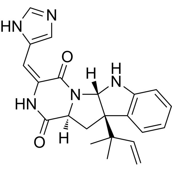
Roquefortine C is a mycotoxin that can be isolated from Penicillium species. Roquefortine C is an agonist of P-gp and an inhibitor of P450 3A and P450 1A. Roquefortine C can inhibit Gram-positive bacteria and also has certain neurotoxicity. Additionally, Roquefortine C can exert antitumor activity.
For research use only. We do not sell to patients.
- Purity : 98.03%
- CAS No.: 58735-64-1
- Formula: C22H23N5O2
- Molecular Weight:389.45
-
Storage:
-20°C, protect from light, stored under nitrogen
* In solvent : -80°C, 6 months; -20°C, 1 month (protect from light, stored under nitrogen)

To place orders, for customer services and technical support, please contact: MedChemExpress USA
Tel: 609-228-6898 E-mail: [email protected] [email protected]


-
 Biological Activity
-
Chemical Information
-
Solvent & Solubility
- Protocols
- Purity & Documentation
- References
-
 Help & FAQs


-
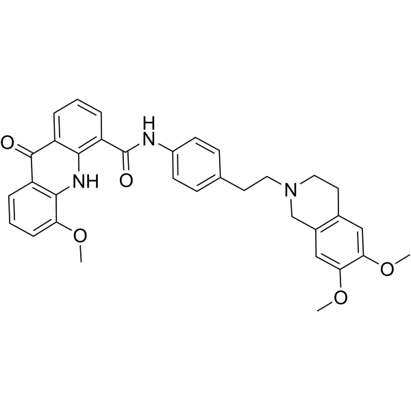
HY-50879
ElacridarP-gp And BCRP Dual-Inhibitor -

HY-50879R
Elacridar (Standard)P-gp And BCRP Dual-Inhibitor -
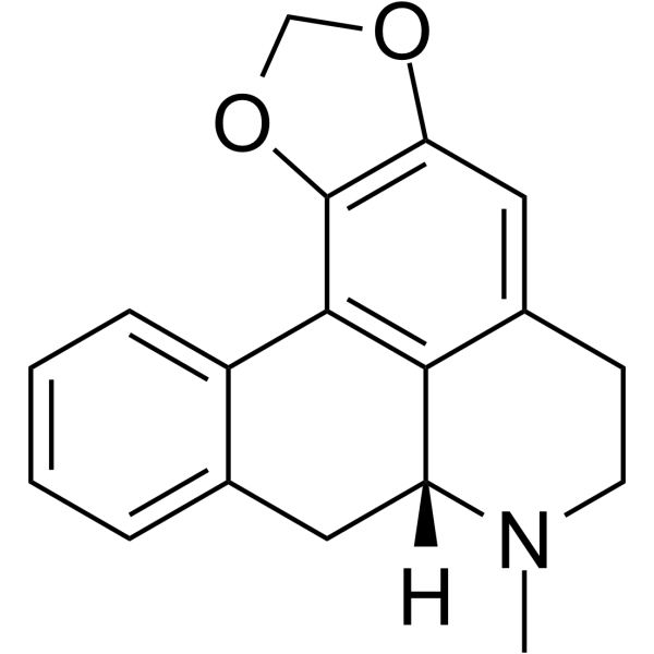
HY-121793
RoemerineEndogenous Metabolite -
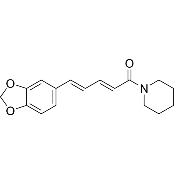
HY-N0144
PiperineAutophagy Inducer -
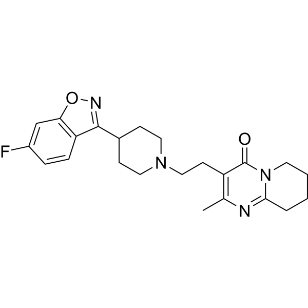
HY-11018
Risperidone5-HT2 Receptor Blocker -
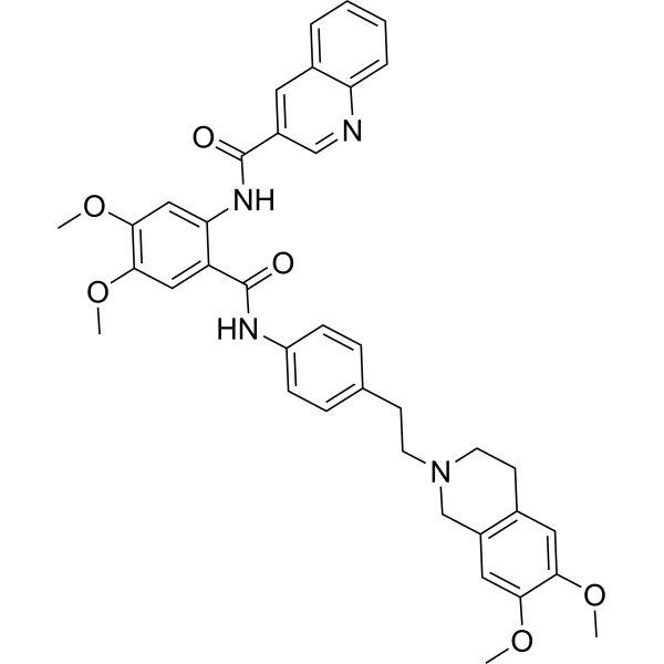
HY-10550
TariquidarP-gp Inhibitor -

HY-N0144R
Piperine (Standard)Autophagy Inducer -
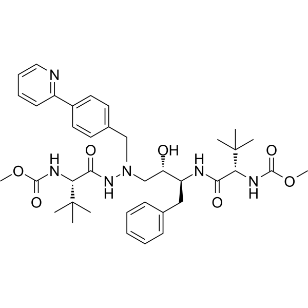
HY-17367
AtazanavirHIV Protease Inhibitor -
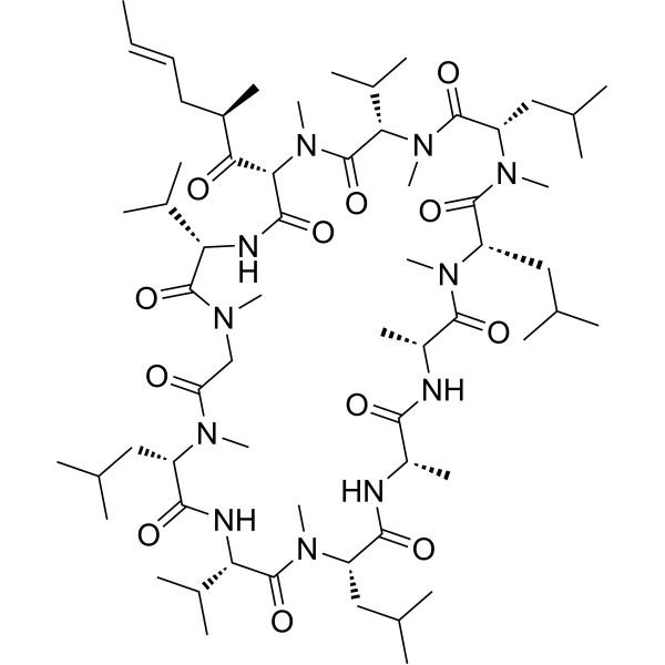
HY-17384
ValspodarP-glycoprotein Inhibitor -
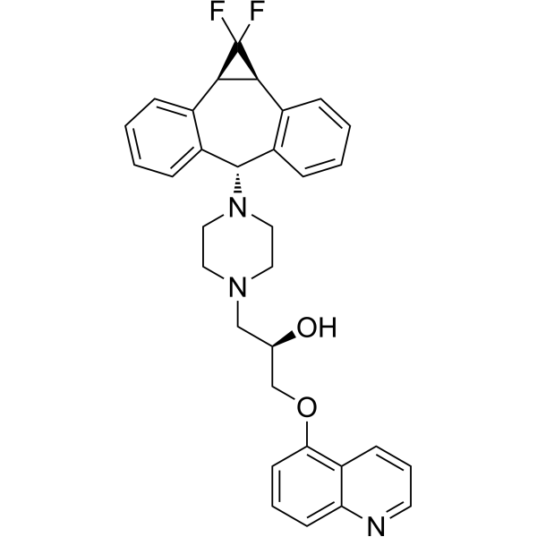
HY-15255
ZosuquidarP-gp Inhibitor -
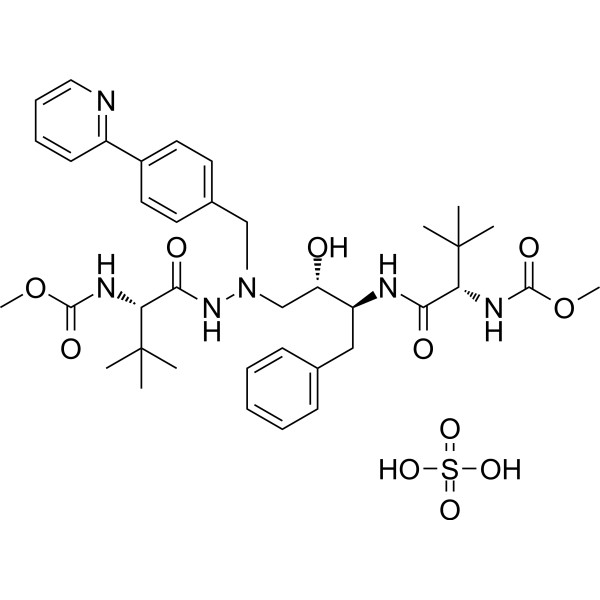
HY-17367A
Atazanavir sulfateHIV Protease Inhibitor -
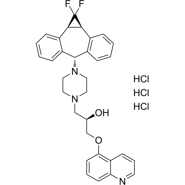
HY-50671
Zosuquidar trihydrochlorideP-gp Inhibitor
Publications Citing Use of MedChemExpress (MCE) Roquefortine C
More

Biological Activity
Description
Cellular Effect
|
Cell Line

|
Type | Value | Description | References |
|---|---|---|---|---|
| BT-474 | IC50 |
29.6 μM
Compound: 2; Roq C
|
Antiproliferative activity against human BT-474 cells expressing c-MET assessed as reduction in cell viability incubated for 72 hrs by MTT assay
Antiproliferative activity against human BT-474 cells expressing c-MET assessed as reduction in cell viability incubated for 72 hrs by MTT assay
|
[PMID: 26692349] |
| MCF7 | IC50 |
21.7 μM
Compound: 2; Roq C
|
Antiproliferative activity against human MCF7 cells expressing c-MET assessed as reduction in cell viability incubated for 72 hrs by MTT assay
Antiproliferative activity against human MCF7 cells expressing c-MET assessed as reduction in cell viability incubated for 72 hrs by MTT assay
|
[PMID: 26692349] |
| MDA-MB-231 | IC50 |
23.5 μM
Compound: 2; Roq C
|
Antiproliferative activity against human MDA-MB-231 cells expressing c-MET assessed as reduction in cell viability incubated for 72 hrs by MTT assay
Antiproliferative activity against human MDA-MB-231 cells expressing c-MET assessed as reduction in cell viability incubated for 72 hrs by MTT assay
|
[PMID: 26692349] |
| MDA-MB-468 | IC50 |
28.6 μM
Compound: 2; Roq C
|
Antiproliferative activity against human MDA-MB-468 cells expressing c-MET assessed as reduction in cell viability incubated for 72 hrs by MTT assay
Antiproliferative activity against human MDA-MB-468 cells expressing c-MET assessed as reduction in cell viability incubated for 72 hrs by MTT assay
|
[PMID: 26692349] |
| SK-BR-3 | IC50 |
23.4 μM
Compound: 2; Roq C
|
Antiproliferative activity against human SK-BR-3 cells expressing c-MET assessed as reduction in cell viability incubated for 72 hrs by MTT assay
Antiproliferative activity against human SK-BR-3 cells expressing c-MET assessed as reduction in cell viability incubated for 72 hrs by MTT assay
|
[PMID: 26692349] |
In Vitro
Roquefortine C (72 h) exhibits certain antiproliferative activity in c-Met-dependent human breast cancer cell lines, with an IC50 range of 21.7-29.6 μM[1].
Roquefortine C (5 μM; 1 h) can inhibit the phosphorylation of wild-type c-Met, with inhibition rates of 39.7% (5 μM) and 59.7% (10 μM)[1].
Roquefortine C (15-30 μM; 24 h) can inhibit HGF-induced invasion of MDA-MB-231 breast cancer cells[1].
Roquefortine C (>10 μM) exhibits cytotoxic effects towards human neuroblastoma cell lines[2].
MedChemExpress (MCE) has not independently confirmed the accuracy of these methods. They are for reference only. Further protocols information, click here.
-
Cell Line:c-Met-dependent human breast cancer cell lines
-
Concentration:/
-
Incubation Time:72 h
-
Result:Inhibited the cell viability, with IC50 values of 21.7, 24.4, 29.6, 23.4, 23.5, 28.6 μM for MCF7, MCF7-dox, BT-474, SKBR-3, MDA-MB-231, MDA-MB-468, respectively.


Chemical Information
-
CAS No. 58735-64-1
-
Appearance Solid
-
Molecular Weight 389.45
-
Formula C22H23N5O2
-
Color White to off-white
-
SMILES
O=C([C@](N1C/2=O)([H])C[C@]3(C(C)(C)C=C)[C@@]1([H])NC4=C3C=CC=C4)NC2=C\C5=CN=CN5
-
Structure Classification
-
Initial Source
Penicillium roqueforti
-
Shipping
Room temperature in continental US; may vary elsewhere.
-
Storage
-20°C, protect from light, stored under nitrogen
* In solvent : -80°C, 6 months; -20°C, 1 month (protect from light, stored under nitrogen)


Publications (1)
-
Journal Impact Factor
-
Most Recent
-
Cell Signal
Hsa-miR-34a-5p reverses multidrug resistance in gastric cancer cells by targeting the 3'-UTR of SIRT1 and inhibiting its expression. [Abstract]2021 Aug:84:110016. PMID: 33894312


Solvent & Solubility
In Vitro:
DMSO : 10 mg/mL (25.68 mM; Need ultrasonic and warming; Hygroscopic DMSO has a significant impact on the solubility of product, please use newly opened DMSO)
Please refer to the solubility information to select the appropriate solvent. Once prepared, please aliquot and store the solution to prevent product inactivation from repeated freeze-thaw cycles.
Storage method and period of stock solution: -80°C, 6 months; -20°C, 1 month (protect from light, stored under nitrogen). When stored at -80°C, please use it within 6 months. When stored at -20°C, please use it within 1 month.
Please refer to the solubility information to select the appropriate solvent. Once prepared, please aliquot and store the solution to prevent product inactivation from repeated freeze-thaw cycles.
Storage method and period of stock solution: -80°C, 6 months; -20°C, 1 month (protect from light, stored under nitrogen). When stored at -80°C, please use it within 6 months. When stored at -20°C, please use it within 1 month.
Concentration (start) × Volume (start) = Concentration (final) × Volume (final)

Protocols
-
Neurotoxicity Study
This protocol assesses in vitro neurotoxicity by combining neuronal viability, mitochondrial/metabolic activity, neurite outgrowth, and optional neuronal network function readouts. Calcein-AM or resazurin/PrestoBlue readouts estimate viable or metabolically active cells; βIII-tubulin immunofluorescence detects neuronal morphology and neurite networks; TMRE detects mitochondrial membrane potential; and MEA recordings detect functional changes in neuronal network activity.
-
Gram Staining of Tissue Sections
Gram staining of tissue sections is a histochemical technique used to differentiate Gram-positive and Gram-negative bacteria within histological specimens based on differences in bacterial cell wall structure and dye retention, adapted from classical bacteriological Gram staining into tissue-compatible “histological Gram stain” variants. In tissue applications, modifications of the Brown-Hopps and Brown-Brenn methods are commonly used to improve differentiation of microorganisms embedded within host connective tissue and to reduce overstaining or loss of Gram-negative signal, which are known limitations of earlier approaches. The principle relies on crystal violet-iodine complex retention in Gram-positive organisms and subsequent decolorization and counterstaining steps that allow contrast visualization of Gram-negative organisms against tissue background.
-
Subcutaneous Cell-Line-Derived Xenograft
Subcutaneous cell-line-derived xenograft (CDX) models are established by implanting cultured human cancer cell lines into immunodeficient mice, where the injected cells form localized tumors that can be monitored in vivo as a measure of tumorigenic potential, growth kinetics, and treatment response. These models are widely used in oncology research because they allow reproducible tumor formation and enable comparative assessment of tumor growth between different cell lines or genetic manipulations in a controlled in vivo microenvironment. Subcutaneous implantation of cancer cells in immunodeficient mice is a standard approach for evaluating tumor growth behavior and therapeutic response across multiple cancer types, including prostate, esophageal, pancreatic, and colon cancer models.


Purity & Documentation
-
Data Sheet (285 KB)
-
SDS (481 KB)
- English - EN (481 KB)
- Français - FR (481 KB)
- Deutsch - DE (481 KB)
- Norwegian - NO (481 KB)
- Español - ES (481 KB)
- Swedish - SV (481 KB)
- Italian - IT (481 KB)
- Korean - KR (481 KB)
- Portuguese - PT (481 KB)
-
Handling Instructions (2659 KB)
References
[1]. Mady MS, et al. The indole alkaloid meleagrin, from the olive tree endophytic fungus Penicillium chrysogenum, as a novel lead for the control of c-Met-dependent breast cancer proliferation, migration and invasion. Bioorg Med Chem. 2016 Jan 15;24(2):113-22. [Content Brief]
[2]. Aninat C, et al. Oxidative metabolism by P450 and function coupling to efflux systems: modulation of mycotoxin toxicity. Food Addit Contam. 2005 Apr;22(4):361-8. [Content Brief]
[3]. García-Rico RO, et al. Effect of a heterotrimeric G protein alpha subunit on conidia germination, stress response, and roquefortine C production in Penicillium roqueforti. Food Addit Contam. 2005 Apr;22(4):361-8. [Content Brief]
[4]. Tiwary AK, et al. Using roquefortine C as a biomarker for penitrem A intoxication. J Vet Diagn Invest. 2009 Mar;21(2):237-9. [Content Brief]
[5]. Deng XJ, et al. Hsa-miR-34a-5p reverses multidrug resistance in gastric cancer cells by targeting the 3'-UTR of SIRT1 and inhibiting its expression. Cell Signal. 2021 Aug;84:110016. [Content Brief]


Complete Stock Solution Preparation Table
Please refer to the solubility information to select the appropriate solvent. Once prepared, please aliquot and store the solution to prevent product inactivation from repeated freeze-thaw cycles.
Storage method and period of stock solution: -80°C, 6 months; -20°C, 1 month (protect from light, stored under nitrogen). When stored at -80°C, please use it within 6 months. When stored at -20°C, please use it within 1 month.
| Optional Solvent | Concentration Solvent Mass | 1 mg | 5 mg | 10 mg | 25 mg |
|---|---|---|---|---|---|
| DMSO | 1 mM | 2.5677 mL | 12.8386 mL | 25.6772 mL | 64.1931 mL |
| 5 mM | 0.5135 mL | 2.5677 mL | 5.1354 mL | 12.8386 mL | |
| 10 mM | 0.2568 mL | 1.2839 mL | 2.5677 mL | 6.4193 mL | |
| 15 mM | 0.1712 mL | 0.8559 mL | 1.7118 mL | 4.2795 mL | |
| 20 mM | 0.1284 mL | 0.6419 mL | 1.2839 mL | 3.2097 mL | |
| 25 mM | 0.1027 mL | 0.5135 mL | 1.0271 mL | 2.5677 mL |


Roquefortine C Related Classifications
HY-N6748 Related Classifications
 Powered by Bioz