1243 Results for "

fluorescence

" in MedChemExpress (MCE) Product Catalog:
Products (1243)

1243 Results for "fluorescence" in MCE Product Catalog:

Cat. No.: HY-D0233R
CAS No.: 603-48-5
Leucocrystal violet (Standard) is the analytical standard of Leucocrystal violet (HY-D0233). This product is intended for research and analytical applications. Leucocrystal violet is a cationic triarylmethane dye. Leucocrystal violet generates a colored purple product in the presence of hydrogen peroxide and horseradish peroxidase, or when oxidized by periodate under the catalysis of horseradish peroxidase. Leucocrystal violet serves as a chromogenic reagent for the determination of submicrogram-level hydrogen peroxide, with no interference from tryptophan, persulfate or chromate. Leucocrystal violet reacts with blood to produce a purple/violet color in the presence of hydrogen peroxide; it forms a very weak fluorophore when acting with whole blood in an unbuffered solution, with a maximum absorption wavelength of 630 nm and a maximum fluorescence emission wavelength of 665 nm upon excitation at 630 nm. Leucocrystal violet is widely used for staining blood residues on porous and non-porous materials.
loading...
    loading...
Cat. No.: HY-D2763
CAS No.: 890928-51-5
BP Fluor 532 maleimide is a photostable, bright yellow-fluorescent dye with pH insensitive emission from pH 4 to pH 10. The excitation of BP Fluor 532 is ideally suited for the frequency-doubled Nd:YAG laser line. BP Fluor 532 dye can be conjugated to a variety of antibodies, peptides, proteins, tracers, and amplification substrates, and is often used for the generation of stable signals in imaging and flow cytometry. Maleimide is the most popular sulfhydryl-reactive group for conjugating the dye to a thiol group on a protein, oligonucleotide thiophosphate, or low molecular weight ligand. The maleimide group specifically and efficiently reacts with reduced thiols (sulfhydryl groups, –SH) at pH 6.5 to 7.5 to form a stable thioether bond. The resulting conjugates exhibit brighter fluorescence and greater photostability than the conjugates of many other spectrally similar fluorophores.
loading...
    loading...
Cat. No.: HY-D2773
CAS No.: 219729-26-7
5-Carboxyrhodamine 110 NHS Ester is the nonsulfonated analog of the BP Fluor 488 dye. The amine-reactive 5(6)-Carboxyrhodamine 110 NHS Ester can be used to create bright and photostable green-fluorescent bioconjugates with excitation/emission maxima ~502/527 nm. For many applications, the dye is preferred over 5-(6)-carboxyfluorescein NHS ester or FITC because of its exceptional photostability and fluorescence insensitivity to pH (4-9). Although the mixed isomers of Carboxyrhodamine 110 NHS Ester preferred, routinely used fluorescent dye for labeling proteins, peptides and nucleotides, purification of peptide and nucleotides labeled with 5(6) isomers might be troublesome due to significant signal broadening in HPLC purification. Peptides and nucleotides labeled with a single isomer usually give better resolution in HPLC purification that is often required in the conjugation processes.
loading...
    loading...
Cat. No.: HY-D3073
CAS No.: 2477607-64-8
Target:  

Fluorescent Dye

Research Areas:  

Others

DXB-NIR is a push-pull dioxaboron fluorescent probe with solvatochromic properties, featuring high fluorescence brightness, photostability, and a large two-photon absorption cross-section. DXB-NIR exhibits Ex/Em of 550/620–780 nm (with two-photon excitation at 930 nm). DXB-NIR quantifies local polarity via the intensity ratio I (>640)/I (<640) between the far-red channel (<640 nm) and the near-infrared channel (>640 nm), and it can simultaneously label the plasma membrane, endoplasmic reticulum, and lipid droplets in live cells. DXB-NIR monitors the increase in local polarity of various cell compartments under conditions of cholesterol depletion, starvation, and oxidative stress. DXB-NIR can be used for polarity imaging of biological membranes and lipid droplets, as well as studies related to cellular stress .
loading...
    loading...
Cat. No.: HY-D3085
CAS No.: 2882924-23-2
SSR-LDs is a fluorescent probe for lipid droplet polarity detection and lipid droplet imaging. SSR-LDs exhibits polarity-dependent fluorescence: it produces intense short-wavelength emission in low-polarity environments, while generating weak long-wavelength emission in high-polarity environments. SSR-LDs enters cells via free diffusion, specifically targets lipid droplets, and is unaffected by intracellular viscosity or pH values ranging from 4.5 to 9.0. SSR-LDs shows variable Ex/Em wavelengths in different solvents, including 590/663 nm in THF, 600/660 nm in mouse tissue and in vivo imaging, and an excitation wavelength of 580 nm with an emission wavelength range of 610-750 nm in cell imaging. SSR-LDs can be used for research related to fatty liver, liver injury and hepatitis .
loading...
    loading...
Cat. No.: HY-D3086
CAS No.: 1668565-72-7
Target:  

Fluorescent Dye

Research Areas:  

Cancer

Mito-polarity is a mitochondria-targeted polarity probe. Mito-polarity can be used to detect mitochondrial polarity. The detection mechanism of Mito-polarity is based on an intramolecular charge transfer (ICT) system designed with a donor-π-bridge-acceptor (D-π-A) structure. Among them, the 467 nm green emission generated by the Coumarin (HY-N0709) structure is extremely sensitive to polarity changes, while the 642 nm red emission produced by the extended π-conjugation and ICT effect between the Coumarin and benzothiazine structures shows only a weak response, thus forming a ratiometric fluorescence response that is linearly correlated with solvent polarity. When Mito-polarity is excited at 405 nm in methanol, its excitation/emission wavelengths are Ex/Em = 405/467 nm and Ex/Em = 405/642 nm, with maximum absorption wavelengths of 426 nm and 561 nm .
loading...
    loading...
Cat. No.: HY-D3391
RMR-Tre is a fluorescent probe targeting the mycobacterial acyltransferase Ag85. Under the catalysis of Ag85, RMR-Tre undergoes 6-position mycoloylation and anchors to the mycobacterial membrane, while achieving fluorescence activation by inhibiting the intramolecular twisted charge transfer state transition. RMR-Tre can distinguish live mycobacteria from dead ones through metabolism-driven labeling, enabling rapid, wash-free, low-background detection of viable bacteria. RMR-Tre reports the drug resistance of Mycobacterium tuberculosis via the trehalose catalytic shift activity readout associated with TreS. In addition, RMR-Tre can be combined with flow cytometry or high-content imaging techniques to visualize and quantitatively analyze the metabolic heterogeneity of Mycobacterium tuberculosis related to persistence and drug resistance. RMR-Tre is widely used in tuberculosis-related research .
loading...
    loading...
Cat. No.: HY-D3419
Neuro-DiO 4-chlorobenzenesulfonate is a hydrophobic C18 alkyl chain carbocyanine dye with green fluorescence, commonly used as a vascular marker, cell internalizer and deposition agent. Neuro-DiO 4-chlorobenzenesulfonate inserts its alkyl chain into the endothelial plasma membrane via liposome-mediated perfusion to achieve vascular labeling. Neuro-DiO chlorobenzenesulfonate can also stain the cell membrane and cytoplasm of cancer cells to assist in confocal microscopy observations. Neuro-DiO chlorobenzenesulfonate can be released from nanosponges and accumulate on the surface of mouse retina, then internalize into retinal ganglion cells, which is applicable to researches related to glaucoma and other diseases. It should be noted that during liposome-mediated vascular staining in mice, Neuro-DiO 4-chlorobenzenesulfonate may cause leakage of airway lavage fluid .
loading...
    loading...
Cat. No.: HY-D3428
CAS No.: 2863678-02-6
LJ-2P is an NIR-II small-molecule fluorescent probe with fibrin/fibrinogen affinity. LJ-2P can bind coagulation-related proteins and restrict dye molecular motion, thereby enhancing local fluorescence for high-contrast imaging of bleeding and blood clots in vivo. LJ-2P can also co-precipitate with Ca 2+ to form LJ-2P nanoparticles, significantly enhancing NIR-II luminescence through the precipitation-enhanced emission (PEE) mechanism. LJ-2P can be used for research on bleeding disorders and breast cancer. LJ-2P exhibits dual-modal emission properties (Ex/Em ≈740/945 nm or 740/1066 nm) .
loading...
    loading...
Cat. No.: HY-D3577
Target:  

Fluorescent Dye

Research Areas:  

Others

AF555 Picolyl Azide is a Fluorescent dye that enables site-specific fluorescent labeling via click chemistry. AF555 Picolyl Azide conjugates with the non-canonical amino acid N-propargyl-L-lysine introduced through ochre stop codon suppression via the copper (I)-catalyzed azide-alkyne cycloaddition (CuAAC) reaction, achieving labeling of hyperpolarization-activated cyclic nucleotide-gated HCN channels; its picolyl group accelerates the CuAAC reaction rate, thus completing labeling rapidly at room temperature and minimizing copper-induced cytotoxicity. AF555 Picolyl Azide forms covalent bonds with the alkynyl groups of nucleotide analogs through its azide moiety, labeling viral genomes containing 5-ethynyl-2'-deoxyuridine for visualization via fluorescence microscopy .
loading...
    loading...
Cat. No.: HY-DY1097
CAS No.: 107091-89-4
Thiazole Orange (solution) is an asymmetric anthocyanin dye that can be coupled with oligonucleotides (ONs) to prepare fluorescent hybridization probes. Thiazole Orange has been widely used in biomolecular detection and staining of DNA/ RNA in gels and can be used for reticulocyte analysis. Thiazole orange generates a significant fluorescence enhancement and high quantum yield when it binds with nucleic acids, especially RNA. Thiazole orange can permeate living cell membranes. Thiazole orange can use UV light for detection, but can also be detected with blue light. The excitation and emission of Thiazole orange are λex = 510 nm (488 nm and 470 nm also show strong excitation) and λem = 527 nm, respectively .
Solvent and concentration: DMSO: 10 mM
The 1 mL volume is defined as the base specification. All larger sizes correspond to incremental volumes of this base.
loading...
    loading...
Cat. No.: HY-DY2041
Target:  

Fluorescent Dye

Research Areas:  

Infection

Callose Staining Solution (Aniline Blue, Bright-Field Method) is a fluorescent dye used for detecting callose in plant tissues, green algal cell walls, plant cells, and protoplasts. Callose Staining Solution (Aniline Blue, Bright-Field Method) specifically binds to callose, a β-(1,3)-D-glucan polymer, and exhibits blue-green fluorescence under alkaline conditions with reduced background staining and non-toxicity to living cells. Callose Staining Solution (Aniline Blue, Bright-Field Method) is suitable for observation under UV or epifluorescence microscopy (Ex/Em = 365-370/397-509 nm). Callose Staining Solution (Aniline Blue, Bright-Field Method) can be used in studies related to the visualization of callose deposition in plants .
loading...
    loading...
Cat. No.: HY-NP229
DNP-BSA is a multivalent antigen and protein-binding reagent. DNP-BSA binds to subdomain IIA (Site I) of bovine serum albumin via predominantly hydrophobic forces, with spontaneous complex formation via combined dynamic and static quenching. DNP-BSA induces cross-linking of IgE-bound FceRI, triggering tyrosine phosphorylation of FceRI β and γ subunits. DNP-BSA binds to anti-DNP IgE via its DNP groups, forming cross-links between IgE-FceRI complexes, with most DNP groups initially unavailable but transiently exposed for binding. DNP-BSA induces FITC fluorescence quenching in FITC-labeled anti-DNP IgE, proportional to occupied IgE Fab binding sites. DNP-BSA can be used for the research of allergy, inflammation, and contact sensitivity (allergic contact dermatitis) .
loading...
    loading...
Cat. No.: HY-P4551
CAS No.: 31373-65-6
Synonyms: N-Benzoyl-Gly-His-Leu
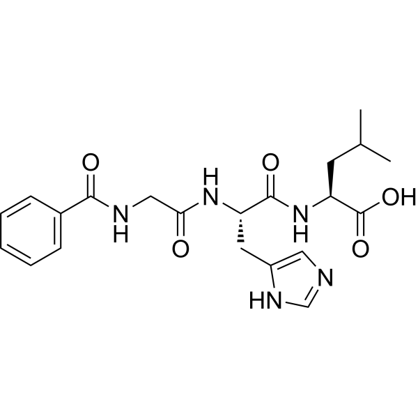
Hippuryl-His-Leu-OH (N-Benzoyl-Gly-His-Leu) is a specific substrate for angiotensin-converting enzyme (ACE I) and a molecular tool used for ACE activity detection in in vitro experiments. Hippuryl-His-Leu-OH is hydrolyzed by ACE through competitive binding. Under ACE catalysis, Hippuryl-His-Leu-OH undergoes hydrolysis to produce hippuric acid (HA). The amount of HA produced can be used to quantitatively assess ACE activity or screen for ACE inhibitors. The released His-Leu can also react with o-phthalaldehyde or Fluorescamine (HY-D0715) for fluorescence detection. Hippuryl-His-Leu-OH can be applied to the in vitro screening of ACE inhibitors for hypertension and cardiovascular diseases, and is also used in the study of ACE activity changes in physiological and pathological processes such as renal compensatory hypertrophy .
loading...
    loading...
Cat. No.: HY-W004588
CAS No.: 72914-19-3
Research Areas:  

Others Cancer

tBu2bpy is a bidentate nitrogen ligand that coordinates with Pt (II) and Pt (IV) to form functional complexes. The tBu2bpy Pt (II) bis-crown ether complex selectively recognizes Mg 2+ and Zn 2+; both ions significantly enhance the fluorescence of the complex and cause a blue shift of the absorption peak. Its six-coordinate Pt (IV) complex [Pt (X)2Me2 ( tBu2bpy)] inhibits the proliferation of tumor cells. tBu2bpy Pt (IV) complexes bind to DNA through multiple interactions including partial intercalation, groove binding, and electrostatic interaction, and can competitively displace ethidium bromide and covalently bind to purine nucleotides. tBu2bpy can be used in relevant research in fields such as organic synthesis .
loading...
    loading...
Cat. No.: HY-W014394R
CAS No.: 82654-98-6
Protoporphyrin IX (Standard) is the analytical standard of Protoporphyrin IX. This product is intended for research and analytical applications. Protoporphyrin IX is a final intermediate in the heme biosynthetic pathway, which acts as a radiation sensitizer enhancing ROS generation even in a hypoxic state and inducing DNA damage. Protoporphyrin IX also acts as a photo sensitizer undergoing photobleaching that occurs through direct degradation by light irradiation. Protoporphyrin IX is formed and accumulated following 5-aminolevulinic acid (5-ALA) (HY-W000450) administration in the tumor cells of rats. Protoporphyrin IX causes selective improvement of basal cell carcinoma when activated red fluorescence of a peak wavelength at 405 nm. Protoporphyrin IX is promising for research of sonodynamic and photodynamic agents for a wide range of cancers, such as bladder cancer and nodular basal cell carcinoma .
loading...
    loading...
Cat. No.: HY-137875
CAS No.: 207386-83-2
Purity:  98.60%
Synonyms: N-Benzoyl-Gly-His-Leu hydrate
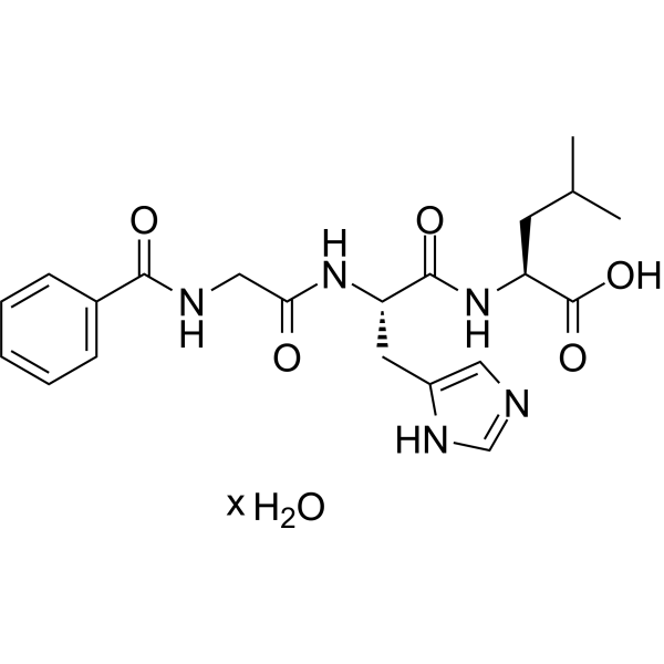
Hippuryl-His-Leu-OH (N-Benzoyl-Gly-His-Leu) hydrate is a specific substrate for angiotensin-converting enzyme ACE I and is a molecular tool used for ACE activity detection in in vitro experiments. Hippuryl-His-Leu-OH hydrate is hydrolyzed by ACE through competitive binding. Under ACE catalysis, Hippuryl-His-Leu-OH hydrate undergoes hydrolysis to produce hippuric acid (HA). The amount of HA produced can be used to quantitatively assess ACE activity or screen for ACE inhibitors. The His-Leu released from Hippuryl-His-Leu-OH hydrate can also react with o-phthalaldehyde or Fluorescamine (HY-D0715) for fluorescence detection. Hippuryl-His-Leu-OH hydrate can be applied to the in vitro screening of ACE inhibitors for hypertension and cardiovascular diseases, and is also used in the study of changes in ACE activity during physiological and pathological processes such as renal compensatory hypertrophy .
loading...
    loading...
Cat. No.: HY-D0996
CAS No.: 181885-68-7
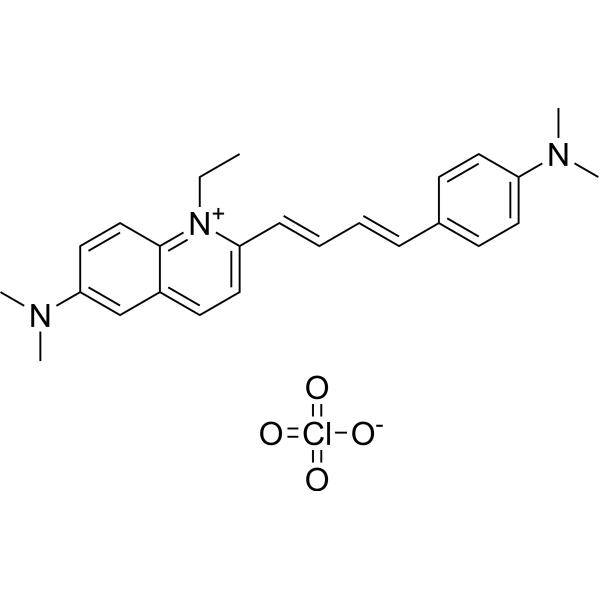
Lds-751 is a nucleic acid stain that mainly detects DNA. Lds-751 is a nucleic acid stain that mainly detects DNA. Lds-751 has a high affinity for DNA and fluorescence is enhanced after binding, but the maximum emission wavelength is 670nm. Lds-751 and Thiazole orange can be used for the differentiation of red blood cells, platelets, reticulocytes, and nucleated cells and can be stimulated at 488nm. Studies have shown that LDS-751 binds almost exclusively to mitochondria when incubated with nucleated living cells. After nucleated Acridine Orange (HY-101879) staining and LDS-751 treatment of cells, confocal microscopy revealed almost no co-location of the cells. Staining with Rhodamine 123 (HY-D0816), a dye known to bind polarized mitochondria, was almost identical to the pattern observed with LDS-751 .
loading...
    loading...
Cat. No.: HY-D1072
CAS No.: 53213-81-3
Synonyms: 3,3′-Dipentyloxacarbocyanine iodide
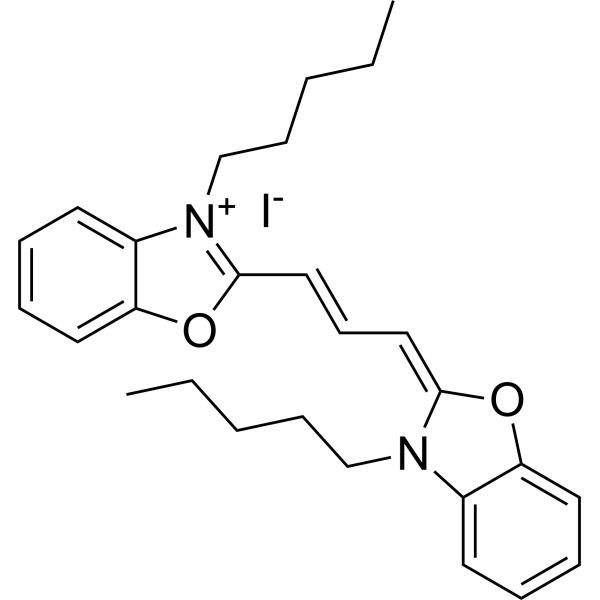
DiOC5(3) (3,3′-Dipentyloxacarbocyanine iodide) is a mitochondrial Fluorescent dye that alters membrane potential, and it also acts as an inhibitor of NADH-ubiquinone reductase (respiratory complex I). DiOC5(3) increases the NADH/NAD ratio and induces excessive production of ROS. DiOC5(3) induces Apoptosis. DiOC5(3) accumulates in the mitochondria of leukemia stem-like cells via organic anion-transporting polypeptides, specifically labels these cells, and inhibits their proliferation. DiOC5(3) enhances the cellular uptake of Hoechst 33342 (HY-15559), reduces Hoechst 33342-associated cytotoxicity, and improves the resolution of DNA content analysis by flow cytometry. DiOC5(3) shows no detectable fluorescence signal when excited in the 400-500 nm ultraviolet light range. DiOC5(3) is applicable to leukemia-related research .
loading...
    loading...
Cat. No.: HY-D1737
RADA is a fluorescent D-amino acid (FDAA) with high photostability and thermostability, which emits yellow-to-orange fluorescence. RADA shows low outer membrane permeability in wild-type Gram-negative Escherichia coli, but it targets penicillin-binding proteins and L,D-transpeptidases, mimics the interaction between acyl acceptors and enzyme intermediates, and integrates into peptidoglycan during biosynthesis. As a peptidoglycan labeling reagent, RADA metabolically integrates into the nascent peptidoglycan of live bacterial cells, labels the peptidoglycan at the poles and lateral walls of mycobacteria, and enables visualization of peptidoglycan synthesis and remodeling processes. RADA serves as a non-specific stain for fixed cells, is non-toxic to bacterial cells, and its red-shifted excitation/emission spectra reduce phototoxicity. RADA also supports virtual pulse-chase labeling experiments and stochastic optical reconstruction microscopy for sub-diffraction-limited imaging of bacterial cell walls .
loading...
    loading...

