



SIAIS164018 hydrochloride

Based on 1 Customer Validation
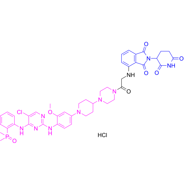
SIAIS164018 hydrochloride is a multi-kinase (ALK, EGFR, FAK, PYK2, PTK6) PROTAC degrader, with IC50 values of 2.5 nM and 6.6 nM against ALK and ALK (G1202R), respectively. SIAIS164018 hydrochloride promotes the ubiquitination and degradation of ALK, EGFR, FAK, PYK2 and PTK6. SIAIS164018 hydrochloride induces cell cycle arrest at the G1 phase and triggers cell Apoptosis. SIAIS164018 hydrochloride exhibits preferential inhibitory activity against FER kinase. SIAIS164018 hydrochloride can be used in research related to non-small cell lung cancer, ovarian cancer and triple-negative breast cancer.
(Pink: Anaplastic lymphoma kinase (ALK) and EGFR and FAK and PYK2 and PTK6 ligand (HY-150910); Blue: Cereblon ligand (HY-14658); Black: linker).
연구목적의 판매만을 진행합니다. 환자를 대상으로 한 판매는 하지 않습니다.
- Purity : 98.06%
- 화학식: C43H49Cl2N10O7P
- 분자량:919.79
-
보관:
4°C, sealed storage, away from moisture
* In solvent : -80°C, 6 months; -20°C, 1 month (sealed storage, away from moisture)

To place orders, for customer services and technical support, please contact: MedChemExpress USA
Tel: 609-228-6898 E-mail: [email protected] [email protected]


-
 Biological Activity
-
Chemical Information
-
용액&용해도
- Protocol
- 순도&문서
- References
-
 Help & FAQs


-
HY-P3212
Allo-acaLeptin Receptor Antagonist Peptide -

HY-162874
diABZI-V/C-DBCOSTING Agonist -
HY-105155
LobradimilBradykinin B2 Receptor Agonist -
HY-139100B
m7G(5')ppp(5')A diammoniumCap Analog -
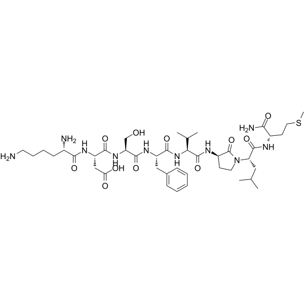
HY-P1278
GR 64349NK2 Receptor Agonist -
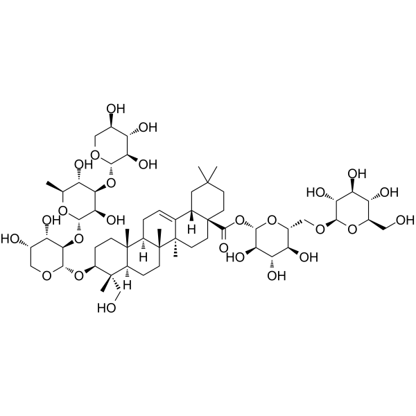
HY-N9319
Fulvotomentoside ATriterpenoid Saponin -

HY-141466A
Crotonyl-CoA tetrasodiumDerivative of CoA -
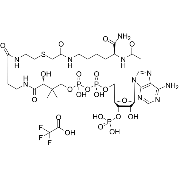
HY-131035
Ac-Lys(CoA)-NH2 TFAp300 HAT Inhibitor -
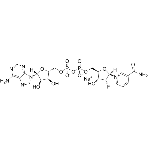
HY-134358A
Ara-F-NAD+ sodiumCD38 Inhibitor -

HY-P3134A
CPN-267 TFANMUR1 Agonist -

HY-P6374
IIQLPEIVVV TFADrp1-Mff Interaction Inhibitor -
HY-P2616A
Mca-YVADAP-Lys(Dnp)-OH TFACaspase1/ACE2 Substrate


Biological Activity
제품 설명
IC50 & Target
|
Cereblon |
In Vitro
SIAIS164018 (0-1 μM; 16 h) hydrochloride significantly inhibits the proliferation of SR cells[2].
SIAIS164018 hydrochloride preferentially inhibits FER kinase activity over ALK kinase activity in cell-free KINOMEScan kinase profiling assays[3].
SIAIS164018 (0-100 nM; 72 h) hydrochloride exhibits better cell proliferation inhibitory activity than Brigatinib (HY-12857) in ALK (G1202R)-overexpressing 293T cells and EGFR-expressing H1975 cell lines[2].
SIAIS164018 (100 nM; 24-48 h) hydrochloride induces significant G1 cell cycle arrest in ALK-negative Calu-1 and MDA-MB-231 cells[2].
SIAIS164018 (<10 nM-100 nM) hydrochloride degrades ALK at concentrations of <10 nM and degrades EGFR at concentrations of 100 nM[4].
SIAIS164018 hydrochloride inhibits the proliferation of 293T cells overexpressing ALKG1202R, with an IC50 of 21 nM[4].
SIAIS164018 (0.01-1000 nM; 16 h) hydrochloride downregulates the protein levels of FAK, PYK2, FER, RSK1 and GAK in ALK-positive SR cell lines and ALK-negative Calu-1 cell lines[2].
MedChemExpress (MCE) has not independently confirmed the accuracy of these methods. They are for reference only. Further protocols information, click here.
-
Cell Line:SR cells
-
Concentration:0-1 μM
-
Incubation Time:16 h
-
Result:Significantly inhibited SR cell proliferation with an IC50 value of 2 nM.
-
Cell Line:SR and Calu-1 cells
-
Concentration:0.01, 0.1, 1, 10, 100 and 1000 nM
-
Incubation Time:16 h
-
Result:Down-regulated the protein level of FAK, PYK2, FER, RSK1, and GAK.
-
Cell Line:ALK-negative Calu-1 and MDA-MB-231 cells
-
Concentration:100 nM
-
Incubation Time:24 h, 48 h
-
Result:Induced a significant G1 cell cycle arrest in ALK-negative Calu-1 and MDA-MB-231 cells.
In Vivo
MedChemExpress (MCE) has not independently confirmed the accuracy of these methods. They are for reference only.


Chemical Information
-
Appearance Solid
-
분자량 919.79
-
화학식 C43H49Cl2N10O7P
-
Color Light brown to brown
-
SMILES
O=C1NC(C(CC1)N2C(C3=C(C2=O)C(NCC(N4CCN(C5CCN(CC5)C6=CC=C(C(OC)=C6)NC7=NC=C(C(NC8=C(P(C)(C)=O)C=CC=C8)=N7)Cl)CC4)=O)=CC=C3)=O)=O.Cl
-
선적
Room temperature in continental US; may vary elsewhere.
-
보관
4°C, sealed storage, away from moisture
* In solvent : -80°C, 6 months; -20°C, 1 month (sealed storage, away from moisture)


용액&용해도
In Vitro:
DMSO : 75 mg/mL (81.54 mM; Need ultrasonic; Hygroscopic DMSO has a significant impact on the solubility of product, please use newly opened DMSO)
Please refer to the solubility information to select the appropriate solvent. Once prepared, please aliquot and store the solution to prevent product inactivation from repeated freeze-thaw cycles.
Storage method and period of stock solution: -80°C, 6 months; -20°C, 1 month (sealed storage, away from moisture). When stored at -80°C, please use it within 6 months. When stored at -20°C, please use it within 1 month.
Please refer to the solubility information to select the appropriate solvent. Once prepared, please aliquot and store the solution to prevent product inactivation from repeated freeze-thaw cycles.
Storage method and period of stock solution: -80°C, 6 months; -20°C, 1 month (sealed storage, away from moisture). When stored at -80°C, please use it within 6 months. When stored at -20°C, please use it within 1 month.
Concentration (start) × Volume (start) = Concentration (final) × Volume (final)

Protocol
-
Apoptosis
Apoptosis, also called programmed cell death, is generally characterized by distinct morphological characteristics.
-
TUNEL staining for apoptotic DNA fragmentation
TUNEL staining detects DNA strand breaks by using terminal deoxynucleotidyl transferase to add labeled nucleotides to exposed 3′-OH DNA termini, generating either microscopic staining in fixed cells or tissue sections, or fluorescence/cytometric signal in cell suspensions. TUNEL positivity reflects DNA fragmentation but should not be interpreted alone as definitive apoptosis, because TUNEL can also label necrotic, autolytic, mechanically damaged, or DNA-repair-associated DNA breaks.
-
Cell Cytotoxicity Assay
Cytotoxicity assays are usually based on the assessment of cell membrane damage, which can also be indirectly detected by measuring cell viability. Detection methods include MTT assay, CKK-8 assay, LDH assay and ATP assay, etc.
-
Flow cytometric DNA-content cell-cycle staining
Flow cytometric DNA-content cell-cycle staining measures the fluorescence intensity of DNA-bound fluorochromes in single cells or nuclei to estimate DNA content distributions, allowing assignment of populations to G0/G1, S, and G2/M phases by DNA histogram deconvolution. Propidium iodide (PI) intercalates into DNA, and PI fluorescence is proportional to cellular DNA content when staining is performed under conditions that make DNA accessible and minimize non-DNA signal. Cells with G2/M DNA content are expected to show approximately twice the fluorescence intensity of G0/G1 cells, while S-phase cells occupy intermediate fluorescence values. PI-based DNA-content analysis can also detect cells with fractional DNA content, often reported as sub-G1, when DNA fragmentation and extraction during staining reduce retained DNA signal in apoptotic cells. DAPI is an alternative DNA fluorochrome for univariate DNA-content analysis, while bivariate approaches combining DNA content with proliferation
-
Annexin V plus membrane-impermeant dye apoptosis staining
Annexin V-based apoptosis assays rely on the detection of phosphatidylserine (PS) externalization from the inner leaflet of the plasma membrane to the outer leaflet, an early biochemical hallmark of apoptosis. Fluorescently labeled Annexin V binds PS in a calcium-dependent manner, enabling identification of early apoptotic cells by flow cytometry or fluorescence microscopy. When combined with a membrane-impermeant DNA-binding dye (e. g. , propidium iodide), this approach allows discrimination between viable (Annexin V−/dye−), early apoptotic (Annexin V+/dye−), and late apoptotic or necrotic (Annexin V+/dye+) cell populations by assessing membrane integrity and PS exposure.
-
BrdU Incorporation Assay
Bromodeoxyuridine (BrdU) incorporation assay is based on the principle that BrdU, a thymidine analog, is incorporated into newly synthesized DNA during the S phase of the cell cycle, thereby serving as a marker of DNA replication and cellular proliferation. Incorporated BrdU can be detected using anti-BrdU antibodies following DNA denaturation, enabling visualization or quantification of proliferating cells through immunochemical detection methods such as immunofluorescence or immunohistochemistry.
-
Apoptosis Solutions
Apoptosis is a regulated, generally non-lytic cell-death pathway that removes unwanted, damaged, infected, or abnormal cells through coordinated morphological changes, caspase activation, DNA fragmentation, and membrane remodeling. The intrinsic apoptosis pathway is controlled mainly by mitochondrial outer membrane permeabilization, BCL-2 family proteins, cytochrome c release, apoptosome formation, caspase-9 activation, and downstream executioner caspase-3/7 activation. The extrinsic apoptosis pathway is initiated by death receptors such as Fas, TNFR, and TRAIL receptors, which recruit adaptor proteins and activate caspase-8 before engaging executioner caspases or mitochondrial amplification through BID cleavage. Apoptosis is linked to many phenotypes, including cancer cell killing, tissue homeostasis, immune regulation, neurodegeneration, infection response, and treatment-induced cytotoxicity; unresolved questions include how apoptosis interacts with necroptosis, pyroptosis, ferroptos
-
Protocol for Cell Cycle
Cell-cycle analysis by flow cytometry measures DNA content in single cells to estimate the fraction of cells in G0/G1, S, and G2/M phases. Propidium iodide intercalates into DNA, and after RNA removal with RNase, fluorescence intensity reflects cellular DNA content: 2N cells are assigned to G0/G1, cells between 2N and 4N to S phase, and 4N cells to G2/M. DNA-content analysis alone cannot reliably separate G0 from G1 or G2 from M. Ki-67 can distinguish quiescent G0 cells from cycling cells, EdU or BrdU incorporation marks active DNA synthesis in S phase, and phospho-histone H3 staining identifies mitotic cells within the 4N population.
-
Breast Cancer Modeling
Breast cancer is a heterogeneous cancer, and it has been distinguished into four subtypes: luminal A, luminal B, HER2-positive and basal-like. Molecular mutations, epigenetic alterations, hormone exposure and immune microenvironment are related to the progression of breast cancer.


순도&문서
-
Data Sheet (281 KB)
-
SDS (252 KB)
- English - EN (252 KB)
- Français - FR (252 KB)
- Deutsch - DE (252 KB)
- Norwegian - NO (252 KB)
- Español - ES (252 KB)
- Swedish - SV (252 KB)
- Italian - IT (252 KB)
- Korean - KR (252 KB)
- Portuguese - PT (252 KB)
-
Handling Instructions (2659 KB)
References
[1]. Kossakowski K, et al. FDA-approved kinase inhibitors in PROTAC design, development and synthesis. Journal of enzyme inhibition and medicinal chemistry. 2025 Dec;40(1):2542357. [Content Brief]
[2]. Ren C, et al. Discovery of a Brigatinib Degrader SIAIS164018 with Destroying Metastasis-Related Oncoproteins and a Reshuffling Kinome Profile. Journal of medicinal chemistry. 2021 Jul 08;64(13):9152-9165. [Content Brief]
[3]. Zhang Y, et al. Development of the nonreceptor tyrosine kinase FER-targeting PROTACs as a potential strategy for antagonizing ovarian cancer cell motility and invasiveness. The Journal of biological chemistry. 2023 Jun;299(6):104825. [Content Brief]
[4]. Rallabandi NC, et al. EGFR molecular degraders: preclinical successes and the road ahead. Future Med Chem. 2025 Mar;17(6):633-636. [Content Brief]


Complete Stock Solution Preparation Table
Please refer to the solubility information to select the appropriate solvent. Once prepared, please aliquot and store the solution to prevent product inactivation from repeated freeze-thaw cycles.
Storage method and period of stock solution: -80°C, 6 months; -20°C, 1 month (sealed storage, away from moisture). When stored at -80°C, please use it within 6 months. When stored at -20°C, please use it within 1 month.
| Optional Solvent | Concentration Solvent Mass | 1 mg | 5 mg | 10 mg | 25 mg |
|---|---|---|---|---|---|
| DMSO | 1 mM | 1.0872 mL | 5.4360 mL | 10.8720 mL | 27.1801 mL |
| 5 mM | 0.2174 mL | 1.0872 mL | 2.1744 mL | 5.4360 mL | |
| 10 mM | 0.1087 mL | 0.5436 mL | 1.0872 mL | 2.7180 mL | |
| 15 mM | 0.0725 mL | 0.3624 mL | 0.7248 mL | 1.8120 mL | |
| 20 mM | 0.0544 mL | 0.2718 mL | 0.5436 mL | 1.3590 mL | |
| 25 mM | 0.0435 mL | 0.2174 mL | 0.4349 mL | 1.0872 mL | |
| 30 mM | 0.0362 mL | 0.1812 mL | 0.3624 mL | 0.9060 mL | |
| 40 mM | 0.0272 mL | 0.1359 mL | 0.2718 mL | 0.6795 mL | |
| 50 mM | 0.0217 mL | 0.1087 mL | 0.2174 mL | 0.5436 mL | |
| 60 mM | 0.0181 mL | 0.0906 mL | 0.1812 mL | 0.4530 mL | |
| 80 mM | 0.0136 mL | 0.0680 mL | 0.1359 mL | 0.3398 mL |


SIAIS164018 hydrochloride Related Classifications
HY-147219A Related Classifications
 Powered by Bioz