



SK1-I
Based on 1 publication(s) in Google Scholar
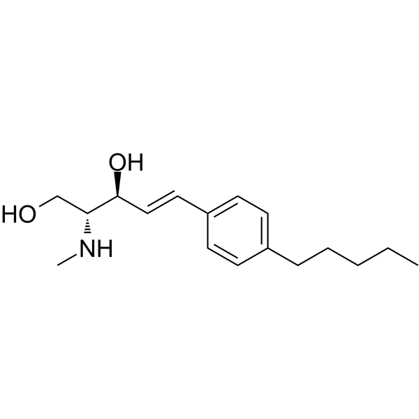
SK1-I (BML-258), an analog of sphingosine, is an isozyme-specific competitive SPHK1 inhibitor with a Ki value of 10 μM. SK1-I shows no activity at SPHK1 PKCα, PKCδ, PKA, AKT1, ERK1, EGFR, CDK2, IKKβ or CamK2β. SK1-I enhances autophagy and has antitumor activity.
For research use only. We do not sell to patients.
- Purity : 98.83%
- CAS No.: 1072443-89-0
- Formula: C17H27NO2
- Molecular Weight:277.40
-
Storage:Powder -20°C, 3 years , 4°C, 2 years ; In solvent -80°C, 6 months , -20°C, 1 month

To place orders, for customer services and technical support, please contact: MedChemExpress USA
Tel: 609-228-6898 E-mail: [email protected] [email protected]


-
 Biological Activity
-
Chemical Information
-
Solvent & Solubility
- Protocols
- Purity & Documentation
- References
-
 Help & FAQs


-
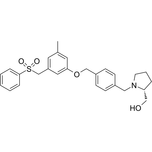
HY-15425
PF-543SPHK1 Inhibitor -
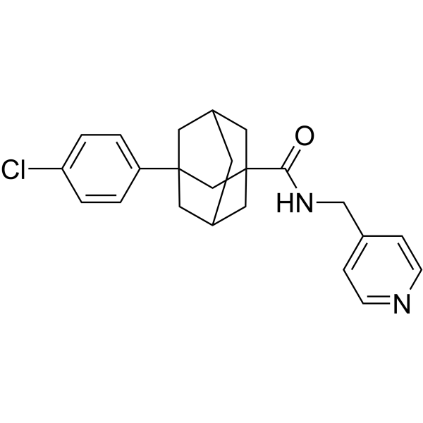
HY-16015
Opaganib -
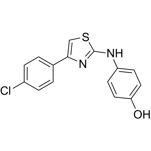
HY-13822
SKI IISphK/DES1 Inhibitor -
HY-101805
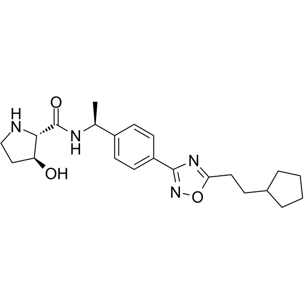
SK1-IN-1SPHK1 Inhibitor -
HY-15779A
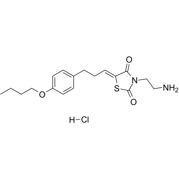
K145 hydrochlorideSphK2 Inhibitor -

HY-18739
Phorbol 12-myristate 13-acetatePKC Activator -
HY-12892
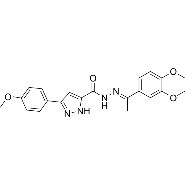
SKI-178SphK1/SphK2 Inhibitor -
HY-108491
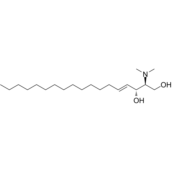
N,N-DimethylsphingosineSphingosine Kinase Inhibitor -

HY-16015R
Opaganib (Standard) -

HY-18739R
Phorbol 12-myristate 13-acetate (Standard)PKC Activator -
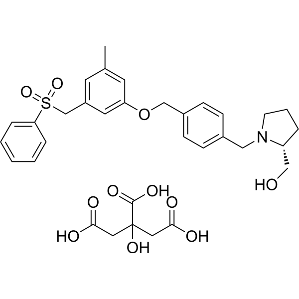
HY-15425A
PF-543 CitrateSPHK1 Inhibitor -
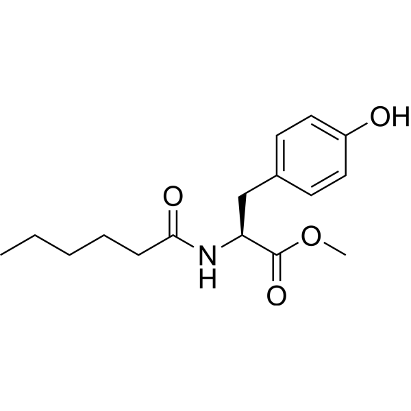
HY-101572
MHPSPHK1 Activator
Publications Citing Use of MedChemExpress (MCE) SK1-I
More

Biological Activity
Description
IC50 & Target
|
SphK1 |
Cellular Effect
|
Cell Line

|
Type | Value | Description | References |
|---|---|---|---|---|
| HEK293 | IC50 |
10 μM
Compound: 7, SK1-I
|
Competitive inhibition of V5-His-tagged human SphK2 expressed in HEK293 cells assessed as S1P production using sphingosine and [gamma-32P]ATP as substrate by thin layer chromatography
Competitive inhibition of V5-His-tagged human SphK2 expressed in HEK293 cells assessed as S1P production using sphingosine and [gamma-32P]ATP as substrate by thin layer chromatography
|
[PMID: 24471412] |
In Vitro
SK1-I (0-10 μM; 24 hours) attenuates cancer cell growth and survival in a TP53-dependent manner in HCT116 cells and HCT116 cells bearing TP53 null cancer[2].
SK1-I (0-20 μM; 12 hours) induces more CASP3 cleavage in HCT116 cells, compared to HCT116 cells lacking TP53, leading to a hallmark of apoptosis[2].
MedChemExpress (MCE) has not independently confirmed the accuracy of these methods. They are for reference only. Further protocols information, click here.
-
Cell Line:HCT116 cells and HCT116 cells bearing TP53 null cancer
-
Concentration:0 µM, 2.5 µM, 5 µM, 7.5 µM, 10 µM
-
Incubation Time:24 hours
-
Result:Decreased cancer cell growth and survival.
-
Cell Line:HCT116 cells and HCT116 cells bearing TP53 null cancer
-
Concentration:0 µM, 5 µM, 10 µM, 20 µM
-
Incubation Time:12 hours
-
Result:Induced more CASP3 cleavage in HCT116 cells, compared to HCT116 cells lacking TP53.
In Vivo
MedChemExpress (MCE) has not independently confirmed the accuracy of these methods. They are for reference only.
-
Animal Model:Male C57BL/6 mice (24 ± 3.5 g) [3]
-
Dosage:75 mg/kg
-
Administration:Intraperitoneal (i.p.) injection; once; 24 hours prior to baseline MAP measurement
-
Result:Significantly lowered baseline mean arterial blood pressure (MAP).


Chemical Information
-
CAS No. 1072443-89-0
-
Appearance Solid
-
Molecular Weight 277.40
-
Formula C17H27NO2
-
Color Off-white to light yellow
-
SMILES
OC[C@H]([C@H](/C=C/C1=CC=C(C=C1)CCCCC)O)NC
-
Synonyms
BML-258
-
Shipping
Room temperature in continental US; may vary elsewhere.
-
Storage
Powder -20°C 3 years 4°C 2 years In solvent -80°C 6 months -20°C 1 month


Publications (1)
-
Journal Impact Factor
-
Most Recent
-
PLoS Pathog
Enzymatic independent role of sphingosine kinase 2 in regulating the expression of type I interferon during influenza A virus infection. [Abstract]2022 Sep 7;18(9):e1010794. PMID: 36070294


Solvent & Solubility
In Vitro:
DMSO : 100 mg/mL (360.49 mM; Need ultrasonic; Hygroscopic DMSO has a significant impact on the solubility of product, please use newly opened DMSO)
Please refer to the solubility information to select the appropriate solvent. Once prepared, please aliquot and store the solution to prevent product inactivation from repeated freeze-thaw cycles.
Storage method and period of stock solution: -80°C, 6 months; -20°C, 1 month. When stored at -80°C, please use it within 6 months. When stored at -20°C, please use it within 1 month.
Please refer to the solubility information to select the appropriate solvent. Once prepared, please aliquot and store the solution to prevent product inactivation from repeated freeze-thaw cycles.
Storage method and period of stock solution: -80°C, 6 months; -20°C, 1 month. When stored at -80°C, please use it within 6 months. When stored at -20°C, please use it within 1 month.
Concentration (start) × Volume (start) = Concentration (final) × Volume (final)

Protocols
-
Autophagy
Autophagy is a process in which eukaryotic cells use lysosomes to degrade their own cytoplasmic proteins and damaged organelles under the regulation of autophagy related gene (Atg). Microtubule-associated proteins light chain 3 (LC3) is recognized as autophagy marker, which transfers from cytoplasmic LC3 (LC3-I) to membrane type (LC3-II). LC3-II/I ratio could be detected by Western Blot and fluorescence microscopy.
-
Lysosome and acidic-vesicle live-cell staining
Lysosome and acidic-vesicle live-cell staining detects acidic intracellular compartments by using membrane-permeant acidotropic probes that accumulate in low-pH vesicles, including lysosomes, late endosomes, autolysosomes, and acidic phagosomes. LysoTracker staining is commonly used as an intensity-based readout of acidic lysosomal compartment abundance or enlargement, while acridine orange produces green fluorescence in less concentrated compartments and red fluorescence after concentration-dependent accumulation in acidic vesicular organelles. Loss or reduction of acridine-orange red signal can be used as a readout of lysosomal membrane permeabilization or reduced acidic-vesicle integrity. This protocol is designed for live cultured cells and can be adapted for fluorescence microscopy, high-content imaging, plate-reader readout, or flow cytometry when the selected literature supports the readout. Because these dyes report acidotropic accumulation rather than lysosome identity alone,
-
Subcutaneous Cell-Line-Derived Xenograft
Subcutaneous cell-line-derived xenograft (CDX) models are established by implanting cultured human cancer cell lines into immunodeficient mice, where the injected cells form localized tumors that can be monitored in vivo as a measure of tumorigenic potential, growth kinetics, and treatment response. These models are widely used in oncology research because they allow reproducible tumor formation and enable comparative assessment of tumor growth between different cell lines or genetic manipulations in a controlled in vivo microenvironment. Subcutaneous implantation of cancer cells in immunodeficient mice is a standard approach for evaluating tumor growth behavior and therapeutic response across multiple cancer types, including prostate, esophageal, pancreatic, and colon cancer models.
-
Macroautophagy Solutions
Macroautophagy is a conserved lysosome-dependent degradation pathway in which cytoplasmic material is sequestered into double-membrane autophagosomes and delivered to lysosomes for degradation and recycling. The pathway supports cellular homeostasis during nutrient limitation, organelle stress, protein-aggregate accumulation, infection, differentiation, and tissue remodeling by coupling cargo sequestration, autophagosome maturation, lysosomal fusion, and degradation of cargo-derived macromolecules. The core molecular sequence includes initiation by nutrient- and stress-regulated autophagy machinery, autophagosome nucleation, LC3/ATG8-family conjugation to autophagosomal membranes, cargo selection through receptors such as SQSTM1/p62, autophagosome-lysosome fusion, and lysosomal degradation. LC3 was identified as a mammalian homolog of yeast Atg8 that localizes to autophagosomal membranes after processing, and p62/SQSTM1 was shown to connect ubiquitinated cargo with autophagic degradati


Purity & Documentation
-
Data Sheet (271 KB)
-
SDS (251 KB)
- English - EN (251 KB)
- Français - FR (251 KB)
- Deutsch - DE (251 KB)
- Norwegian - NO (251 KB)
- Español - ES (251 KB)
- Swedish - SV (251 KB)
- Italian - IT (251 KB)
- Korean - KR (251 KB)
- Portuguese - PT (251 KB)
-
Handling Instructions (2659 KB)
References
[1]. Melissa R Pitman, et al. Inhibitors of the sphingosine kinase pathway as potential therapeutics. Curr Cancer Drug Targets. 2010 Jun;10(4):354-67. [Content Brief]
[2]. Santiago Lima, et al. TP53 is required for BECN1- and ATG5-dependent cell death induced by sphingosine kinase 1 inhibition. Autophagy. 2018;14(6):942-957. [Content Brief]
[3]. Fiona H Greig, et al. Requirement for sphingosine kinase 1 in mediating phase 1 of the hypotensive response to anandamide in the anaesthetised mouse. Eur J Pharmacol. 2019 Jan 5;842:1-9. [Content Brief]


Complete Stock Solution Preparation Table
Please refer to the solubility information to select the appropriate solvent. Once prepared, please aliquot and store the solution to prevent product inactivation from repeated freeze-thaw cycles.
Storage method and period of stock solution: -80°C, 6 months; -20°C, 1 month. When stored at -80°C, please use it within 6 months. When stored at -20°C, please use it within 1 month.
| Optional Solvent | Concentration Solvent Mass | 1 mg | 5 mg | 10 mg | 25 mg |
|---|---|---|---|---|---|
| DMSO | 1 mM | 3.6049 mL | 18.0245 mL | 36.0490 mL | 90.1226 mL |
| 5 mM | 0.7210 mL | 3.6049 mL | 7.2098 mL | 18.0245 mL | |
| 10 mM | 0.3605 mL | 1.8025 mL | 3.6049 mL | 9.0123 mL | |
| 15 mM | 0.2403 mL | 1.2016 mL | 2.4033 mL | 6.0082 mL | |
| 20 mM | 0.1802 mL | 0.9012 mL | 1.8025 mL | 4.5061 mL | |
| 25 mM | 0.1442 mL | 0.7210 mL | 1.4420 mL | 3.6049 mL | |
| 30 mM | 0.1202 mL | 0.6008 mL | 1.2016 mL | 3.0041 mL | |
| 40 mM | 0.0901 mL | 0.4506 mL | 0.9012 mL | 2.2531 mL | |
| 50 mM | 0.0721 mL | 0.3605 mL | 0.7210 mL | 1.8025 mL | |
| 60 mM | 0.0601 mL | 0.3004 mL | 0.6008 mL | 1.5020 mL | |
| 80 mM | 0.0451 mL | 0.2253 mL | 0.4506 mL | 1.1265 mL | |
| 100 mM | 0.0360 mL | 0.1802 mL | 0.3605 mL | 0.9012 mL |


SK1-I Related Classifications
HY-119016 Related Classifications
 Powered by Bioz