



Soyasaponin Ba
Based on 1 publication(s) in Google Scholar
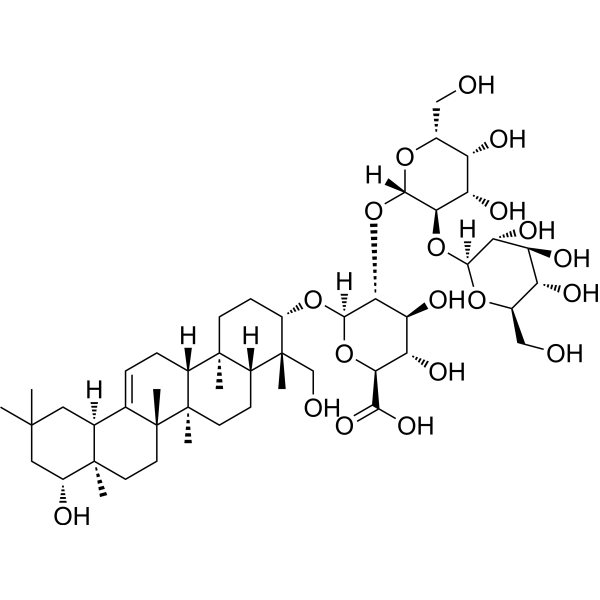
Soyasaponin Ba is a soyasaponin that can be isolated from Phaseolus vulgaris, acts as an aldose reductase inhibitor (ARI). Soyasaponin Ba activates Akt/GSK3β/β-catenin signaling pathway, reduces lipid accumulation, lowers ROS generation, improves mitochondrial membrane potential, ATP levels, and morphology, and inhibits apoptosis. Soyasaponin Ba can be used for the research of lipid accumulation and secondary diabetic complications.
For research use only. We do not sell to patients.
- Purity : 99.47%
- CAS No.: 114590-20-4
- Formula: C48H78O19
- Molecular Weight:959.12
-
Storage:
4°C, protect from light
* In solvent : -80°C, 6 months; -20°C, 1 month (protect from light)

To place orders, for customer services and technical support, please contact: MedChemExpress USA
Tel: 609-228-6898 E-mail: [email protected] [email protected]


-
 Biological Activity
-
Chemical Information
-
Solvent & Solubility
- Protocols
- Purity & Documentation
- References
-
 Help & FAQs


-
HY-N0310
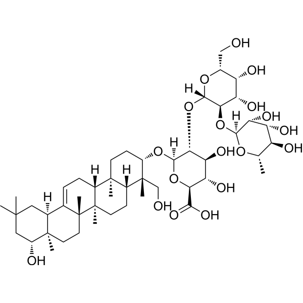
Soyasaponin BbAldose Reductase Inhibitor -
HY-N6074
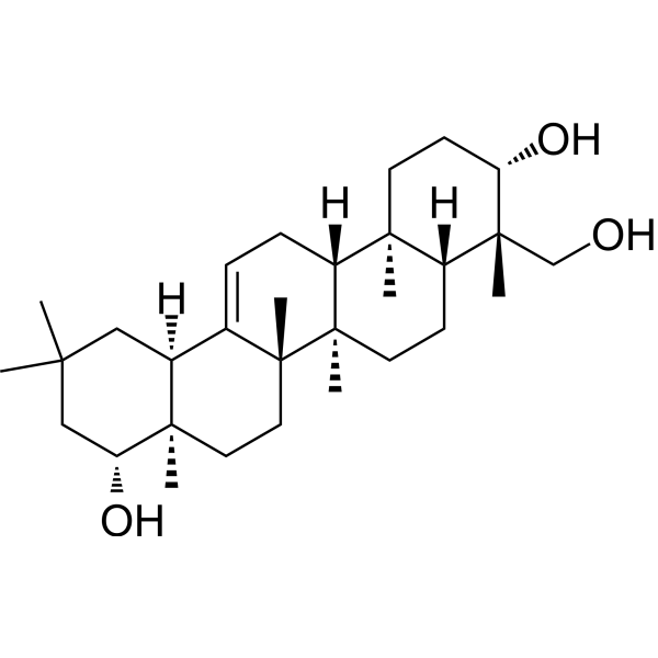
Soyasapogenol BAutophagy Inducer -
HY-N6073
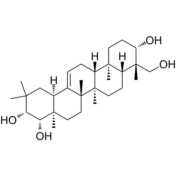
Soyasapogenol ATriterpenoid Aglycone -

HY-N0309R
Soyasaponin Ba (Standard)Aldose Reductase Inhibitor -
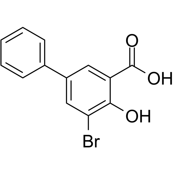
HY-114009
AKR1C1-IN-1AKR1C1 Inhibitor -
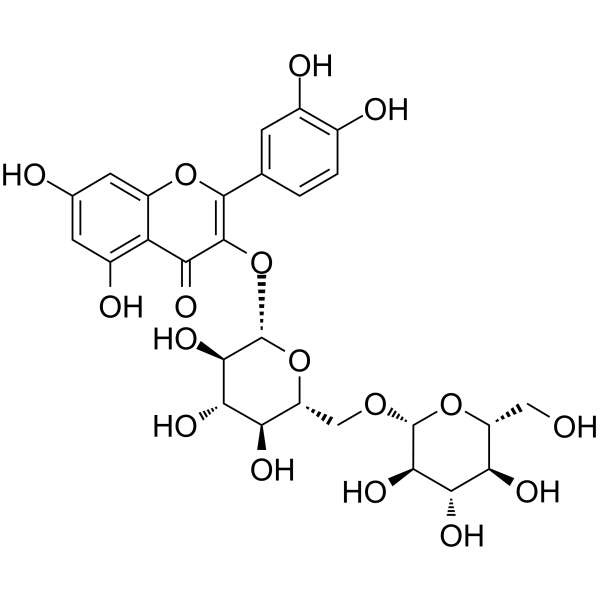
HY-N4089
Quercetin 3-gentiobiosideAromatase Inhibitor -
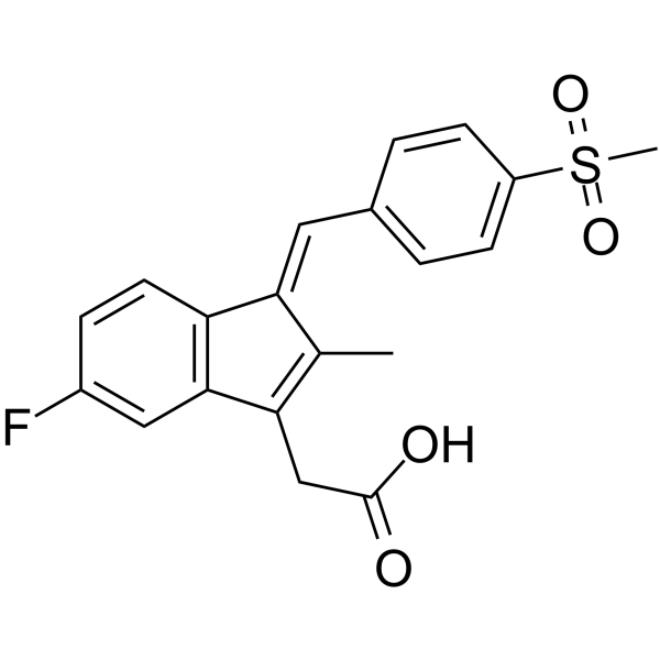
HY-13633
ExisulindAldose Reductase Inhibitor -
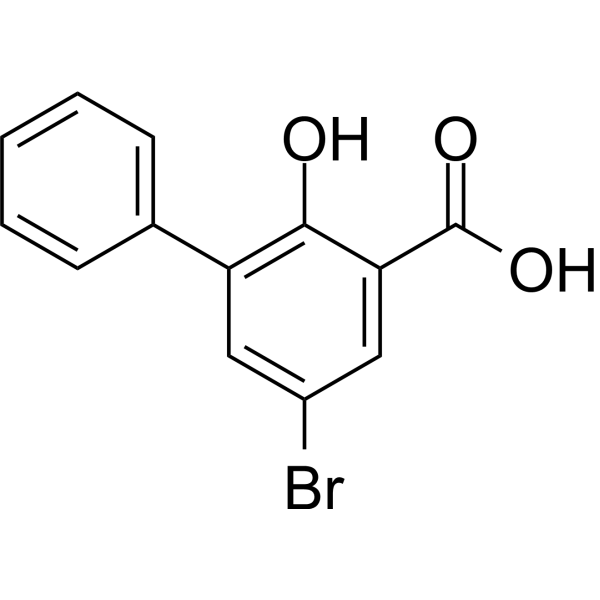
HY-118819
5-Bromo-3-phenyl salicylic acidAKR1C1 Inhibitor -
HY-N2461
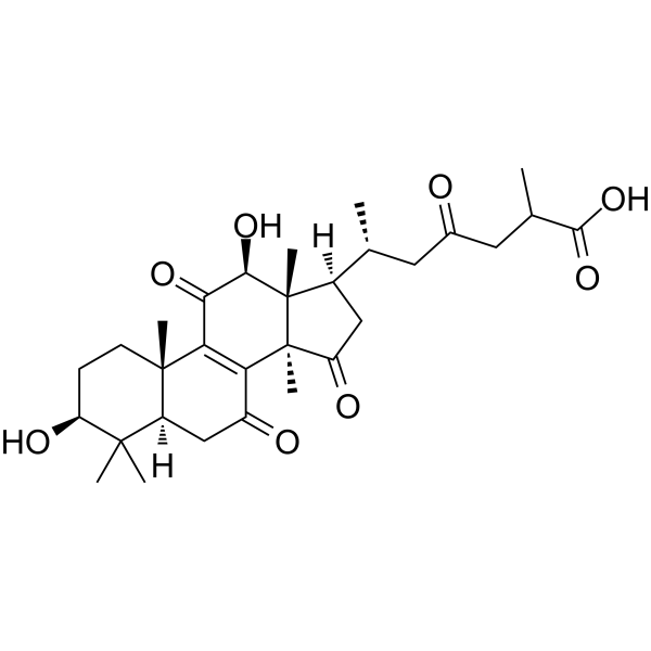
Ganoderic acid C6Aldose reductase Inhibitor -

HY-W319094
4′-HydroxyflavoneAldose Reductase Inhibitors -

HY-N4089R
Quercetin 3-gentiobioside (Standard)Standard of Quercetin 3-gentiobioside
Publications Citing Use of MedChemExpress (MCE) Soyasaponin Ba
More

Biological Activity
Description
In Vitro
Soyasaponin Ba (0.78-25 μM; 0-600 s) directly binds to purified AKT1 protein with a Kd of 4.19 μM[1].
Soyasaponin Ba (4-32 μM; 24 h) dose-dependently alleviates FFA-induced intracellular ROS accumulation, and improves FFA-induced mitochondrial dysfunction in HepG2 cells[1].
Soyasaponin Ba (4-32 μM; 24 h) dose-dependently alleviates FFA-induced apoptosis in HepG2 cells[1].
Soyasaponin Ba (16-32 μM; 24 h) activates the Akt/GSK3β/β-catenin signaling pathway in FFA-induced HepG2 cells[1].
Soyasaponin Ba (8-32 μM; 24 h) dose-dependently alleviates FFA-induced lipid accumulation in THLE-2 cells[1].
Soyasaponin Ba (16-32 μM; 24 h) dose-dependently alleviates FFA-induced ROS accumulation, mitochondrial dysfunction, and apoptosis in THLE-2 cells[1].
MedChemExpress (MCE) has not independently confirmed the accuracy of these methods. They are for reference only. Further protocols information, click here.
-
Cell Line:HepG2 cells
-
Concentration:4; 8; 16; 32 μM
-
Incubation Time:24 h
-
Result:Reduced FFA-induced apoptosis in a concentration-dependent manner, as shown by decreased green fluorescence intensity (cleaved caspase-3 activity) in single-cell imaging, normalized relative fluorescent units (RFU) in bulk culture, and reduced Annexin V-mCherry staining.
Significantly decreased caspase-3 activity at 16 and 32 μM, with effects comparable to 20 μM atorvastatin.
-
Cell Line:HepG2 cells
-
Concentration:16; 32 μM
-
Incubation Time:24 h
-
Result:Restored phosphorylated Akt and phosphorylated GSK3β levels reduced by FFA treatment at 32 μM.
Increased intracellular β-catenin and c-Myc levels reduced by FFA treatment in a concentration-dependent manner at 16 and 32 μM.
-
Cell Line:HepG2 cells
-
Concentration:4; 8; 16; 32 μM
-
Incubation Time:24 h
-
Result:Dose-dependently alleviates FFA-induced lipid accumulation in HepG2 cells.
Showed significant effects at all tested concentrations.
In Vivo
MedChemExpress (MCE) has not independently confirmed the accuracy of these methods. They are for reference only.
-
Animal Model:wild-type N2 elegans[1]
-
Dosage:16; 32; 64; 128 μM
-
Administration:incorporated into nematode growth medium; every 48 hours; 3-4 total doses; from L4 stage
-
Result:Dose-dependently reduced high-glucose induced lipid accumulation.
At 128 μM, reduced Nile Red fluorescence intensity significantly compared to the high-glucose untreated group.
Dose-dependently reduced high-glucose induced ROS accumulation.


Chemical Information
-
CAS No. 114590-20-4
-
Appearance Solid
-
Molecular Weight 959.12
-
Formula C48H78O19
-
Color White to off-white
-
SMILES
O[C@H]([C@H]1O[C@@]2([H])[C@H](O[C@]3([H])O[C@H](CO)[C@@H](O)[C@H](O)[C@H]3O)[C@@H](O)[C@@H](O)[C@@H](CO)O2)[C@H](O)[C@@H](C(O)=O)O[C@@]1([H])O[C@@H]4[C@@](CO)(C)[C@]5([H])CC[C@@]6(C)[C@]7(C)CC[C@@]8(C)[C@H](O)CC(C)(C)C[C@@]8([H])C7=CC[C@]6([H])[C@@]5(C)CC4
-
Structure Classification
-
Initial Source
-
Shipping
Room temperature in continental US; may vary elsewhere.
-
Storage
4°C, protect from light
* In solvent : -80°C, 6 months; -20°C, 1 month (protect from light)


Publications (1)
-
Journal Impact Factor
-
Most Recent
-
Phytopathology
Untargeted metabolomic analysis reveals a potential role of saponins in the partial resistance of pea (Pisum sativum) against a root rot pathogen, Aphanomyces euteiches. [Abstract]2024 Dec;114(12):2502-2514. PMID: 39186063


Solvent & Solubility
In Vitro:
DMSO : 250 mg/mL (260.66 mM; Need ultrasonic; Hygroscopic DMSO has a significant impact on the solubility of product, please use newly opened DMSO)
Please refer to the solubility information to select the appropriate solvent. Once prepared, please aliquot and store the solution to prevent product inactivation from repeated freeze-thaw cycles.
Storage method and period of stock solution: -80°C, 6 months; -20°C, 1 month (protect from light). When stored at -80°C, please use it within 6 months. When stored at -20°C, please use it within 1 month.
Please refer to the solubility information to select the appropriate solvent. Once prepared, please aliquot and store the solution to prevent product inactivation from repeated freeze-thaw cycles.
Storage method and period of stock solution: -80°C, 6 months; -20°C, 1 month (protect from light). When stored at -80°C, please use it within 6 months. When stored at -20°C, please use it within 1 month.
Concentration (start) × Volume (start) = Concentration (final) × Volume (final)

Protocols
-
3T3-L1 preadipocyte-to-adipocyte differentiation
3T3-L1 preadipocytes are induced to differentiate after growth arrest using adipogenic media containing insulin, dexamethasone, and IBMX; differentiation is assessed by lipid-droplet accumulation, triglyceride increase, Oil Red O staining, and adipocyte-marker induction such as PPARγ and C/EBPα.
-
Mitochondrial membrane-potential fluorescent assay
Mitochondrial membrane potential fluorescent assays estimate ΔΨm in living cells using lipophilic cationic dyes such as TMRM, TMRE, rhodamine 123, and JC-1, which accumulate in mitochondria according to membrane polarization; loss of signal after FCCP or CCCP treatment is interpreted as mitochondrial depolarization. TMRM/TMRE and rhodamine 123 are commonly used for semi-quantitative live-cell microscopy or flow cytometry, while JC-1 can report a shift from red aggregate fluorescence to green monomer fluorescence during depolarization; interpretation requires controls because dye concentration, quenching mode, cell type, dye efflux, and mitochondrial mass can affect fluorescence independently of ΔΨm.
-
Apoptosis
Apoptosis, also called programmed cell death, is generally characterized by distinct morphological characteristics.
-
TUNEL staining for apoptotic DNA fragmentation
TUNEL staining detects DNA strand breaks by using terminal deoxynucleotidyl transferase to add labeled nucleotides to exposed 3′-OH DNA termini, generating either microscopic staining in fixed cells or tissue sections, or fluorescence/cytometric signal in cell suspensions. TUNEL positivity reflects DNA fragmentation but should not be interpreted alone as definitive apoptosis, because TUNEL can also label necrotic, autolytic, mechanically damaged, or DNA-repair-associated DNA breaks.
-
Annexin V plus membrane-impermeant dye apoptosis staining
Annexin V-based apoptosis assays rely on the detection of phosphatidylserine (PS) externalization from the inner leaflet of the plasma membrane to the outer leaflet, an early biochemical hallmark of apoptosis. Fluorescently labeled Annexin V binds PS in a calcium-dependent manner, enabling identification of early apoptotic cells by flow cytometry or fluorescence microscopy. When combined with a membrane-impermeant DNA-binding dye (e. g. , propidium iodide), this approach allows discrimination between viable (Annexin V−/dye−), early apoptotic (Annexin V+/dye−), and late apoptotic or necrotic (Annexin V+/dye+) cell populations by assessing membrane integrity and PS exposure.
-
ROS/oxidative-stress fluorescent staining
ROS/oxidative-stress fluorescent staining uses cell-permeant fluorogenic probes that become fluorescent after oxidation inside cells or tissues; commonly used examples include DCFH-DA/DCFDA for broad cellular oxidant detection, DHE for superoxide-related signal detection, MitoSOX for mitochondrial superoxide-related signal detection, and CellROX probes for oxidative-stress-associated fluorescence readouts. The assay detects probe oxidation rather than a single ROS species unless the probe and analysis method have been chemically validated for that species. DCFH-DA enters cells, is deacetylated by intracellular esterases to DCFH, and produces fluorescent DCF after oxidation, so the readout is used as an operational measure of total cellular oxidative stress rather than a species-specific ROS measurement. DHE and MitoSOX can report superoxide-related oxidation, but red fluorescence alone can include non-specific ethidium-like oxidation products; HPLC or optimized spectral approaches are
-
Fluorescent plasma-membrane potential dye assay
Fluorescent plasma-membrane potential dye assays measure changes in cell membrane potential using voltage-sensitive dyes whose fluorescence changes when cells depolarize or hyperpolarize. Anionic bis-oxonol dyes such as DiBAC4(3) enter depolarized cells more readily and show increased fluorescence after intracellular binding, while hyperpolarization reduces dye accumulation and fluorescence. FMP/FLIPR membrane-potential dyes are used for faster, homogeneous microplate assays of ion-channel or receptor-mediated membrane-potential changes.
-
Mitochondrial membrane-potential and mitochondrial mass staining
Mitochondrial membrane potential staining measures the electrochemical polarization across the mitochondrial inner membrane in live cells using lipophilic cationic fluorescent probes; early rhodamine-based work showed that selective mitochondrial dye accumulation is lost when the mitochondrial transmembrane potential is dissipated. JC-1 reports mitochondrial polarization by shifting from green monomer fluorescence to red J-aggregate fluorescence as dye concentration increases within energized mitochondria; therefore, the red/green fluorescence ratio is used as a relative readout of mitochondrial membrane potential. TMRE or TMRM staining provides a single-channel relative readout because these cationic rhodamine esters accumulate in polarized mitochondria, and lower fluorescence indicates reduced mitochondrial polarization when acquisition and dye-loading conditions are controlled. Mitochondrial mass staining is commonly performed with MitoTracker Green FM or related MitoTracker dyes as
-
Apoptosis Solutions
Apoptosis is a regulated, generally non-lytic cell-death pathway that removes unwanted, damaged, infected, or abnormal cells through coordinated morphological changes, caspase activation, DNA fragmentation, and membrane remodeling. The intrinsic apoptosis pathway is controlled mainly by mitochondrial outer membrane permeabilization, BCL-2 family proteins, cytochrome c release, apoptosome formation, caspase-9 activation, and downstream executioner caspase-3/7 activation. The extrinsic apoptosis pathway is initiated by death receptors such as Fas, TNFR, and TRAIL receptors, which recruit adaptor proteins and activate caspase-8 before engaging executioner caspases or mitochondrial amplification through BID cleavage. Apoptosis is linked to many phenotypes, including cancer cell killing, tissue homeostasis, immune regulation, neurodegeneration, infection response, and treatment-induced cytotoxicity; unresolved questions include how apoptosis interacts with necroptosis, pyroptosis, ferroptos
-
Lipid Droplets: Oil Red O/Sudan Dye Lipid Staining
Lipid droplets are intracellular organelles with a neutral-lipid core that stores triacylglycerols and sterol esters, and Oil Red O or Sudan dyes detect these hydrophobic lipid deposits by partitioning into retained lipids in fresh or frozen specimens. Oil Red O stains neutral triglycerides and lipids in frozen tissue sections or air-dried cytologic preparations, while Sudan Black B has also been used as a histochemical fat stain for lipid-rich tissue structures.


Purity & Documentation
-
Data Sheet (284 KB)
-
SDS (252 KB)
- English - EN (252 KB)
- Français - FR (252 KB)
- Deutsch - DE (252 KB)
- Norwegian - NO (252 KB)
- Español - ES (252 KB)
- Swedish - SV (252 KB)
- Italian - IT (252 KB)
- Korean - KR (252 KB)
- Portuguese - PT (252 KB)
-
Handling Instructions (2659 KB)
References
[1]. Luo J, et al. Soyasaponin Ba Alleviates Lipid Accumulation via Mitochondrial Remodeling: Multiomics Insights. J Agric Food Chem. 2025;73(33):21199-21221. [Content Brief]
[2]. Balestri F, et al. Soyasaponins from Zolfino bean as aldose reductase differential inhibitors. J Enzyme Inhib Med Chem. 2019;34(1):350-360. [Content Brief]


Complete Stock Solution Preparation Table
Please refer to the solubility information to select the appropriate solvent. Once prepared, please aliquot and store the solution to prevent product inactivation from repeated freeze-thaw cycles.
Storage method and period of stock solution: -80°C, 6 months; -20°C, 1 month (protect from light). When stored at -80°C, please use it within 6 months. When stored at -20°C, please use it within 1 month.
| Optional Solvent | Concentration Solvent Mass | 1 mg | 5 mg | 10 mg | 25 mg |
|---|---|---|---|---|---|
| DMSO | 1 mM | 1.0426 mL | 5.2131 mL | 10.4262 mL | 26.0656 mL |
| 5 mM | 0.2085 mL | 1.0426 mL | 2.0852 mL | 5.2131 mL | |
| 10 mM | 0.1043 mL | 0.5213 mL | 1.0426 mL | 2.6066 mL | |
| 15 mM | 0.0695 mL | 0.3475 mL | 0.6951 mL | 1.7377 mL | |
| 20 mM | 0.0521 mL | 0.2607 mL | 0.5213 mL | 1.3033 mL | |
| 25 mM | 0.0417 mL | 0.2085 mL | 0.4170 mL | 1.0426 mL | |
| 30 mM | 0.0348 mL | 0.1738 mL | 0.3475 mL | 0.8689 mL | |
| 40 mM | 0.0261 mL | 0.1303 mL | 0.2607 mL | 0.6516 mL | |
| 50 mM | 0.0209 mL | 0.1043 mL | 0.2085 mL | 0.5213 mL | |
| 60 mM | 0.0174 mL | 0.0869 mL | 0.1738 mL | 0.4344 mL | |
| 80 mM | 0.0130 mL | 0.0652 mL | 0.1303 mL | 0.3258 mL | |
| 100 mM | 0.0104 mL | 0.0521 mL | 0.1043 mL | 0.2607 mL |


Soyasaponin Ba Related Classifications
HY-N0309 Related Classifications
 Powered by Bioz
Keywords
- Soyasaponin Ba
- 114590-20-4
- Aldose Reductase
- Akt
- GSK-3
- β-catenin
- Reactive Oxygen Species (ROS)
- Mitochondrial Metabolism
- Apoptosis
- c-Myc
- Akt/GSK3β/β-catenin signaling pathway
- lipid accumulation
- wild-type N2 C. elegans
- HepG2 cells
- AKT1
- hAKR1B1
- apoptosis
- THLE-2 cells
- mitochondrial function
- reactive oxygen species
- Inhibitor
- inhibitor
- inhibit