



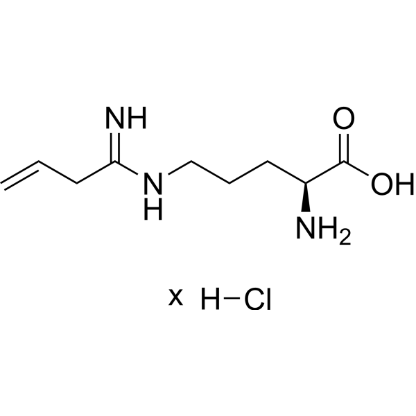
Vinyl-L-NIO hydrochloride

Based on 1 Customer Validation
Vinyl-L-NIO (L-VNIO) hydrochloride is a neuronal nitric oxide synthase (NOS) inhibitor with a rat Ki of 0.10 μM. Vinyl-L-NIO hydrochloride inhibits NADPH oxidase activity, attenuates renal fibrosis, inflammation, oxidative stress indices, and albuminuria. Vinyl-L-NIO hydrochloride can be used for the research of parkinson's disease, migraine headache, and hypertension.
For research use only. We do not sell to patients.
- Purity : 95%
- CAS No.: 728944-69-2
- Formula: C9H17N3O2.xHCl
-
Storage:
-20°C, stored under nitrogen, away from moisture
* In solvent : -80°C, 6 months; -20°C, 1 month (stored under nitrogen, away from moisture)

To place orders, for customer services and technical support, please contact: MedChemExpress USA
Tel: 609-228-6898 E-mail: [email protected] [email protected]


-
 Biological Activity
-
Chemical Information
-
Solvent & Solubility
- Protocols
- Purity & Documentation
- References
-
 Help & FAQs


-
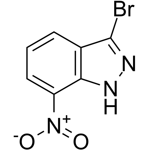
HY-101175
3-Bromo-7-nitroindazolenNOS Inhibitor -
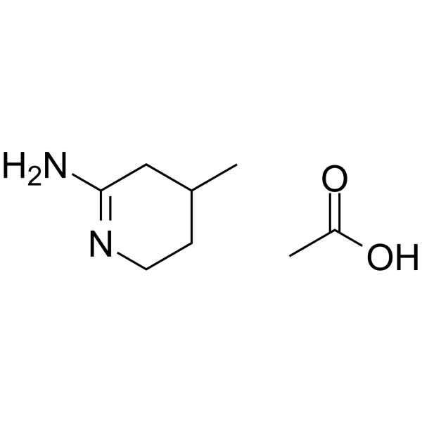
HY-138564
NOS-IN-1NO Synthase Inhibitor -
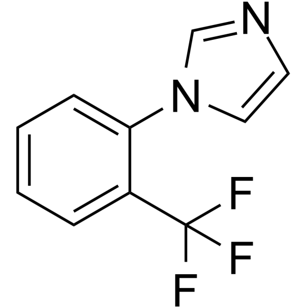
HY-101316
TRIMNOS Inhibitor -
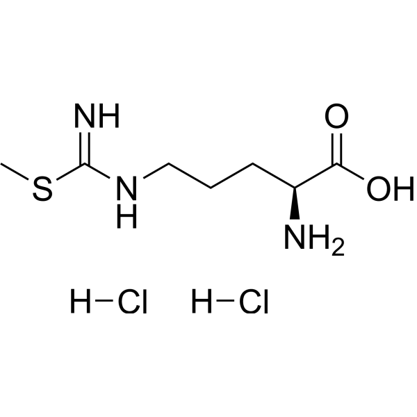
HY-U00432A
S-MTC dihydrochlorideNOS Inhibitor -
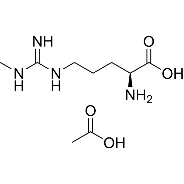
HY-18732A
L-NMMA acetateEndogenous Metabolite -
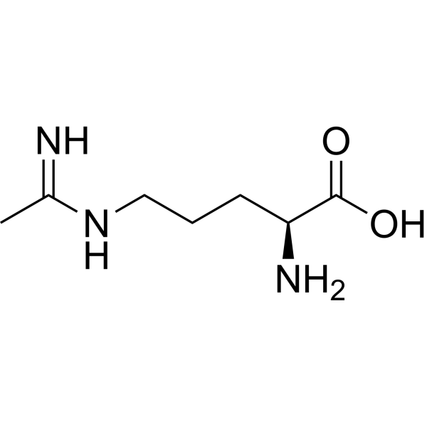
HY-100986B
L-NIONOS Inhibitor -
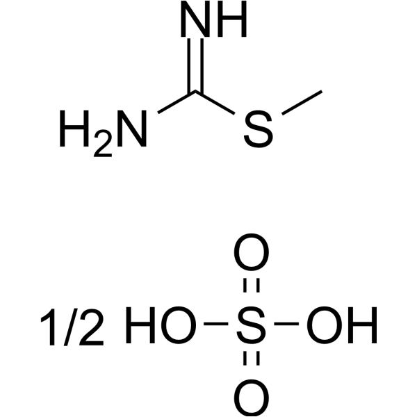
HY-79457
S-Methylisothiourea sulfateiNOS Inhibitor -
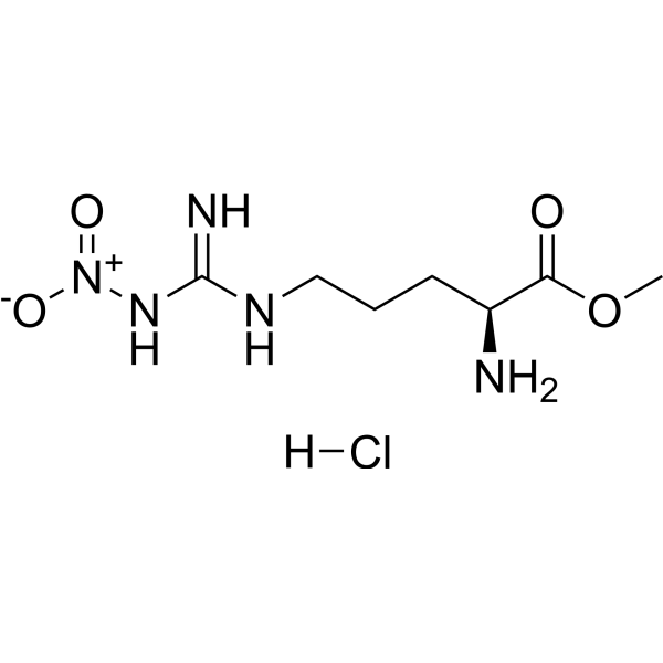
HY-18729A
L-NAME hydrochlorideNO Synthase Inhibitor -
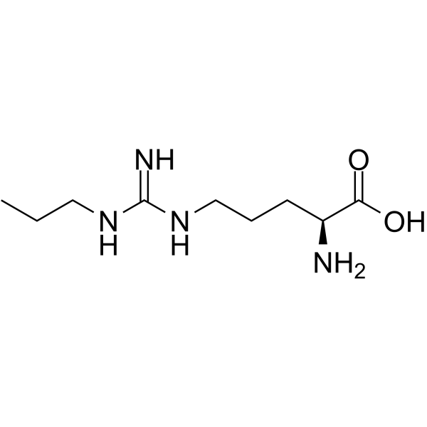
HY-102062
Nω-Propyl-L-argininenNOS Inhibitor -
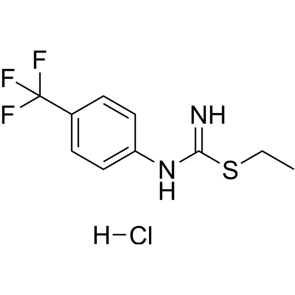
HY-W484263
hnNOS-IN-3nNOS Inhibitor -
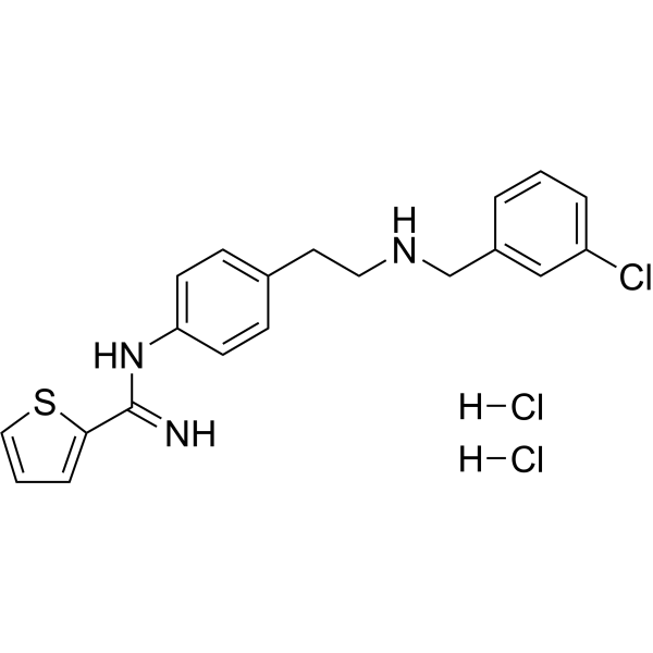
HY-107720
ARL 17477NOS1Inhibitor -
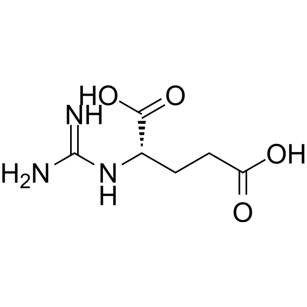
HY-130325
α-Guanidinoglutaric acidGuanidino Compound


Biological Activity
Description
In Vitro
Vinyl-L-NIO (40-160 nM; 4 h) hydrochloride potently and competitively inhibits neuronal NOS (Ki = 100 nM) with 120-fold and 600-fold lower potency against endothelia NOS and inducible NOS, respectively, demonstrating marked neuronal NOS selectivity in reversible binding[1].
Vinyl-L-NIO (0.1-1.0 μM) hydrochloride irreversibly inactivates purified neuronal NOS in a NADPH-, O2-, and Ca2+/calmodulin-dependent manner, with kinact = 0.078/min and Ki = 90 nM[1].
Vinyl-L-NIO (10 μM) hydrochloride potently inhibits the oxygenase domain-dependent NADPH oxidase activity of purified neuronal NOS but does not affect the reductase domain-specific cytochrome c reduction activity[1].
Vinyl-L-NIO (500 μM; 20 minutes) hydrochloride incubation of NOS1+/+ murine left ventricular myocytes significantly prolongs time to 50% relaxation at 1, 3, and 6 Hz, and potentiates the contractile response to β-adrenergic stimulation at 3 and 6 Hz[4].
MedChemExpress (MCE) has not independently confirmed the accuracy of these methods. They are for reference only. Further protocols information, click here.
In Vivo
MedChemExpress (MCE) has not independently confirmed the accuracy of these methods. They are for reference only.
-
Animal Model:mRen2.Lewis (female, 4 weeks old, 90-100 g at study start, high-salt diet induced hypertension and renal injury)[2]
-
Dosage:0.5 mg/kg/day
-
Administration:i.p.; daily; 28 days
-
Result:Significantly reduced systolic blood pressure at week 13 (P < 0.05), with no significant difference at weeks 12, 14, or 15 (week 15: 212 mmHg vs. untreated high-salt group 206 mmHg, P > 0.05).
Reduced albuminuria to 7.9 mg·kg-1·d-1 (vs. untreated high-salt group 109.4 mg·kg-1·d-1, P < 0.05).
Normalized creatinine clearance to 0.9 mL/min (vs. untreated high-salt group 0.4 mL/min, P < 0.05).
Significantly reduced renal injury scores: interstitial fibrosis (56 vs. untreated high-salt group 150, P < 0.05), glomerulosclerosis (6.3 vs. untreated high-salt group 150, P < 0.05), tubular dilation (6.3 vs. untreated high-salt group 125, P < 0.05), and interstitial inflammation (56 vs. untreated high-salt group 150, P < 0.05); vascular smooth muscle cell hyperplasia was unchanged.
Significantly reduced total renal cortical collagen area (collagen/field ratio ~0.007 vs. untreated high-salt group ~0.014, P < 0.05).
Reduced kidney-to-body-weight ratio to 4.4 g (vs. untreated high-salt group 5.3 g, P < 0.05).
Significantly reduced CD68-positive cell abundance to 7.7 cells per 200× area (vs. untreated high-salt group 36.8 cells per 200× area, P < 0.05).
Reduced 4-HNE relative intensity to 0.49 units (vs. untreated high-salt group 0.60 units, P < 0.05).
Reduced 8-OHdG-positive cell count to 432 cells per 200× area (vs. untreated high-salt group 929 cells per 200× area, P < 0.05).


Chemical Information
-
CAS No. 728944-69-2
-
Appearance Oil
-
Formula C9H17N3O2.xHCl
-
Color Off-white to light yellow
-
SMILES
N[C@@H](CCCNC(CC=C)=N)C(O)=O.Cl.[x]
-
Synonyms
L-VNIO hydrochloride
-
Shipping
Room temperature in continental US; may vary elsewhere.
-
Storage
-20°C, stored under nitrogen, away from moisture
* In solvent : -80°C, 6 months; -20°C, 1 month (stored under nitrogen, away from moisture)


Solvent & Solubility
In Vitro:
DMSO : 50 mg/mL (Need ultrasonic and warming; Hygroscopic DMSO has a significant impact on the solubility of product, please use newly opened DMSO)

Protocols
-
Fibrosis/Collagen Morphometry
Fibrosis and collagen morphometry is based on the quantitative visualization of fibrillar collagen deposition in tissue sections using histochemical stains such as Sirius Red (Picrosirius Red) or Masson's trichrome, followed by image-based or polarization-enhanced analysis to estimate collagen proportional area as a surrogate of extracellular matrix accumulation during fibrotic remodeling. Sirius Red combined with polarized light microscopy enhances detection of collagen fibers due to birefringence properties, enabling more specific visualization of collagen type I and III fibrils compared to conventional bright-field histology, while whole-section or region-restricted digital morphometry reduces field-selection bias in fibrosis assessment. Alternative quantitative approaches include second harmonic generation (SHG) and two-photon excited fluorescence microscopy, which enable label-free detection of fibrillar collagen and have been validated against histological staining and biochemica
-
Collagen: Sirius Red Staining
Sirius Red or picrosirius red staining is a histochemical method for visualizing collagen-rich extracellular matrix in tissue sections, and collagen fibers are detected as red-stained structures under bright-field microscopy with enhanced birefringence under polarized light. Picrosirius red is useful for assessing total collagen organization, distribution, and fibrosis burden, but polarized color should not be interpreted as a definitive collagen type I versus type III readout because color is affected by fiber orientation, thickness, and packing.
-
ROS/oxidative-stress fluorescent staining
ROS/oxidative-stress fluorescent staining uses cell-permeant fluorogenic probes that become fluorescent after oxidation inside cells or tissues; commonly used examples include DCFH-DA/DCFDA for broad cellular oxidant detection, DHE for superoxide-related signal detection, MitoSOX for mitochondrial superoxide-related signal detection, and CellROX probes for oxidative-stress-associated fluorescence readouts. The assay detects probe oxidation rather than a single ROS species unless the probe and analysis method have been chemically validated for that species. DCFH-DA enters cells, is deacetylated by intracellular esterases to DCFH, and produces fluorescent DCF after oxidation, so the readout is used as an operational measure of total cellular oxidative stress rather than a species-specific ROS measurement. DHE and MitoSOX can report superoxide-related oxidation, but red fluorescence alone can include non-specific ethidium-like oxidation products; HPLC or optimized spectral approaches are
-
Human pluripotent stem cell midbrain dopaminergic neuron differentiation
Human pluripotent stem cells are directed toward midbrain dopaminergic neurons by first inducing a neural floor-plate-like progenitor state, then patterning cells with ventralizing SHH signaling and midbrain/WNT-FGF cues, and finally maturing progenitors into neurons expressing dopaminergic markers such as TH, NURR1/NR4A2, PITX3, DAT/SLC6A3, VMAT2/SLC18A2, GIRK2/KCNJ6, FOXA2, LMX1A, and EN1. The main readouts are loss of pluripotency, acquisition of FOXA2+/LMX1A+ midbrain floor-plate progenitors, emergence of βIII-tubulin+/MAP2+ neurons, and production of TH+ dopaminergic neurons with molecular, dopamine-release, and electrophysiological features of midbrain dopaminergic identity.
-
Connective Tissue: Masson's Trichrome/Collagen Trichrome Staining
Masson’s Trichrome (collagen/trichrome staining) is a histological technique that differentially stains tissue compartments using sequential acidic dyes to distinguish collagen from muscle and cytoplasmic components based on dye affinity and tissue permeability differences, enabling visualization of fibrosis and connective tissue architecture in histological sections. The classical formulation typically uses Weigert's iron hematoxylin for nuclear staining, Biebrich scarlet-acid fuchsin for cytoplasm and muscle, and aniline blue (or light green variants) for collagen, producing a characteristic blue/green collagen signal contrasted against red cytoplasm and dark nuclei. The staining principle relies on selective displacement of smaller dye molecules by larger anionic dyes in collagen-rich regions under controlled acidified conditions, which enhances collagen-specific dye retention. This property makes the method widely used for fibrosis assessment in organs such as heart, liver, lung, a
-
Research Protocol for Cardiovascular Diseases
Cardiovascular disease can be modeled as maladaptive cardiac remodeling, where ischemic injury or pressure overload activates inflammatory signaling, fibroblast activation, extracellular-matrix deposition, cardiomyocyte hypertrophy, vascular remodeling, and progressive ventricular dysfunction. The TGF-β/SMAD axis is a central profibrotic pathway after myocardial injury and pressure overload, while innate immune and cytokine pathways regulate leukocyte recruitment, scar formation, and adverse remodeling. Key unresolved questions include which inflammatory signals are reparative versus harmful, when fibrosis is protective versus maladaptive, and whether pathway inhibition improves function without weakening necessary infarct healing or compensatory remodeling.
-
Research Protocol for Inflammation-related Diseases
The NLRP3 inflammasome is a cytosolic innate immune signaling platform that integrates priming signals and danger-signal activation to promote caspase-1 activation, maturation of IL-1β and IL-18, and gasdermin D-mediated pyroptotic cell death. The core experimental logic is to determine whether inflammatory disease phenotypes are driven by increased NLRP3 expression, ASC-containing inflammasome assembly, caspase-1 cleavage, GSDMD cleavage, and extracellular release of IL-1β/IL-18 rather than by nonspecific cell injury alone. The pathway is strongly linked to inflammation-related disease phenotypes because monosodium urate crystals activate NALP3/NLRP3 inflammasome signaling in gout-like crystal inflammation, cholesterol crystals activate NLRP3 inflammasomes in atherogenesis models, and DSS-induced intestinal inflammation has been reported to involve NLRP3 inflammasome activity. However, experimental colitis studies also show context-dependent protective effects of NLRP3 inflammasome co


Purity & Documentation
-
Data Sheet (273 KB)
-
SDS (252 KB)
- English - EN (252 KB)
- Français - FR (252 KB)
- Deutsch - DE (252 KB)
- Norwegian - NO (252 KB)
- Español - ES (252 KB)
- Swedish - SV (252 KB)
- Italian - IT (252 KB)
- Korean - KR (252 KB)
- Portuguese - PT (252 KB)
-
Handling Instructions (2659 KB)
References
[1]. Babu BR, et al. N5-(1-Imino-3-butenyl)-L-ornithine. A neuronal isoform selective mechanism-based inactivator of nitric oxide synthase. J Biol Chem. 1998 Apr 10;273(15):8882-9. [Content Brief]
[2]. Yamaleyeva LM, et al. Amelioration of renal injury and oxidative stress by the nNOS inhibitor L-VNIO in the salt-sensitive mRen2.Lewis congenic rat. J Cardiovasc Pharmacol. 2012;59(6):529-538. [Content Brief]
[3]. Hatanaka Y, et al. Neuronal nitric-oxide synthase inhibition facilitates adrenergic neurotransmission in rat mesenteric resistance arteries. J Pharmacol Exp Ther. 2006;316(2):490-497. [Content Brief]
[4]. Ashley EA, et al. Cardiac nitric oxide synthase 1 regulates basal and beta-adrenergic contractility in murine ventricular myocytes. Circulation. 2002;105(25):3011-3016. [Content Brief]

Calculators
Concentration (start) × Volume (start) = Concentration (final) × Volume (final)


Vinyl-L-NIO hydrochloride Related Classifications
HY-130579 Related Classifications
 Powered by Bioz