



ZY-444
Based on 2 publication(s) in Google Scholar
ZY-444 is an anti-cancer agent, targeting pyruvate carboxylase (PC). ZY-444 suppresses the Wnt/β-catenin/Snail signaling pathway by blocking nuclear translocation of β-catenin. ZY-444 selectively inhibits proliferation, migration, and invasion and induces apoptosis in cancer cells. ZY-444 exhibits potent anti-tumor in cancer mouse models. ZY-444 can be used for the study of breast cancer, lung cancer (NSCLC), prostate cancer and iodine-refractory thyroid cancer.
商品は「研究用試薬」です。人や動物の医療用・臨床診断用・食品用の製品ではありません。
研究用途以外に使用した場合、当社は一切の責任を負いかねます。
- 純度 : 99.51%
- CAS 番号: 1802650-31-2
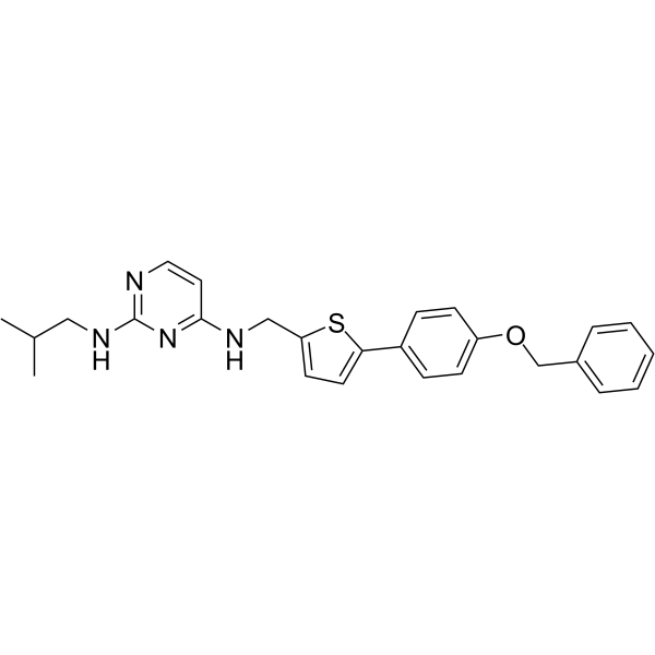
- 分子式: C26H28N4OS
- 分子量:444.59
-
保管条件:Powder -20°C, 3 years , 4°C, 2 years ; In solvent -80°C, 6 months , -20°C, 1 month

To place orders, for customer services and technical support, please contact: MedChemExpress USA
Tel: 609-228-6898 E-mail: [email protected] [email protected]


-
 生物活性
生物活性
-
化学情報
-
溶剤 & 溶解度
- プロトコル
- 純度とドキュメンテーション
- 参考文献
-
 Help & FAQs



MedChemExpress(MCE)の使用を引用している文献 ZY-444
More

生物活性
製品説明
体外実験
ZY-444 (0-10 μM, 48 h) inhibits BCA cell lines (MDA-MB-231 and 4T1) and (PCa cells DU145 and PC3) proliferation, migration, and invasion[1][2].
ZY-444 (0-20 μM, 24 h) significantly induces apoptosis in BCA cells (MDA-MB-231, MCF7, 4T1)[1].
ZY-444 (2-18 μM, 5 h) dose-dependently decreases basal respiration, spare respiratory capacity, and ATP production in BCA cells (MDA-MB-231)[1].
ZY-444 (0-10 μM, 24 h) downregulates total expression of key Wnt/β-catenin/Snail pathway proteins in MDA-MB-231 cells, prevents nuclear translocation of β-catenin, and similar effects are observed in PC-silenced MDA-MB-231 cells[1].
ZY-444 (24-72 h) halves the proliferation ability of TPC-1 and KTC-1 cells, with IC50 values of 3.82 μM (48 h) and 3.34 μM (72 h) for TPC-1 cells, and 3.79 μM (48 h) and 3.69 μM (72 h) for KTC-1 cells[3].
ZY-444 significantly inhibits the activation of the MAPK/ERK signaling pathway in TPC-1 and KTC-1 cells, reducing the expression levels of ERK1/2 and p-ERK1/2[3].
MedChemExpress (MCE) has not independently confirmed the accuracy of these methods. They are for reference only.
-
Cell Line:MDA-MB-231, MCF7, and 4T1cells
-
Concentration:0, 5, 6, 8, 10, 15, 20 μM
-
Incubation Time:24 h
-
Result:Induced apoptosis in BCA cells (MDA-MB-231, MCF7).
Increased apoptotic cells.
Upregulated expression of apoptotic biomarkers (cleaved caspase-3, cleaved PARP).
Downregulated anti-apoptotic biomarker (Bcl-2).
-
Cell Line:MDA-MB-231 cells
-
Concentration:0, 6, 8, 10 μM
-
Incubation Time:24 h
-
Result:Downregulated expression of mesenchymal markers (Snail1, Snail2, Vimentin, N-cadherin) associated with epithelial-mesenchymal transition (EMT).
-
Cell Line:MDA-MB-231, MCF7, and 4T1cells
-
Concentration:MDA-MB-231 cells
-
Incubation Time:24 h
-
Result:Downregulated total expression of β-catenin, c-Myc, and Cyclin D1 in MDA-MB-231 cells.
Prevented nuclear translocation of β-catenin (increased cytosolic β-catenin).
体内実験
ZY-444 (2.5-5 mg/kg, i.p., daily, 24 days) demonstrates excellent anti-prostate cancer efficacy in DU145 xenograft model[2].
ZY-444 (daily, 12 days) exhibits inhibitory effect on the growth of xenograft tumors derived from TPC-1 and KTC-1 PTC cells in 4-week-old female BALB/c nude mice[3].
MedChemExpress (MCE) has not independently confirmed the accuracy of these methods. They are for reference only.
-
Animal Model:4T1 or MDA-MB-231 breast cancer cells were orthotopically implanted into female mice[1]
-
Dosage:2.5, 5 mg/kg
-
Administration:i.p., daily or every 2 days, 26 days
-
Result:Achieved tumor growth inhibition.
Showed no significant changes in body weight.
Upregulated apoptotic biomarkers and downregulated mesenchymal biomarkers in primary tumors.
-
Animal Model:DU145 cells (5 × 10⁷) were subcutaneously implanted into the right flanks of 6-8-week-old male NOD-scid mice[2]
-
Dosage:2.5, 5 mg/kg
-
Administration:i.p., daily, 24 days
-
Result:Achieved tumor growth inhibition.
Showed no significant changes in body weight.
Reduced the Ki67 expression.
Upregulated the expression of TNFAIP3 and IKBα, while downregulated the expression of RIPK1, IKKα, p-IKBα, NF-κB, and p-p65.
- Cytoplasmic-Nuclear Fractionated Protein Extraction
- Detection of Apoptosis
- TUNEL staining for apoptotic DNA fragmentation
- Annexin V plus membrane-impermeant dye apoptosis staining
- Cell migration
- Research Protocol for Endocrine Diseases
- Apoptosis Solutions
- Cell invasion
- Breast Cancer Modeling
- Cell Viability Determination by MTT Colorimetric Assay


化学情報
-
CAS 番号 1802650-31-2
-
性状 Solid
-
分子量 444.59
-
分子式 C26H28N4OS
-
Color Light yellow to yellow
-
SMILES
CC(C)CNC1=NC=CC(NCC2=CC=C(C3=CC=C(OCC4=CC=CC=C4)C=C3)S2)=N1
-
輸送条件
Room temperature in continental US; may vary elsewhere.
-
保管条件
Powder -20°C 3 years 4°C 2 years In solvent -80°C 6 months -20°C 1 month


Publications (2)
-
Journal Impact Factor
-
Most Recent
-
Commun Biol
Pyruvate carboxylase promotes SREBP1a-mediated lipid synthesis in epithelial ovarian cancer. [Abstract]2026 May 14. PMID: 42129486 -


溶剤 & 溶解度
体外:
DMSO : 100 mg/mL (224.93 mM; Need ultrasonic; Hygroscopic DMSO has a significant impact on the solubility of product, please use newly opened DMSO)
Please refer to the solubility information to select the appropriate solvent. Once prepared, please aliquot and store the solution to prevent product inactivation from repeated freeze-thaw cycles.
Storage method and period of stock solution: -80°C, 6 months; -20°C, 1 month. When stored at -80°C, please use it within 6 months. When stored at -20°C, please use it within 1 month.
Please refer to the solubility information to select the appropriate solvent. Once prepared, please aliquot and store the solution to prevent product inactivation from repeated freeze-thaw cycles.
Storage method and period of stock solution: -80°C, 6 months; -20°C, 1 month. When stored at -80°C, please use it within 6 months. When stored at -20°C, please use it within 1 month.
濃度 (開始) × 体積 (開始) = 濃度 (終了) × 体積 (終了)
体内:
Select the appropriate dissolution method based on your experimental animal and administration route.
- For the following dissolution methods, please ensure to first prepare a clear stock solution using an In Vitro approach and then sequentially add co-solvents:
- To ensure reliable experimental results, the clarified stock solution can be appropriately stored based on storage conditions. As for the working solution for In Vivo experiments, it is recommended to prepare freshly and use it on the same day.
- The percentages shown for the solvents indicate their volumetric ratio in the final prepared solution. If precipitation or phase separation occurs during preparation, heat and/or sonication can be used to aid dissolution.
In Vivo Dissolution Calculator
Please enter the basic information of animal experiments:
-
-
-
-
Recommended: Prepare an additional quantity of animals to account for potential losses during experiments.
Please enter your animal formula composition:
-
%DMSO +
Recommended: Keep the proportion of DMSO in working solution below 2% if your animal is weak.
-
%+
-
+%Tween-80 + +
-
%Saline +
The co-solvents required include: DMSO, . All of co-solvents are available by MedChemExpress (MCE). , Tween 80. All of co-solvents are available by MedChemExpress (MCE).
Working solution concentration: 0.22 mg/mL
Method for preparing stock solution: mg drug dissolved in μL DMSO. Stock solution concentration: mg/mL.
1. Take μL DMSO stock solution;
2. Add μL .
μL , mix evenly;
3. Then add μL Tween 80, mix evenly;
4. Then add μL
Please ensure that the stock solution in the first step is dissolved to a clear state, and add co-solvents in sequence. You can use ultrasonic heating (ultrasonic cleaner, recommended frequency 20-40 kHz), vortexing, etc. to assist dissolution.


純度とドキュメンテーション
-
データシート (278 KB)
-
SDS (251 KB)
- English - EN (251 KB)
- Français - FR (251 KB)
- Deutsch - DE (251 KB)
- Norwegian - NO (251 KB)
- Español - ES (251 KB)
- Swedish - SV (251 KB)
- Italian - IT (251 KB)
- Korean - KR (251 KB)
- Portuguese - PT (251 KB)
-
取扱説明書 (2659 KB)
参考文献
[1]. Lin Q, et al. Targeting Pyruvate Carboxylase by a Small Molecule Suppresses Breast Cancer Progression. Adv Sci (Weinh). 2020 Mar 12;7(9):1903483. [Content Brief]
[2]. Han MT, et al. ZY-444 inhibits the growth and metastasis of prostate cancer by targeting TNFAIP3 through TNF signaling pathway. Am J Cancer Res. 2023 Apr 15;13(4):1533-1546. [Content Brief]
[3]. Liu Y, et al. Pyruvate carboxylase promotes malignant transformation of papillary thyroid carcinoma and reduces iodine uptake. Cell Death Discov. 2022 Oct 20;8(1):423. [Content Brief]


Complete Stock Solution Preparation Table
Please refer to the solubility information to select the appropriate solvent. Once prepared, please aliquot and store the solution to prevent product inactivation from repeated freeze-thaw cycles.
Storage method and period of stock solution: -80°C, 6 months; -20°C, 1 month. When stored at -80°C, please use it within 6 months. When stored at -20°C, please use it within 1 month.
| Optional Solvent | Concentration Solvent Mass | 1 mg | 5 mg | 10 mg | 25 mg |
|---|---|---|---|---|---|
| DMSO | 1 mM | 2.2493 mL | 11.2463 mL | 22.4926 mL | 56.2316 mL |
| 5 mM | 0.4499 mL | 2.2493 mL | 4.4985 mL | 11.2463 mL | |
| 10 mM | 0.2249 mL | 1.1246 mL | 2.2493 mL | 5.6232 mL | |
| 15 mM | 0.1500 mL | 0.7498 mL | 1.4995 mL | 3.7488 mL | |
| 20 mM | 0.1125 mL | 0.5623 mL | 1.1246 mL | 2.8116 mL | |
| 25 mM | 0.0900 mL | 0.4499 mL | 0.8997 mL | 2.2493 mL | |
| 30 mM | 0.0750 mL | 0.3749 mL | 0.7498 mL | 1.8744 mL | |
| 40 mM | 0.0562 mL | 0.2812 mL | 0.5623 mL | 1.4058 mL | |
| 50 mM | 0.0450 mL | 0.2249 mL | 0.4499 mL | 1.1246 mL | |
| 60 mM | 0.0375 mL | 0.1874 mL | 0.3749 mL | 0.9372 mL | |
| 80 mM | 0.0281 mL | 0.1406 mL | 0.2812 mL | 0.7029 mL | |
| 100 mM | 0.0225 mL | 0.1125 mL | 0.2249 mL | 0.5623 mL |


ZY-444 関連分類
HY-142870 関連分類
 Powered by Bioz