



4-Hydroxychalcone
Based on 2 publication(s) in Google Scholar
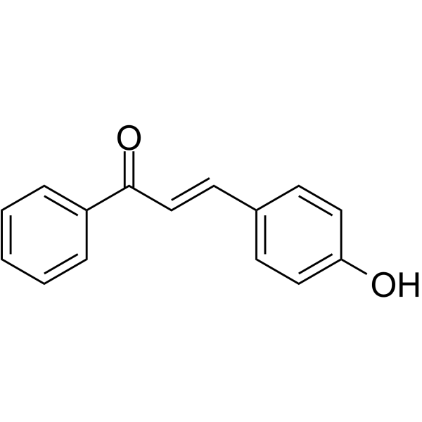
4-Hydroxychalcone is an orally active flavonoid precursor. 4-Hydroxychalcone inhibits VEGF- and bFGF-induced phosphorylation of ERK1/2 and Akt. 4-Hydroxychalcone suppresses resistant hypertension by alleviating hyperaldosteronism, inflammation and renal injury in cryptochrome gene knockout mice. 4-Hydroxychalcone possesses anti-angiogenic activity.
For research use only. We do not sell to patients.
- Purity : 99.29%
- CAS No.: 20426-12-4
- Formula: C15H12O2
- Molecular Weight:224.25
-
Storage:
4°C, protect from light
* In solvent : -80°C, 6 months; -20°C, 1 month (protect from light)

To place orders, for customer services and technical support, please contact: MedChemExpress USA
Tel: 609-228-6898 E-mail: [email protected] [email protected]


-
 Biological Activity
-
Chemical Information
-
Solvent & Solubility
- Protocols
- Purity & Documentation
- References
-
 Help & FAQs


-
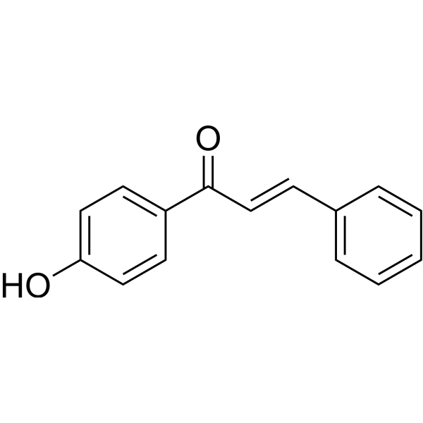
HY-N7056
4'-HydroxychalconeChalcone -
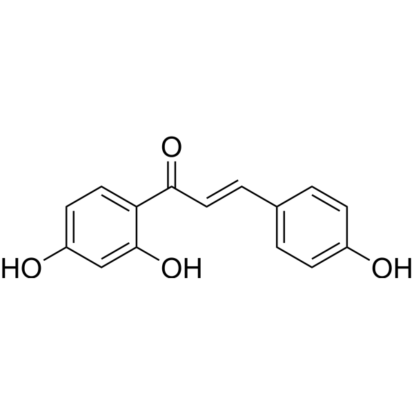
HY-N0102
IsoliquiritigeninAutophagy Inducer -
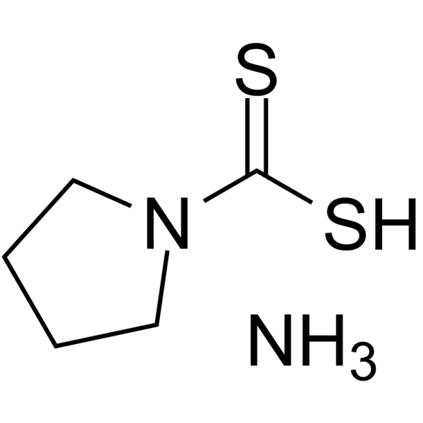
HY-18738
Pyrrolidinedithiocarbamate ammoniumSelective NF-κB Inhibitor -
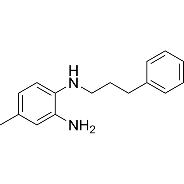
HY-13982
JSH-23NF-κB Inhibitor -
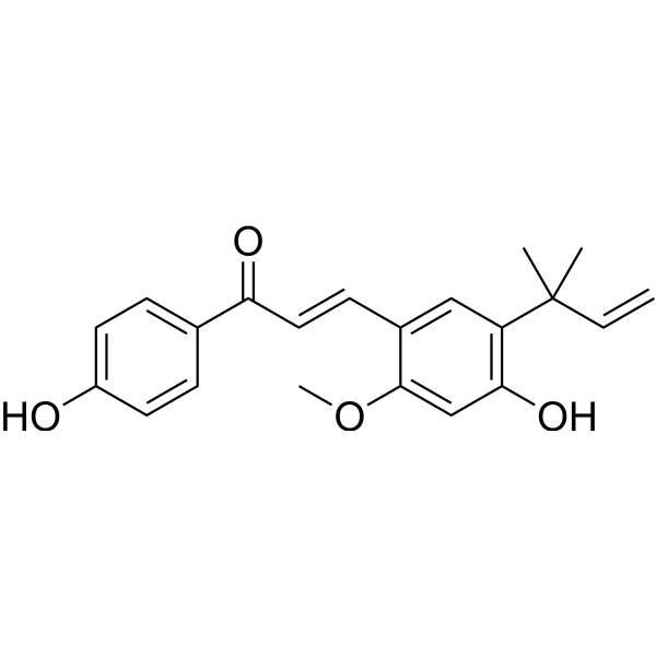
HY-N0372
Licochalcone AUGTs inhibitor -
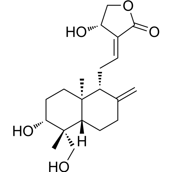
HY-N0191
AndrographolideNF-κB Inhibitor -
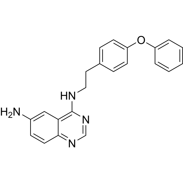
HY-13812
QNZTNF Receptor Inhibitor -
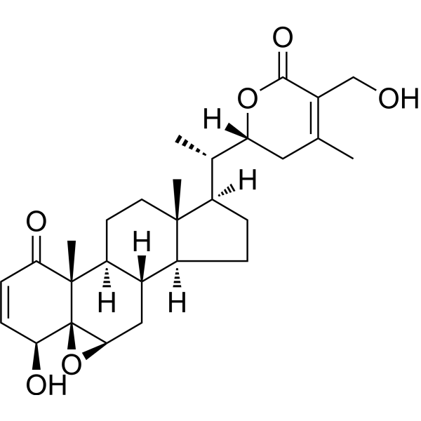
HY-N2065
Withaferin ANF-κB Inhibitor -

HY-P0151
SN50NF-κB Translocation Inhibitor -
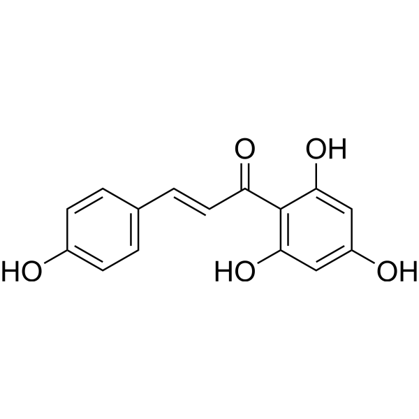
HY-N3007
Naringenin chalconeFlavonol Intermediate -
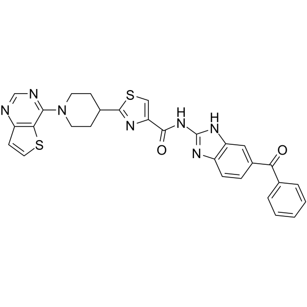
HY-10496
SC75741NF-κB Inhibitor -
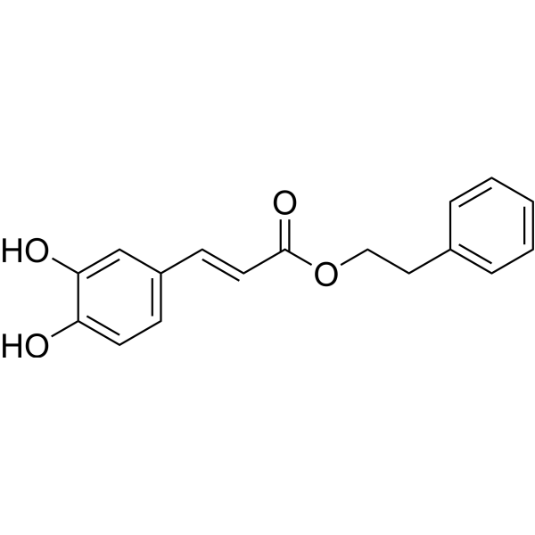
HY-N0274
Caffeic acid phenethyl esterApoptosis Inducer
Publications Citing Use of MedChemExpress (MCE) 4-Hydroxychalcone
More

Biological Activity
Description
IC50 & Target
[1]|
ERK1 |
ERK2 |
Cellular Effect
|
Cell Line

|
Type | Value | Description | References |
|---|---|---|---|---|
| A549 | IC50 |
>20 μM
Compound: 9e
|
Inhibition of TNFalpha induced NF-kappaB activation in human A549 cells by luciferase reporter gene assay
Inhibition of TNFalpha induced NF-kappaB activation in human A549 cells by luciferase reporter gene assay
|
[PMID: 19883086] |
| A549 | IC50 |
26.6 μM
Compound: 2
|
Antiproliferative activity against human A549 cells assessed as cell growth inhibition measured after 48 hrs by CellTiter Aqueous One Solution MTS assay
Antiproliferative activity against human A549 cells assessed as cell growth inhibition measured after 48 hrs by CellTiter Aqueous One Solution MTS assay
|
[PMID: 37666364] |
| CCRF-CEM | IC50 |
9.31 μM
Compound: 32
|
Concentration required to inhibit 50% growth of Human CEM T-lymphocytes cells
Concentration required to inhibit 50% growth of Human CEM T-lymphocytes cells
|
[PMID: 9544201] |
| DLD-1 | EC50 |
51 μM
Compound: 8
|
Cytotoxicity against human DLD-1 cells assessed as reduction in cell viability incubated for 48 hrs by AlamarBlue-based assay
Cytotoxicity against human DLD-1 cells assessed as reduction in cell viability incubated for 48 hrs by AlamarBlue-based assay
|
[PMID: 36356534] |
| FHC | CC50 |
>100 μM
Compound: 8
|
Cytotoxicity against human FHC cells assessed as reduction in cell viability incubated for 48 hrs by AlamarBlue-based assay
Cytotoxicity against human FHC cells assessed as reduction in cell viability incubated for 48 hrs by AlamarBlue-based assay
|
[PMID: 36356534] |
| HCT-116 | EC50 |
32 μM
Compound: 8
|
Cytotoxicity against human HCT-116 cells assessed as reduction in cell viability incubated for 48 hrs by AlamarBlue-based assay
Cytotoxicity against human HCT-116 cells assessed as reduction in cell viability incubated for 48 hrs by AlamarBlue-based assay
|
[PMID: 36356534] |
| HCT-116 | EC50 |
32 μM
Compound: 8
|
Cytotoxicity against p53-/- human HCT-116 cells assessed as reduction in cell viability incubated for 48 hrs by AlamarBlue-based assay
Cytotoxicity against p53-/- human HCT-116 cells assessed as reduction in cell viability incubated for 48 hrs by AlamarBlue-based assay
|
[PMID: 36356534] |
| HEK293 | IC50 |
>50 μM
Compound: 2
|
Blockade of human K+ channel in HEK293 cells assessed as inhibition of outward delayed rectifying K+ current
Blockade of human K+ channel in HEK293 cells assessed as inhibition of outward delayed rectifying K+ current
|
[PMID: 18032041] |
| HT-29 | EC50 |
39 μM
Compound: 8
|
Cytotoxicity against human HT-29 cells assessed as reduction in cell viability incubated for 48 hrs by AlamarBlue-based assay
Cytotoxicity against human HT-29 cells assessed as reduction in cell viability incubated for 48 hrs by AlamarBlue-based assay
|
[PMID: 36356534] |
| HT-29 | IC50 |
24.6 μM
Compound: 16
|
Cytotoxicity against Homo sapiens (human) HT-29 cells after 72 hr by MTT assay
Cytotoxicity against Homo sapiens (human) HT-29 cells after 72 hr by MTT assay
|
10.1007/s00044-011-9549-9 |
| K562 | IC50 |
24 μM
Compound: 4-Hydroxychalcone
|
Inhibition of NF-kappaB transactivation in TNF-alpha-stimulated human K562 cells preincubated for 2 hrs followed by TNF-alpha challenge measured after 6 hrs by dual luciferase reporter gene assay
Inhibition of NF-kappaB transactivation in TNF-alpha-stimulated human K562 cells preincubated for 2 hrs followed by TNF-alpha challenge measured after 6 hrs by dual luciferase reporter gene assay
|
[PMID: 24775915] |
| L1210 | IC50 |
37.9 μM
Compound: 32
|
Concentration required to inhibit 50% growth of L1210 leukemia cells
Concentration required to inhibit 50% growth of L1210 leukemia cells
|
[PMID: 9544201] |
| Neutrophil | IC50 |
11.6 μM
Compound: 25
|
Inhibition of PMA-induce ROS/RNS generation in human neutrophils measured up to 30 mins in presence of 30 mM glucose by luminol-amplified chemiluminescence method
Inhibition of PMA-induce ROS/RNS generation in human neutrophils measured up to 30 mins in presence of 30 mM glucose by luminol-amplified chemiluminescence method
|
[PMID: 33006891] |
| Neutrophil | IC50 |
8.8 μM
Compound: 25
|
Inhibition of PMA-induce ROS/RNS generation in human neutrophils measured up to 30 mins in presence of 5.5 mM glucose by luminol-amplified chemiluminescence method
Inhibition of PMA-induce ROS/RNS generation in human neutrophils measured up to 30 mins in presence of 5.5 mM glucose by luminol-amplified chemiluminescence method
|
[PMID: 33006891] |
| NIH3T3 | IC50 |
>20 μM
Compound: 1i
|
Inhibition of cobalt chloride-induced HIF-1 activation expressed in mouse NIH3T3 cells after 8 hrs by luciferase reporter gene assay
Inhibition of cobalt chloride-induced HIF-1 activation expressed in mouse NIH3T3 cells after 8 hrs by luciferase reporter gene assay
|
[PMID: 21112783] |
| P388 | IC50 |
14.3 μM
Compound: 32
|
Inhibition of proliferation of murine P388 cells
Inhibition of proliferation of murine P388 cells
|
[PMID: 9544201] |
| SGC-7901 | IC50 |
13.74 μM
Compound: 16
|
Cytotoxicity against Homo sapiens (human) SGC7901 cells after 72 hr by MTT assay
Cytotoxicity against Homo sapiens (human) SGC7901 cells after 72 hr by MTT assay
|
10.1007/s00044-011-9549-9 |
In Vitro
4-hydroxychalcone (22 µg/mL; 6 days) potently inhibits VEGF/TNF-α- and bFGF/TNF-α-induced capillary-like tube formation of hMVEC in 3D fibrin matrices, with no cytotoxicity to endothelial monolayers[1].
4-hydroxychalcone (0.22-22 µg/mL; 24-144 h) exhibits no cytotoxicity toward confluent HUVEC and hMVEC monolayers[1].
4-hydroxychalcone (0.22-22 µg/mL; 48-72 h) exerts a cytostatic effect on the proliferation of HUVEC, but shows no cytostatic effect on HeLa, MCF-7 or A549 tumor cell lines even at a concentration as high as 22 µg/mL with a 72 h treatment[1].
MedChemExpress (MCE) has not independently confirmed the accuracy of these methods. They are for reference only. Further protocols information, click here.
-
Cell Line:human umbilical vein endothelial cells (HUVEC)
-
Concentration:2.2-22 µg/mL
-
Incubation Time:1 h pre-incubation
-
Result:Significantly inhibited both bFGF- and VEGF-induced phosphorylation of Akt and ERK1/2 in HUVEC at 22 µg/mL.
Significantly inhibited VEGF-induced phosphorylation of ERK1/2, but not Akt, in HUVEC at 2.2 µg/mL.
-
Cell Line:human umbilical vein endothelial cells (HUVEC)
-
Concentration:22 µg/mL
-
Incubation Time:1 h pre-incubation, followed by 1 h TNF-α stimulation
-
Result:Had no effect on TNF-α-induced nuclear translocation of the p65 subunit of NF-κB in HUVEC.
In Vivo
4-Hydroxychalcone (10-40 mg/kg; p.o.; twice daily; for consecutive 35 days) exerts dose-dependent protective effects against refractory hypertension, hyperaldosteronism, inflammation and renal injury in cryptochrome-knockout mice treated with high salt[2].
MedChemExpress (MCE) has not independently confirmed the accuracy of these methods. They are for reference only.
-
Animal Model:Cryptochrome-null mice (male/female unspecified; high-salt diet-induced hypertension, hyperaldosteronism, inflammation, renal injury model)[2]
-
Dosage:10 mg/kg; 20 mg/kg; 40 mg/kg
-
Administration:p.o.; twice daily; 35 days
-
Result:Produced no significant change in systolic blood pressure compared to untreated cryptochrome-null mice; significantly reduced serum IL-1β levels compared to untreated cryptochrome-null mice (P < 0.05); did not significantly reduce serum TNF-α levels, serum aldosterone levels, renal NF-κB activation, or renal injury.
Produced no significant change in systolic blood pressure compared to untreated cryptochrome-null mice; significantly reduced serum IL-1β levels compared to untreated cryptochrome-null mice (P < 0.01); significantly reduced serum aldosterone levels compared to untreated cryptochrome-null mice (P < 0.05); did not significantly reduce serum TNF-α levels, renal NF-κB activation, or renal injury.
Significantly lowered systolic blood pressure in cryptochrome-null mice compared to untreated controls (P < 0.05); significantly reduced serum aldosterone levels compared to untreated cryptochrome-null mice (P < 0.01); significantly reduced serum IL-1β levels compared to untreated cryptochrome-null mice (P < 0.01); significantly suppressed serum TNF-α production compared to untreated cryptochrome-null mice (P < 0.05); reduced renal NF-κB protein activation in kidney cell nuclei compared to untreated cryptochrome-null mice; dramatically reduced severe glomerulosclerosis, tubulointerstitial injury, and inflammatory cell infiltration in kidney tissue compared to untreated cryptochrome-null mice, resulting in more normal histology.


Chemical Information
-
CAS No. 20426-12-4
-
Appearance Solid
-
Molecular Weight 224.25
-
Formula C15H12O2
-
Color Light yellow to yellow
-
SMILES
O=C(C1=CC=CC=C1)/C=C/C2=CC=C(O)C=C2
-
Structure Classification
-
Initial Source
-
Shipping
Room temperature in continental US; may vary elsewhere.
-
Storage
4°C, protect from light
* In solvent : -80°C, 6 months; -20°C, 1 month (protect from light)


Publications (2)
-
Journal Impact Factor
-
Most Recent
-
Food Chem
Effects of sun drying combined with baking processes on the flavor quality of Chongqing Tuocha raw tea. [Abstract]2025 Dec 30:497:146992. PMID: 41285060 -
Food Chem
Flavonoid-mediated metabolic underpinning quality variation in red bud-sport pear mutants. [Abstract]2025 Oct 15:489:144992. PMID: 40466530


Solvent & Solubility
In Vitro:
DMSO : 100 mg/mL (445.93 mM; Need ultrasonic; Hygroscopic DMSO has a significant impact on the solubility of product, please use newly opened DMSO)
Please refer to the solubility information to select the appropriate solvent. Once prepared, please aliquot and store the solution to prevent product inactivation from repeated freeze-thaw cycles.
Storage method and period of stock solution: -80°C, 6 months; -20°C, 1 month (protect from light). When stored at -80°C, please use it within 6 months. When stored at -20°C, please use it within 1 month.
Please refer to the solubility information to select the appropriate solvent. Once prepared, please aliquot and store the solution to prevent product inactivation from repeated freeze-thaw cycles.
Storage method and period of stock solution: -80°C, 6 months; -20°C, 1 month (protect from light). When stored at -80°C, please use it within 6 months. When stored at -20°C, please use it within 1 month.
Concentration (start) × Volume (start) = Concentration (final) × Volume (final)
In Vivo:
Select the appropriate dissolution method based on your experimental animal and administration route.
- For the following dissolution methods, please ensure to first prepare a clear stock solution using an In Vitro approach and then sequentially add co-solvents:
- To ensure reliable experimental results, the clarified stock solution can be appropriately stored based on storage conditions. As for the working solution for In Vivo experiments, it is recommended to prepare freshly and use it on the same day.
- The percentages shown for the solvents indicate their volumetric ratio in the final prepared solution. If precipitation or phase separation occurs during preparation, heat and/or sonication can be used to aid dissolution.
Add each solvent one by one: 10% DMSO 40% PEG300 5% Tween-80 45% Saline
Solubility: ≥ 2.08 mg/mL (9.28 mM); Clear solution
This protocol yields a clear solution of ≥ 2.08 mg/mL (saturation unknown).
Taking 1 mL working solution as an example, add 100 μL DMSO stock solution (20.8 mg/mL) to 400 μL PEG300, and mix evenly; then add 50 μL Tween-80 and mix evenly; then add 450 μL Saline to adjust the volume to 1 mL.
Preparation of Saline: Dissolve 0.9 g sodium chloride in ddH₂O and dilute to 100 mL to obtain a clear Saline solution.
Add each solvent one by one: 10% DMSO 90% (20% SBE-β-CD in Saline)
Solubility: ≥ 2.08 mg/mL (9.28 mM); Clear solution
This protocol yields a clear solution of ≥ 2.08 mg/mL (saturation unknown).
Taking 1 mL working solution as an example, add 100 μL DMSO stock solution (20.8 mg/mL) to 900 μL 20% SBE-β-CD in Saline, and mix evenly.
Preparation of 20% SBE-β-CD in Saline (4°C, storage for one week): 2 g SBE-β-CD powder is dissolved in 10 mL Saline, completely dissolve until clear.
In Vivo Dissolution Calculator
Please enter the basic information of animal experiments:
-
-
-
-
Recommended: Prepare an additional quantity of animals to account for potential losses during experiments.
Please enter your animal formula composition:
-
%DMSO +
Recommended: Keep the proportion of DMSO in working solution below 2% if your animal is weak.
-
%+
-
+%Tween-80 + +
-
%Saline +
The co-solvents required include: DMSO, . All of co-solvents are available by MedChemExpress (MCE). , Tween 80. All of co-solvents are available by MedChemExpress (MCE).
Working solution concentration: 0.22 mg/mL
Method for preparing stock solution: mg drug dissolved in μL DMSO. Stock solution concentration: mg/mL. * In solvent : -80°C, 6 months; -20°C, 1 month (protect from light)
1. Take μL DMSO stock solution;
2. Add μL .
μL , mix evenly;
3. Then add μL Tween 80, mix evenly;
4. Then add μL
Please ensure that the stock solution in the first step is dissolved to a clear state, and add co-solvents in sequence. You can use ultrasonic heating (ultrasonic cleaner, recommended frequency 20-40 kHz), vortexing, etc. to assist dissolution.

Protocols
-
Kinase activity and phosphorylation assays
Kinase activity assays measure the ability of kinases to transfer phosphate groups from ATP to specific substrates, while phosphorylation assays detect the presence and levels of phosphorylated proteins. Common methods include radiolabeled ATP incorporation (e. g. ,), ADP release detection via bioluminescence (e. g. ,[3]), enzyme-linked immunosorbent assays (ELISA) for phospho-specific epitopes (e. g. ,[6]), and microtiter-based formats for high-throughput screening (e. g. ,[8]). The ADP-Glo assay quantifies kinase activity by measuring ADP produced during phosphorylation using a luciferase-based system. Radiometric assays involve autoradiography or scintillation counting after incorporation of 32P-labeled ATP into substrate proteins. ELISA-based approaches rely on phospho-specific antibodies to detect activated kinases in cell lysates or purified samples.
-
Gene Editing
Gene editing modify specific sites within the genome through gene deletions, insertions or conversions to study functionally unknown genes or conduct gene therapy. It is also used to change the biological traits of organisms to establish new varieties. Gene editing techniques include zinc finger nuclease (ZFN), transcription activator-like effector nuclease (TALEN), and clustered regularly interspaced short palindromic repeats (CRISPR)/CRISPR-associated protein 9 (Cas 9) (CRISPR/Cas9).
-
Western Blot
Western blotting (WB) is a commonly used experimental method in molecular biology, biochemistry, and immunogenetics for identifying and quantifying target proteins. It combines gel electrophoresis with immunoassay, enabling researchers to analyze protein expression, post-translational modifications, and molecular weight.
-
CRISPR-Cas9 knockout in cultured mammalian cells
CRISPR-Cas9 knockout in cultured mammalian cells uses an sgRNA to direct Cas9 to a complementary genomic sequence adjacent to a compatible PAM; Cas9 creates a targeted DNA double-strand break, and repair by non-homologous end joining can introduce insertions or deletions that disrupt the coding sequence or functional genomic element. The readout of knockout is detection of edited alleles and loss of gene product or phenotype, commonly by PCR/Sanger-sequence trace decomposition, targeted sequencing, immunoblotting, immunostaining, or flow cytometry when the target protein is detectable at the cell surface.
-
Nephrotoxicity Study
This protocol assesses nephrotoxicity by combining functional kidney injury readouts, urinary/tissue injury biomarkers, and renal histopathology. Serum creatinine and BUN reflect impaired kidney function, while KIM-1, NGAL, clusterin, osteopontin, IL-18, cystatin C, nephrin, Oat5, urinary protein, glucose, and alkaline phosphatase have been used to detect tubular injury in cisplatin-, gentamicin-, and acetaminophen-induced nephrotoxicity models.
-
CRISPR-Cas9 zebrafish embryo editing
CRISPR-Cas9 zebrafish embryo editing introduces targeted double-strand breaks in genomic DNA by delivering Cas9 nuclease with a guide RNA into one-cell-stage embryos; repair by endogenous DNA-repair pathways produces indels or donor-mediated insertions that can be detected by phenotype, PCR-based genotyping, heteroduplex assays, Sanger sequencing, or amplicon sequencing. The readout reflects the frequency and spectrum of edited alleles in mosaic F0 embryos or transmitted F1 animals; because injected embryos can carry multiple alleles, founder screening and sequence confirmation are required before establishing stable mutant lines.
-
Constitutive Germline Knockout Model
Constitutive germline knockout models are generated by producing a heritable loss-of-function allele in the mouse genome, typically through complete gene disruption in embryonic stem (ES) cells followed by germline transmission or through CRISPR/Cas-mediated editing of zygotes, resulting in offspring that carry a stable null allele in all tissues. Classical approaches rely on homologous recombination in ES cells to introduce targeted gene disruptions, which are then transmitted through chimeric mice to the germline. More recent genome editing strategies use CRISPR/Cas systems to induce double-strand breaks and non-homologous end joining (NHEJ), frequently generating frameshift mutations that abolish gene function, enabling faster generation of knockout alleles directly in embryos. Germline transmission or direct germline editing ensures that the mutation is present in all cells of the resulting animal, allowing systemic functional analysis of gene loss.
-
Endothelial Tube Formation Assay
Endothelial tube formation assay evaluates the ability of endothelial cells to attach, migrate, align, and organize into capillary-like networks when cultured on gelled basement membrane extract or Matrigel; the readout is the morphology and quantity of tube-like networks, which reflects an in vitro endothelial morphogenesis step related to angiogenesis. Basement membrane extract/Matrigel provides laminin-rich extracellular matrix cues that support endothelial differentiation into capillary-like structures, but it can contain biologically active growth factors, so growth-factor-reduced matrix is preferred when testing defined angiogenic stimulators or inhibitors.
-
Research Protocol for Cardiovascular Diseases
Cardiovascular disease can be modeled as maladaptive cardiac remodeling, where ischemic injury or pressure overload activates inflammatory signaling, fibroblast activation, extracellular-matrix deposition, cardiomyocyte hypertrophy, vascular remodeling, and progressive ventricular dysfunction. The TGF-β/SMAD axis is a central profibrotic pathway after myocardial injury and pressure overload, while innate immune and cytokine pathways regulate leukocyte recruitment, scar formation, and adverse remodeling. Key unresolved questions include which inflammatory signals are reparative versus harmful, when fibrosis is protective versus maladaptive, and whether pathway inhibition improves function without weakening necessary infarct healing or compensatory remodeling.
-
Protocol for Kinase activity and phosphorylation assays
Kinase activity assays measure transfer of phosphate from ATP to a protein or peptide substrate, generating phosphorylated substrate, ADP, or incorporated radiolabeled phosphate as the readout; phosphorylation assays measure site-specific phosphorylation in cells or tissues as a proxy for kinase-pathway activation, inhibition, or substrate regulation. Phosphorylation can be detected by phospho-specific Western blot, immunoprecipitation kinase assay, phospho-immunofluorescence, phospho-flow cytometry, luminescent ADP detection, radiolabeled ATP incorporation, or reporter-based pathway assays, and these readouts can be applied to cancer cells, primary neurons, mouse tumors, organoids, inflammatory macrophages, ferroptosis studies, and mitophagy studies when the kinase target is biologically relevant.
-
Research Protocol for Inflammation-related Diseases
The NLRP3 inflammasome is a cytosolic innate immune signaling platform that integrates priming signals and danger-signal activation to promote caspase-1 activation, maturation of IL-1β and IL-18, and gasdermin D-mediated pyroptotic cell death. The core experimental logic is to determine whether inflammatory disease phenotypes are driven by increased NLRP3 expression, ASC-containing inflammasome assembly, caspase-1 cleavage, GSDMD cleavage, and extracellular release of IL-1β/IL-18 rather than by nonspecific cell injury alone. The pathway is strongly linked to inflammation-related disease phenotypes because monosodium urate crystals activate NALP3/NLRP3 inflammasome signaling in gout-like crystal inflammation, cholesterol crystals activate NLRP3 inflammasomes in atherogenesis models, and DSS-induced intestinal inflammation has been reported to involve NLRP3 inflammasome activity. However, experimental colitis studies also show context-dependent protective effects of NLRP3 inflammasome co
-
CRISPR/Cas9 Knockout Animal Model
CRISPR/Cas9 knockout animal modeling uses guide RNA to direct Cas9 to a genomic target, where Cas9 creates a DNA double-strand break; repair by error-prone non-homologous end joining generates insertions or deletions that can disrupt coding sequence and produce knockout alleles. Classic animal-model workflows deliver Cas9 mRNA or Cas9 protein with sgRNA into fertilized zygotes by microinjection or electroporation, then transfer edited embryos into pseudopregnant recipients and genotype founders for target-site mutations.


Purity & Documentation
-
Data Sheet (277 KB)
-
SDS (538 KB)
- English - EN (538 KB)
- Français - FR (538 KB)
- Deutsch - DE (538 KB)
- Norwegian - NO (538 KB)
- Español - ES (538 KB)
- Swedish - SV (538 KB)
- Italian - IT (538 KB)
- Korean - KR (538 KB)
- Portuguese - PT (538 KB)
-
Handling Instructions (2659 KB)
References
[1]. Varinska L, et al. Anti-angiogenic activity of the flavonoid precursor 4-hydroxychalcone. Eur J Pharmacol. 2012;691(1-3):125-133. [Content Brief]
[2]. Qu Q, et al. 4-Hydroxychalcone attenuates hyperaldosteronism, inflammation, and renal injury in cryptochrome-null mice. Biomed Res Int. 2014;2014:603415. [Content Brief]


Complete Stock Solution Preparation Table
Please refer to the solubility information to select the appropriate solvent. Once prepared, please aliquot and store the solution to prevent product inactivation from repeated freeze-thaw cycles.
Storage method and period of stock solution: -80°C, 6 months; -20°C, 1 month (protect from light). When stored at -80°C, please use it within 6 months. When stored at -20°C, please use it within 1 month.
| Optional Solvent | Concentration Solvent Mass | 1 mg | 5 mg | 10 mg | 25 mg |
|---|---|---|---|---|---|
| DMSO | 1 mM | 4.4593 mL | 22.2965 mL | 44.5931 mL | 111.4827 mL |
| 5 mM | 0.8919 mL | 4.4593 mL | 8.9186 mL | 22.2965 mL | |
| 10 mM | 0.4459 mL | 2.2297 mL | 4.4593 mL | 11.1483 mL | |
| 15 mM | 0.2973 mL | 1.4864 mL | 2.9729 mL | 7.4322 mL | |
| 20 mM | 0.2230 mL | 1.1148 mL | 2.2297 mL | 5.5741 mL | |
| 25 mM | 0.1784 mL | 0.8919 mL | 1.7837 mL | 4.4593 mL | |
| 30 mM | 0.1486 mL | 0.7432 mL | 1.4864 mL | 3.7161 mL | |
| 40 mM | 0.1115 mL | 0.5574 mL | 1.1148 mL | 2.7871 mL | |
| 50 mM | 0.0892 mL | 0.4459 mL | 0.8919 mL | 2.2297 mL | |
| 60 mM | 0.0743 mL | 0.3716 mL | 0.7432 mL | 1.8580 mL | |
| 80 mM | 0.0557 mL | 0.2787 mL | 0.5574 mL | 1.3935 mL | |
| 100 mM | 0.0446 mL | 0.2230 mL | 0.4459 mL | 1.1148 mL |


4-Hydroxychalcone Related Classifications
HY-107818 Related Classifications
 Powered by Bioz