



Amprolium
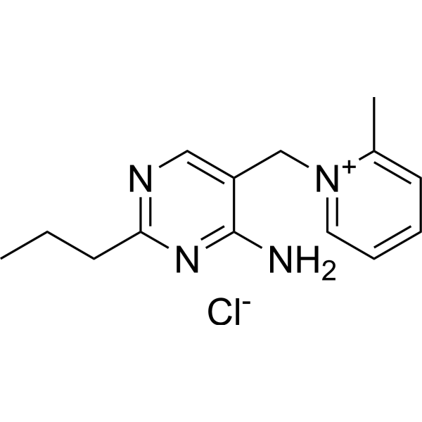
Amprolium is a thiamine transporter inhibitor and antiparasitic agent with oral activity and blood-brain barrier penetration. Amprolium blocks thiamine transport at the blood-brain barrier and intestine, and competitively inhibits thiamine pyrophosphokinase, inducing thiamine pyrophosphate deficiency and impairing carbohydrate synthesis. Amprolium induces oxidative stress, alters antioxidant enzyme activity, and inhibits acetylcholinesterase (AchE) activity. Amprolium disrupts reproductive organs and gametogenesis, and causes histopathological and ultrastructural damage to multiple organs. Amprolium induces thiamine deficiency, reduces body weight gain in mice, and causes psychomotor behavioral changes and nephropathy. Amprolium exhibits molluscicidal activity against the terrestrial snail Eobania vermiculata, and reduces the shedding of Eimeria meleagrimitis oocysts in feces and litter. Amprolium can be used for research on thiamine deficiency and coccidiosis.
For research use only. We do not sell to patients.
- CAS No.: 121-25-5
- Formula: C14H19ClN4
- Molecular Weight:278.78
-
Storage:
Please store the product under the recommended conditions in the Certificate of Analysis.

To place orders, for customer services and technical support, please contact: MedChemExpress USA
Tel: 609-228-6898 E-mail: [email protected] [email protected]


-
 Biological Activity
-
Chemical Information
- Protocols
- Purity & Documentation
- References
-
 Help & FAQs


All Parasite Isoforms
More

Biological Activity
Description
IC50 & Target
[1]|
Coccidia |
AChE |
In Vivo
Amprolium (16.175 mg/mL; petri dish exposure; 7 days) induces significant biochemical alterations and oxidative stress, leading to severe histopathological and ultrastructural damage in multiple organs of Eobania vermiculata[1].
Amprolium (100 g/L; spray application; single dose) reduces Eobania vermiculata populations by 84% after 14 days in field trials when applied as a spray at 100 g/L[1].
MedChemExpress (MCE) has not independently confirmed the accuracy of these methods. They are for reference only.
-
Animal Model:Adult (average length of 26.1 mm, width of 14.1 mm, weight of 2.5 g)[1]
-
Dosage:10, 30, 50, 70, 90, and 100 mg/mL
-
Administration:contact method; single exposure; 7 days
-
Result:Achieved a 3-day LC50 of 101.804 mg/mL and a 7-day LC50 of 32.35 mg/mL.
Induced 100% mortality at 100 mg/mL after 3 days and at 70, 90, and 100 mg/mL after 7 days.
Caused mortality rates of 80.0% at 50 mg/mL, 40.0% at 30 mg/mL, and 20.0% at 10 mg/mL after 7 days.
-
Animal Model:Adult (average length of 26.1 mm, width of 14.1 mm, weight of 2.5 g)[1]
-
Dosage:16.175 mg/mL
-
Administration:exposure in petri dish; 7 days
-
Result:Increased ALP to 151.29 U/L, TL to 146.16 mg/dl, urea to 73.52, creatinine to 1.03 mg/dl, MDA to 111.28, CAT to 158.61 U/g, and NO to 44.20 μmol/L compared to controls.
Decreased AChE to 59.66 U/mg, TP to 56.60 g/mL, and GSH to 69.43 mg/g.
Induced severe histopathological and ultrastructural damage in the digestive gland, hermaphrodite gland, kidney, mucus gland, and cerebral ganglia, and damaged tegumental structures of the mantle-foot region.
-
Animal Model:Adult[1]
-
Dosage:100 g/L
-
Administration:spray application; single dose
-
Result:Reduced the snail population by 39% on Day 1, 50% on Day 3, 75% on Day 7, and 84% on Day 14.
Decreased the mean number of living snails from 35.00 on Day 0 to 7.00 by Day 14.
-
Animal Model:Swiss (male, 50-day postnatal)[2]
-
Dosage:10 mg/mL (TD-A, 20 days); 20 mg/mL (TD-B, 20 days); 10 mg/mL (TD-A, 80 days); 20 mg/mL (TD-B, 80 days)
-
Administration:p.o. (drinking water); daily; 20 or 80 days
-
Result:Reduced motor activity by 39.27% in TD-B mice treated for 80 days.
Reduced exploratory activity by 20.06% in TD-B mice treated for 80 days.
Increased grooming frequency by 90.83% in TD-B mice treated for 80 days.
Reduced latency to fall in rotarod test to 161.67 s (TD-A) and 156.50 s (TD-B) compared to control 187.00 s after 80 days.
Decreased body weight gain to 1.55 g (TD-A) and 1.22 g (TD-B) versus control 4.33 g after 20 days, and to 15.50 g (TD-A) and 3.36 g (TD-B) versus control 21.43 g after 80 days.
Reduced feed intake by 1.63 g (TD-A) and 2.01 g (TD-B) compared to control -0.27 g after 20 days.
Induced moderate proteinuria in both TD-A and TD-B groups at 20 and 80 days.
Induced moderate cellular swelling in proximal tubules in TD-B group after 80 days.
-
Animal Model:Nicholas genetics (Female; day-of-hatch; challenged with 95,000 sporulated E. meleagrimitis oocysts/mL/poult at day 23; 50% vaccinated at day-of-hatch)[3]
-
Dosage:0.024%
-
Administration:drinking water; daily; 5 days (d10-d14)
-
Result:Reduced E. meleagrimitis oocyst shedding during treatment period (d10-14).
Significantly improved post-challenge body weight gain (BWG of 226.13 g for directs).
Reduced intestinal lesion scores (LS of 1.39 for directs).
Modulated ileal and cecal microbiome, notably increasing Turicibacter abundance (24.6% for directs and contacts).


Chemical Information
-
CAS No. 121-25-5
-
Molecular Weight 278.78
-
Formula C14H19ClN4
-
SMILES
CC1=CC=CC=[N+]1CC2=CN=C(CCC)N=C2N.[Cl-]
-
Shipping
Room temperature in continental US; may vary elsewhere.
-
Storage
Please store the product under the recommended conditions in the Certificate of Analysis.


Purity & Documentation
References

Calculators
Concentration (start) × Volume (start) = Concentration (final) × Volume (final)


Amprolium Related Classifications
HY-B0937 Related Classifications
 Powered by Bioz
Keywords
- Amprolium
- 121-25-5
- Parasite
- Cholinesterase (ChE)
- thiamine pyrophosphate deficiency
- oxidative stress
- acetylcholinesterase activity
- coccidiosis
- synthetic anticoccidial
- Eimeria meleagrimitis
- Eobania vermiculata
- blood-brain barrier penetration
- thiamine pyrophosphokinase
- thiamine transporter inhibitor
- Inhibitor
- inhibitor
- inhibit