



L-Cystine
Based on 5 publication(s) in Google Scholar
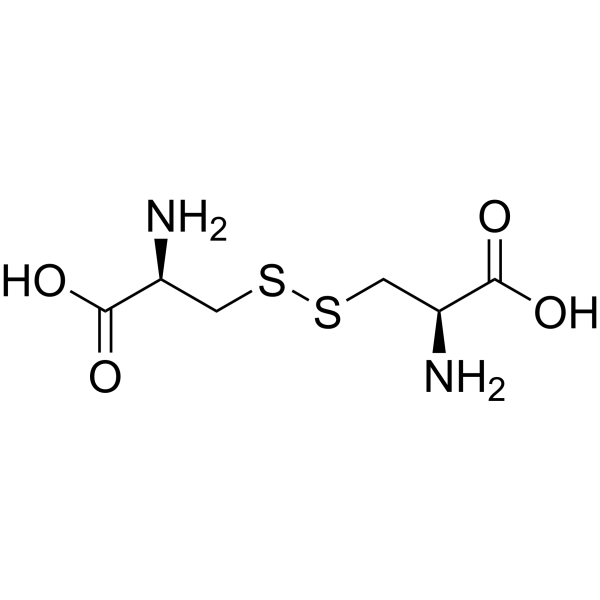
L-Cystine is an orally active extracellular form of L-Cysteine (HY-Y0337), occurring in proteins of plants and animals. L-Cystine elevates Nrf2 protein expression and activates Nrf2 transcription factor. L-cystine reduces ROS generation and protects against oxidant- or Doxorubicin (HY-15142A)-induced apoptosis. L-Cystine combined with L-theanine (HY-15121) enhances the production of antigen-specific IgG by increasing glutathione (GSH) levels and T helper 2 (Th2) mediated responses in mice. L-Cystine is promising for research of cystinuria and kidney stones
For research use only. We do not sell to patients.
- Purity : 99.84%
- CAS No.: 56-89-3
- Formula: C6H12N2O4S2
- Molecular Weight:240.30
-
Storage:Powder -20°C, 3 years , 4°C, 2 years ; In solvent -80°C, 6 months , -20°C, 1 month

To place orders, for customer services and technical support, please contact: MedChemExpress USA
Tel: 609-228-6898 E-mail: [email protected] [email protected]


-
 Biological Activity
-
Chemical Information
-
Solvent & Solubility
- Protocols
- Purity & Documentation
- References
-
 Help & FAQs


-
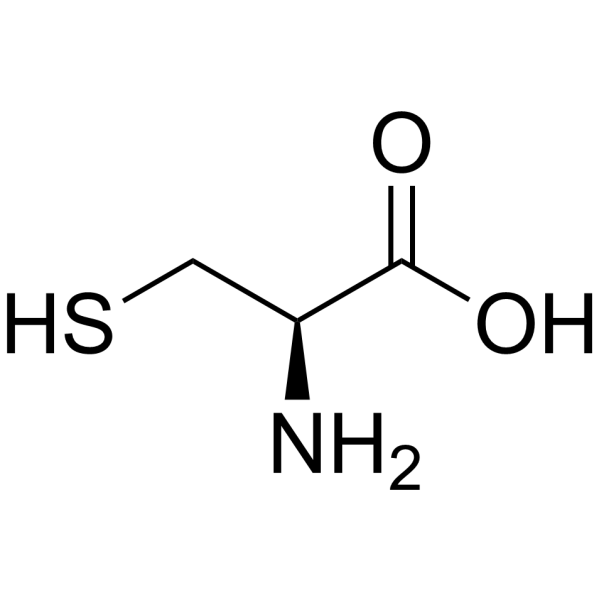
HY-Y0337
L-CysteineAnorectic Agent -
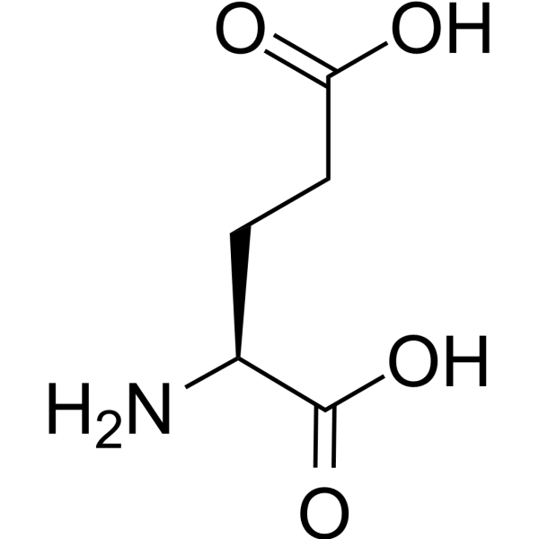
HY-14608
L-Glutamic acidGlutamate Receptors Agonist -
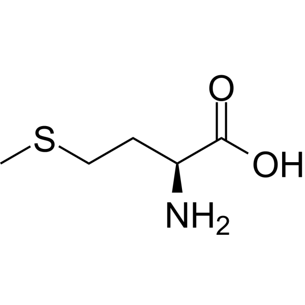
HY-N0326
L-MethionineEndogenous Metabolite -
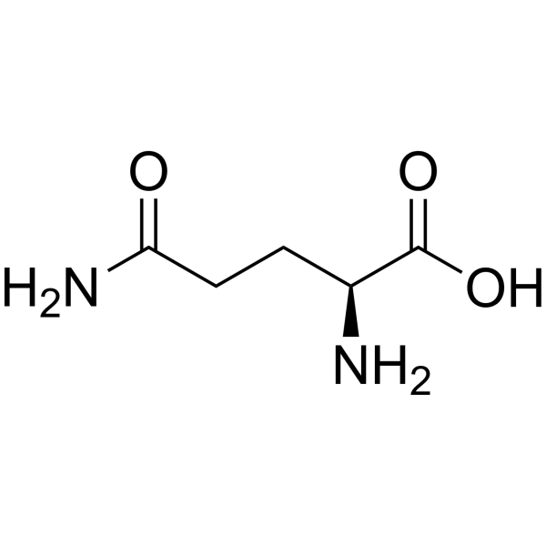
HY-N0390
L-GlutamineEndogenous Metabolite -
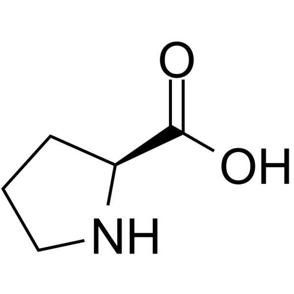
HY-Y0252
L-Proline -
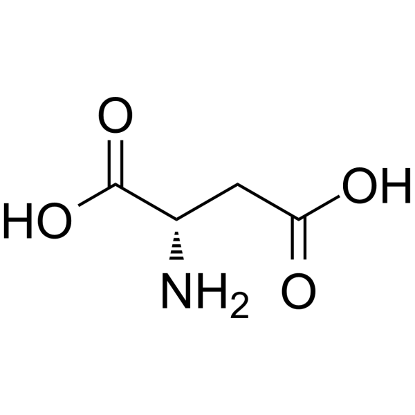
HY-N0666
L-Aspartic acidNMDA Receptor Agonist -
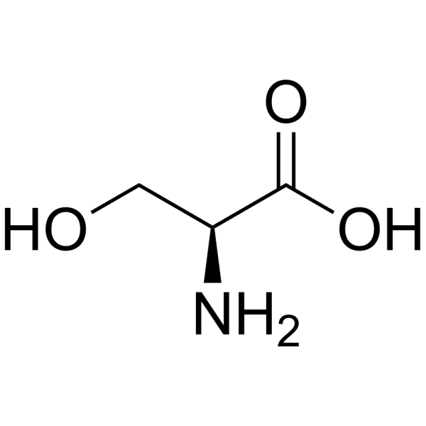
HY-N0650
L-SerineAmino Acid -
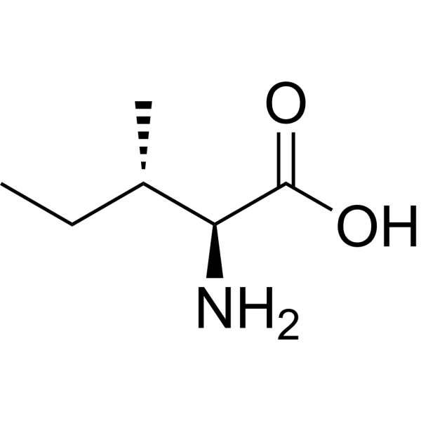
HY-N0771
L-IsoleucineEndogenous Metabolite, L-enantiomer of Isoleucine -
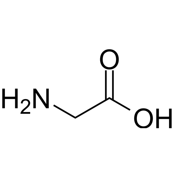
HY-Y0966
GlycineNMDAR Agonist -
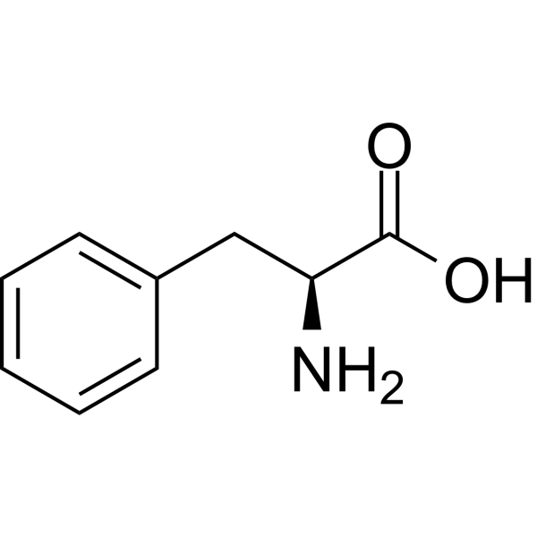
HY-N0215
L-PhenylalanineCalcium Channels Antagonist -
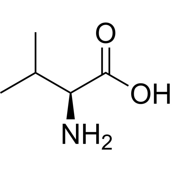
HY-N0717
L-ValineNonlinear Semiorganic Material -
HY-N0486
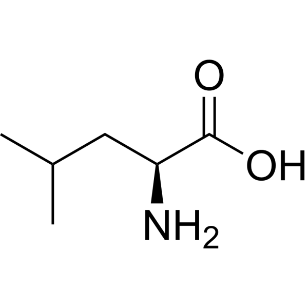
L-LeucinemTOR activator
Publications Citing Use of MedChemExpress (MCE) L-Cystine
More Customer Validation & Images

-
ELISA
-
Histological Imaging/Staining
-
Cell Imaging/Staining
-
WB
All Endogenous Metabolite Isoforms
More

Biological Activity
Description
IC50 & Target
|
Human Endogenous Metabolite |
Cellular Effect
|
Cell Line

|
Type | Value | Description | References |
|---|---|---|---|---|
| KB 3-1 | IC50 |
3.12 mM
Compound: cystine
|
Cytotoxicity against human KB-3-1 cells incubated for 72 hrs by MTT assay
Cytotoxicity against human KB-3-1 cells incubated for 72 hrs by MTT assay
|
[PMID: 21657271] |
| KB-V1 | IC50 |
2.14 mM
Compound: cystine
|
Cytotoxicity against drug-resistant human KBV1 cells expressing P-gp incubated for 72 hrs by MTT assay
Cytotoxicity against drug-resistant human KBV1 cells expressing P-gp incubated for 72 hrs by MTT assay
|
[PMID: 21657271] |
In Vitro
L-Cystine (83 µM or 200 µM, 24 h or 72 h) effectively reverses the growth inhibition of both Sulfasalazine (HY-14655) and Erastin (HY-15763) in Huh6 cells[1]. L-Cystine ( 0.8 mM, 4 h) induces Nrf2 protein expression elevation in a Keap1-dependent manner in HeLa cells[6].
MedChemExpress (MCE) has not independently confirmed the accuracy of these methods. They are for reference only. Further protocols information, click here.
-
Cell Line:Huh6 and Huh7 cells
-
Concentration:83 µM or 200 µM
-
Incubation Time:72 h or 24 h
-
Result:The degree of growth inhibition by Sulfasalazine (HY-14655) or Erastin (HY-15763) was greater in medium containing a physiological level of 83 μM L-cystine than in medium containing 200 μM L-cystine in Huh6 and Huh7 cells.
-
Cell Line:HeLa cells
-
Concentration:0.2 mM
-
Incubation Time:4 h
-
Result:Was sufficient for producing Nrf2 protein elevation at 0.2 mM in HeLa cells.
-
Cell Line:HeLa cells
-
Concentration:0.8 mM
-
Incubation Time:4 h
-
Result:Induced Nrf2 in a Keap1-dependent manner in HeLa cells.
In Vivo
MedChemExpress (MCE) has not independently confirmed the accuracy of these methods. They are for reference only.
-
Animal Model:Nine-week-old female BALB/c mice[2]
-
Dosage:140, 280 and 560 mg/kg
-
Administration:p.o. administration for 11 days
-
Result:Significantly increased IgG and IgM antibody production administrated with L-theanine (HY-15121) after DNP-dextran and DNP-KLH antigenic stimulation in mice.


Chemical Information
-
CAS No. 56-89-3
-
Appearance Solid
-
Molecular Weight 240.30
-
Formula C6H12N2O4S2
-
Color White to off-white
-
SMILES
N[C@@H](CSSC[C@@H](C(O)=O)N)C(O)=O
-
Structure Classification
-
Initial Source
-
Shipping
Room temperature in continental US; may vary elsewhere.
-
Storage
Powder -20°C 3 years 4°C 2 years In solvent -80°C 6 months -20°C 1 month


Publications (5)
-
Journal Impact Factor
-
Most Recent
-
Nature
2025 Jul;643(8070):192-200. PMID: 39695227 -
J Nanobiotechnology
Dual metabolic intervention nanoplatform co-delivering BAY-876 and L-cystine for Wilms tumor therapy via disulfidptosis-associated cytoskeletal collapse. [Abstract]2026 Jun 10. PMID: 42271349 -
Mater Today Bio
CD24-targeted cystine and glucose oxidase cascade catalytic nanosystem triggers disulfidptosis in neuroblastoma. [Abstract]2025 Nov 1:35:102496. PMID: 41281650 -
Mol Psychiatry
Correction of eIF4E overactivation rescues translatome imbalance and core ASD-like behaviors in valproic acid-induced offspring mice. [Abstract]2026 Mar 7. PMID: 41795048 -
Biochim Biophys Acta Mol Basis Dis
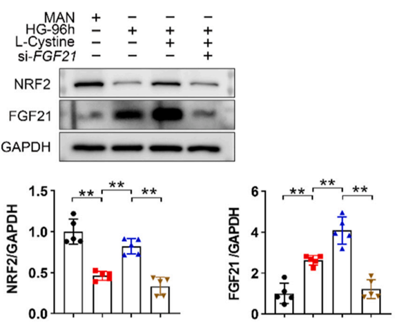
Mitigation of gestational diabetes-induced endothelial dysfunction through FGF21-NRF2 pathway activation involving L-Cystine. [Abstract]2024 Oct;1870(7):167329. PMID: 38960053
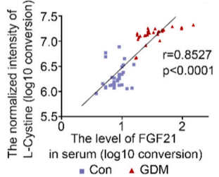
L-Cystine purchased from MedChemExpress. Usage Cited in: Biochim Biophys Acta Mol Basis Dis. 2024 Oct;1870(7):167329. [Abstract]
HUVECs in the presence of 5.5 or 35 mM d-Glucose with or without Si-Scr, Si-FGF21, with or without L-Cystine (0.8 mM) for 96 h. The correlation between L-Cystine and FGF21 expression levels. The X-axis represents the concentration of FGF21 measured via ELISA assay (Log10 conversion). The Y-axis represents the intensity of L-Cystine mass spectrometry signal (Log10 conversion).
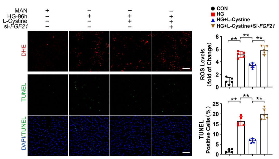
L-Cystine purchased from MedChemExpress. Usage Cited in: Biochim Biophys Acta Mol Basis Dis. 2024 Oct;1870(7):167329. [Abstract]
HUVECs in the presence of 5.5 or 35 mM d-Glucose with or without Si-Scr, Si-FGF21, with or without L-Cystine (0.8 mM) for 96 h. DHE staining and TUNEL staining of HUVECs. Scale bar: 100 μm. The right bar chart explored the quantitative analysis the ROS level and the number of TUNEL positive HUVECs.
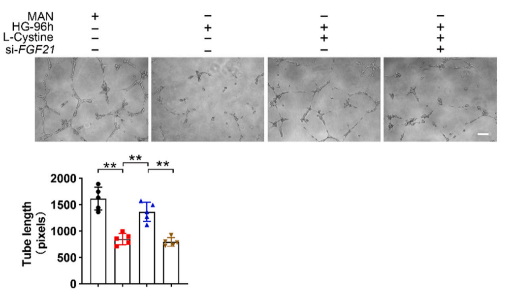
L-Cystine purchased from MedChemExpress. Usage Cited in: Biochim Biophys Acta Mol Basis Dis. 2024 Oct;1870(7):167329. [Abstract]
HUVECs in the presence of 5.5 or 35 mM d-Glucose with or without Si-Scr, Si-FGF21, with or without L-Cystine (0.8 mM) for 96 h. A tube formation assay was performed to assess the angiogenesis ability of HUVECs after being treated as mentioned above. Scale bar: 100 μm. The tube length was quantified and is shown in right.
L-Cystine purchased from MedChemExpress. Usage Cited in: Biochim Biophys Acta Mol Basis Dis. 2024 Oct;1870(7):167329. [Abstract]
HUVECs in the presence of 5.5 or 35 mM d-Glucose with or without Si-Scr, Si-FGF21, with or without L-Cystine (0.8 mM) for 96 h. A tube formation assay was performed to assess the angiogenesis ability of HUVECs after being treated as mentioned above. Scale bar: 100 μm. The tube length was quantified and is shown in right.


Solvent & Solubility
In Vitro:
H2O : 33.33 mg/mL (138.70 mM; ultrasonic and adjust pH to 11 with 1 M NaOH)
0.1 M NaOH : 3.33 mg/mL (13.86 mM; ultrasonic and warming and adjust pH to 11 with NaOH and heat to 70°C)
0.1 M HCl : 2.5 mg/mL (10.40 mM; ultrasonic and adjust pH to 2 with HCl)
DMSO : < 1 mg/mL (insoluble or slightly soluble)
Please refer to the solubility information to select the appropriate solvent. Once prepared, please aliquot and store the solution to prevent product inactivation from repeated freeze-thaw cycles.
Storage method and period of stock solution: -80°C, 6 months; -20°C, 1 month. When stored at -80°C, please use it within 6 months. When stored at -20°C, please use it within 1 month.
* Note: If you choose water as the stock solution, please dilute it to the working solution, then filter and sterilize it with a 0.22 μm filter before use.
Please refer to the solubility information to select the appropriate solvent. Once prepared, please aliquot and store the solution to prevent product inactivation from repeated freeze-thaw cycles.
Storage method and period of stock solution: -80°C, 6 months; -20°C, 1 month. When stored at -80°C, please use it within 6 months. When stored at -20°C, please use it within 1 month.
* Note: If you choose water as the stock solution, please dilute it to the working solution, then filter and sterilize it with a 0.22 μm filter before use.
Concentration (start) × Volume (start) = Concentration (final) × Volume (final)

Protocols
-
Nuclear Protein Extraction (High-Salt/Hypotonic Fractionation)
The high-salt/hypotonic fractionation method for nuclear protein extraction is based on the differential solubility of cellular components. Cytoplasmic proteins are extracted first using a hypotonic buffer that causes cell swelling and membrane rupture, followed by centrifugation to separate the cytoplasmic supernatant from the nuclear pellet. The nuclear pellet is then subjected to high-salt extraction (e. g. , 0. 4 M (NH4)2SO4 or 1 M NaCl) to solubilize tightly bound nuclear matrix proteins, including transcription factors, histones, and structural proteins associated with chromatin and the nuclear scaffold. This approach allows for the isolation of both soluble cytoplasmic proteins and salt-resistant nuclear proteins while minimizing cross-contamination.
-
RNA extraction experimental
By lysing cells, releasing RNA, and removing impurities such as proteins and DNA, high-purity RNA products are finally obtained. The commonly used traditional method is the guanidine isothiocyanate/phenol/chloroform method (Trizol), which is suitable for a variety of animal materials including animal tissues, microorganisms, cultured cells, etc., and most plant materials.
-
Western Blot
Western blotting (WB) is a commonly used experimental method in molecular biology, biochemistry, and immunogenetics for identifying and quantifying target proteins. It combines gel electrophoresis with immunoassay, enabling researchers to analyze protein expression, post-translational modifications, and molecular weight.
-
Apoptosis
Apoptosis, also called programmed cell death, is generally characterized by distinct morphological characteristics.
-
TUNEL staining for apoptotic DNA fragmentation
TUNEL staining detects DNA strand breaks by using terminal deoxynucleotidyl transferase to add labeled nucleotides to exposed 3′-OH DNA termini, generating either microscopic staining in fixed cells or tissue sections, or fluorescence/cytometric signal in cell suspensions. TUNEL positivity reflects DNA fragmentation but should not be interpreted alone as definitive apoptosis, because TUNEL can also label necrotic, autolytic, mechanically damaged, or DNA-repair-associated DNA breaks.
-
Annexin V plus membrane-impermeant dye apoptosis staining
Annexin V-based apoptosis assays rely on the detection of phosphatidylserine (PS) externalization from the inner leaflet of the plasma membrane to the outer leaflet, an early biochemical hallmark of apoptosis. Fluorescently labeled Annexin V binds PS in a calcium-dependent manner, enabling identification of early apoptotic cells by flow cytometry or fluorescence microscopy. When combined with a membrane-impermeant DNA-binding dye (e. g. , propidium iodide), this approach allows discrimination between viable (Annexin V−/dye−), early apoptotic (Annexin V+/dye−), and late apoptotic or necrotic (Annexin V+/dye+) cell populations by assessing membrane integrity and PS exposure.
-
Naïve CD4+ T-cell subset differentiation/polarization
Naïve CD4+ T-cell subset differentiation/polarization is an in vitro assay in which purified naïve CD4+ T cells are activated through TCR and CD28 costimulation and cultured with defined cytokines and neutralizing antibodies to generate Th0, Th1, Th2, Th17, or induced Treg-like populations. Differentiation is detected by subset-associated cytokines and transcription factors: IFN-γ/T-bet for Th1, IL-4/GATA3 for Th2, IL-17A/RORγt for Th17, and Foxp3 for induced Treg cells. The assay readout is usually generated by intracellular cytokine staining after restimulation, transcription-factor staining by flow cytometry, ELISA of secreted cytokines, or gene-expression analysis. The result reflects cytokine-directed lineage commitment or polarization rather than antigen-specific immune protection by itself.
-
ROS/oxidative-stress fluorescent staining
ROS/oxidative-stress fluorescent staining uses cell-permeant fluorogenic probes that become fluorescent after oxidation inside cells or tissues; commonly used examples include DCFH-DA/DCFDA for broad cellular oxidant detection, DHE for superoxide-related signal detection, MitoSOX for mitochondrial superoxide-related signal detection, and CellROX probes for oxidative-stress-associated fluorescence readouts. The assay detects probe oxidation rather than a single ROS species unless the probe and analysis method have been chemically validated for that species. DCFH-DA enters cells, is deacetylated by intracellular esterases to DCFH, and produces fluorescent DCF after oxidation, so the readout is used as an operational measure of total cellular oxidative stress rather than a species-specific ROS measurement. DHE and MitoSOX can report superoxide-related oxidation, but red fluorescence alone can include non-specific ethidium-like oxidation products; HPLC or optimized spectral approaches are
-
Apoptosis Solutions
Apoptosis is a regulated, generally non-lytic cell-death pathway that removes unwanted, damaged, infected, or abnormal cells through coordinated morphological changes, caspase activation, DNA fragmentation, and membrane remodeling. The intrinsic apoptosis pathway is controlled mainly by mitochondrial outer membrane permeabilization, BCL-2 family proteins, cytochrome c release, apoptosome formation, caspase-9 activation, and downstream executioner caspase-3/7 activation. The extrinsic apoptosis pathway is initiated by death receptors such as Fas, TNFR, and TRAIL receptors, which recruit adaptor proteins and activate caspase-8 before engaging executioner caspases or mitochondrial amplification through BID cleavage. Apoptosis is linked to many phenotypes, including cancer cell killing, tissue homeostasis, immune regulation, neurodegeneration, infection response, and treatment-induced cytotoxicity; unresolved questions include how apoptosis interacts with necroptosis, pyroptosis, ferroptos
-
Protocol For Protein Expression And Purification
Recombinant protein expression in Escherichia coli followed by purification of a His-tagged soluble protein by immobilized metal affinity chromatography (IMAC), with optional MBP fusion and TEV tag removal when the construct includes these elements. The biological readout is production of the encoded target protein, detected as an inducible band at the expected molecular mass by SDS-PAGE and quantified by total protein assay or chromatographic absorbance; the purification readout is enrichment of the target protein in elution fractions after selective binding of polyhistidine residues to immobilized Ni2+/metal-chelate resin and elution by imidazole-containing buffer. Expression is driven by an inducible bacterial expression system, commonly T7/lac-based, in which IPTG or lactose/auto-induction activates transcription and translation of the cloned gene; lower induction temperature, lower inducer concentration, induction timing, and solubility-enhancing fusion tags can influence the frac


Purity & Documentation
-
Data Sheet (278 KB)
-
SDS (254 KB)
- English - EN (254 KB)
- Français - FR (254 KB)
- Deutsch - DE (254 KB)
- Norwegian - NO (254 KB)
- Español - ES (254 KB)
- Swedish - SV (254 KB)
- Italian - IT (254 KB)
- Korean - KR (254 KB)
- Portuguese - PT (254 KB)
-
Handling Instructions (2659 KB)
References
[1]. Abdullah M, et al. Extracellular Concentration of L-Cystine Determines the Sensitivity to System xc- Inhibitors[J]. Biomol Ther (Seoul). 2022 Mar 1;30(2):184-190. [Content Brief]
[2]. Kurihara S, et al. Enhancement of antigen-specific immunoglobulin G production in mice by co-administration of L-cystine and L-theanine[J]. J Vet Med Sci. 2007 Dec;69(12):1263-70. [Content Brief]
[4]. Greth WE, et al. Cellular accumulation of L-cystine in rat kidney cortex in vivo[J]. J Clin Invest. 1973 Feb;52(2):454-62. [Content Brief]
[5]. Ohtsu I, et al. Uptake of L-cystine via an ABC transporter contributes defense of oxidative stress in the L-cystine export-dependent manner in Escherichia coli. PLoS One. 2015 Apr 2;10(3):e0120619. [Content Brief]
[6]. Dai W, et al. Fresh Medium or L-Cystine as an Effective Nrf2 Inducer for Cytoprotection in Cell Culture[J]. Cells. 2023 Jan 12;12(2):291. [Content Brief]


Complete Stock Solution Preparation Table
Please refer to the solubility information to select the appropriate solvent. Once prepared, please aliquot and store the solution to prevent product inactivation from repeated freeze-thaw cycles.
Storage method and period of stock solution: -80°C, 6 months; -20°C, 1 month. When stored at -80°C, please use it within 6 months. When stored at -20°C, please use it within 1 month.
| Optional Solvent | Concentration Solvent Mass | 1 mg | 5 mg | 10 mg | 25 mg |
|---|---|---|---|---|---|
| 0.1 M HCl / 0.1 M NaOH / H2O | 1 mM | 4.1615 mL | 20.8073 mL | 41.6146 mL | 104.0366 mL |
| 5 mM | 0.8323 mL | 4.1615 mL | 8.3229 mL | 20.8073 mL | |
| 10 mM | 0.4161 mL | 2.0807 mL | 4.1615 mL | 10.4037 mL | |
| H2O | 15 mM | 0.2774 mL | 1.3872 mL | 2.7743 mL | 6.9358 mL |
| 20 mM | 0.2081 mL | 1.0404 mL | 2.0807 mL | 5.2018 mL | |
| 25 mM | 0.1665 mL | 0.8323 mL | 1.6646 mL | 4.1615 mL | |
| 30 mM | 0.1387 mL | 0.6936 mL | 1.3872 mL | 3.4679 mL | |
| 40 mM | 0.1040 mL | 0.5202 mL | 1.0404 mL | 2.6009 mL | |
| 50 mM | 0.0832 mL | 0.4161 mL | 0.8323 mL | 2.0807 mL | |
| 60 mM | 0.0694 mL | 0.3468 mL | 0.6936 mL | 1.7339 mL | |
| 80 mM | 0.0520 mL | 0.2601 mL | 0.5202 mL | 1.3005 mL | |
| 100 mM | 0.0416 mL | 0.2081 mL | 0.4161 mL | 1.0404 mL |
* Note: If you choose water as the stock solution, please dilute it to the working solution, then filter and sterilize it with a 0.22 μm filter before use.


L-Cystine Related Classifications
HY-N0394 Related Classifications
 Powered by Bioz