



MK2-IN-1
Based on 6 publication(s) in Google Scholar
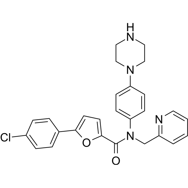
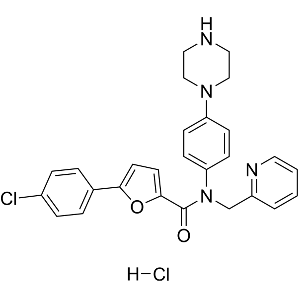
MK2-IN-1 (compound 1) is a potent and selecitve MAPKAPK2 (MK2) inhibitor with an IC50 of 0.11 uM for MK2 and an EC50 of 0.35 uM for pHSP27. MK2-IN-1 impaires the phosphorylation level of serine residues in the Tfcp2l1 protein.
For research use only. We do not sell to patients.
- Purity : 99.04%
- CAS No.: 1314118-92-7
- Formula: C27H25ClN4O2
- Molecular Weight:472.97
-
Storage:Powder -20°C, 3 years , 4°C, 2 years ; In solvent -80°C, 6 months , -20°C, 1 month

To place orders, for customer services and technical support, please contact: MedChemExpress USA
Tel: 609-228-6898 E-mail: [email protected] [email protected]


-
 Biological Activity
-
Chemical Information
- Protocols
- Purity & Documentation
- References
-
 Help & FAQs



Publications Citing Use of MedChemExpress (MCE) MK2-IN-1
More

Biological Activity
Description
Cellular Effect
|
Cell Line

|
Type | Value | Description | References |
|---|---|---|---|---|
| SW1353 | CC50 |
>20 μM
Compound: 25
|
Cytotoxicity against human SW1353 cells after 30 mins by ATP-dependent luciferase luminescence assay
Cytotoxicity against human SW1353 cells after 30 mins by ATP-dependent luciferase luminescence assay
|
[PMID: 24900358] |
| SW1353 | EC50 |
0.35 μM
Compound: 25
|
Inhibition of MK2-mediated HSP27 phosphorylation in IL-1beta stimulated human SW1353 cells incubated for 30 mins prior to IL-1beta-stimulation measured after 27 mins by ELISA
Inhibition of MK2-mediated HSP27 phosphorylation in IL-1beta stimulated human SW1353 cells incubated for 30 mins prior to IL-1beta-stimulation measured after 27 mins by ELISA
|
[PMID: 24900358] |
| SW1353 | IC50 |
5.7 μM
Compound: 25
|
Inhibition of IL1beta-stimulated MMP13 secretion in human SW1353 cells incubated for 30 mins prior to IL1beta-stimulation measured after 24 hrs by ELISA
Inhibition of IL1beta-stimulated MMP13 secretion in human SW1353 cells incubated for 30 mins prior to IL1beta-stimulation measured after 24 hrs by ELISA
|
[PMID: 24900358] |
| THP-1 | EC50 |
0.35 μM
Compound: 1
|
Inhibition of MK2-mediated HSP27 phosphorylation at Ser78 in LPS-stimulated human THP-1 cells pre-incubated for 60 mins before LPS treatment measured after 60 mins
Inhibition of MK2-mediated HSP27 phosphorylation at Ser78 in LPS-stimulated human THP-1 cells pre-incubated for 60 mins before LPS treatment measured after 60 mins
|
[PMID: 22182499] |
| THP-1 | EC50 |
350 nM
Compound: 1
|
Inhibition of MK2 in human THP1 cells assessed as inhibition of LPS-induced HSP27 Ser78 phosphorylation incubated for 60 mins prior to LPS-induction measured after 60 mins
Inhibition of MK2 in human THP1 cells assessed as inhibition of LPS-induced HSP27 Ser78 phosphorylation incubated for 60 mins prior to LPS-induction measured after 60 mins
|
[PMID: 24900435] |
| THP-1 | EC50 |
5 μM
Compound: 2a
|
Inhibition of LPS-stimulated TNFalpha production in human THP1 cells by ELISA
Inhibition of LPS-stimulated TNFalpha production in human THP1 cells by ELISA
|
[PMID: 22169260] |
| THP-1 | IC50 |
4.4 μM
Compound: 25
|
Antiinflammatory activity in human THP1 cells assessed as inhibition of LPS-induced TNFalpha production incubated for 30 mins prior to LPS-challenge measured after 3 hrs by ELISA
Antiinflammatory activity in human THP1 cells assessed as inhibition of LPS-induced TNFalpha production incubated for 30 mins prior to LPS-challenge measured after 3 hrs by ELISA
|
[PMID: 24900358] |
| THP-1 | IC50 |
5.2 μM
Compound: 25
|
Antiinflammatory activity in human THP1 cells assessed as inhibition of LPS-induced IL6 production incubated for 30 mins prior to LPS-challenge measured after 18 hrs by immunoabsorbent-based electrochemical luminescence assay
Antiinflammatory activity in human THP1 cells assessed as inhibition of LPS-induced IL6 production incubated for 30 mins prior to LPS-challenge measured after 18 hrs by immunoabsorbent-based electrochemical luminescence assay
|
[PMID: 24900358] |
In Vitro
MK2-IN-1 (purchased from MCE; 5 μM; 0.5-8 h) gradually increases Tfcp2l1 protein level without a change in the Tfcp2l1 transcript level within 2 h[2].
MK2-IN-1 induces more alkaline phosphatase (AP)-positive colonies than the other factors in a short time[2].
MedChemExpress (MCE) has not independently confirmed the accuracy of these methods. They are for reference only. Further protocols information, click here.
-
Cell Line:46C mouse embryonic stem cells (mESCs)
-
Concentration:5 μM
-
Incubation Time:0.5, 1, 2, 8 h
-
Result:The Tfcp2l1 protein level gradually increased without a change in the Tfcp2l1 transcript level within 2 h.


Chemical Information
-
CAS No. 1314118-92-7
-
Appearance Solid
-
Molecular Weight 472.97
-
Formula C27H25ClN4O2
-
Color White to off-white
-
SMILES
O=C(C1=CC=C(C2=CC=C(Cl)C=C2)O1)N(C3=CC=C(N4CCNCC4)C=C3)CC5=NC=CC=C5
-
Shipping
Room temperature in continental US; may vary elsewhere.
-
Storage
Powder -20°C 3 years 4°C 2 years In solvent -80°C 6 months -20°C 1 month


Publications (6)
-
Journal Impact Factor
-
Most Recent
-
-
Cell Death Dis
Quantitative analysis of phosphoproteome in necroptosis reveals a role of TRIM28 phosphorylation in promoting necroptosis-induced cytokine production. [Abstract]2021 Oct 23;12(11):994. PMID: 34689152 -
Cell Rep
MK2 promotes Tfcp2l1 degradation via β-TrCP ubiquitin ligase to regulate mouse embryonic stem cell self-renewal. [Abstract]2021 Nov 2;37(5):109949. PMID: 34731635 -
J Pharmacol Exp Ther
Clinically Advanced p38 Inhibitors Suppress DUX4 Expression in Cellular and Animal Models of Facioscapulohumeral Muscular Dystrophy. [Abstract]2019 Aug;370(2):219-230. PMID: 31189728 -
Biol Pharm Bull
Stress Response Kinase MK2 Induces Non-canonical Activation of EphA2 in EML4-ALK Lung Cancer Cells. [Abstract]2025;48(2):172-176. PMID: 40024717 -

Protocols
-
Kinase activity and phosphorylation assays
Kinase activity assays measure the ability of kinases to transfer phosphate groups from ATP to specific substrates, while phosphorylation assays detect the presence and levels of phosphorylated proteins. Common methods include radiolabeled ATP incorporation (e. g. ,), ADP release detection via bioluminescence (e. g. ,[3]), enzyme-linked immunosorbent assays (ELISA) for phospho-specific epitopes (e. g. ,[6]), and microtiter-based formats for high-throughput screening (e. g. ,[8]). The ADP-Glo assay quantifies kinase activity by measuring ADP produced during phosphorylation using a luciferase-based system. Radiometric assays involve autoradiography or scintillation counting after incorporation of 32P-labeled ATP into substrate proteins. ELISA-based approaches rely on phospho-specific antibodies to detect activated kinases in cell lysates or purified samples.
-
Western Blot
Western blotting (WB) is a commonly used experimental method in molecular biology, biochemistry, and immunogenetics for identifying and quantifying target proteins. It combines gel electrophoresis with immunoassay, enabling researchers to analyze protein expression, post-translational modifications, and molecular weight.
-
Cell Cytotoxicity Assay
Cytotoxicity assays are usually based on the assessment of cell membrane damage, which can also be indirectly detected by measuring cell viability. Detection methods include MTT assay, CKK-8 assay, LDH assay and ATP assay, etc.
-
Protocol for Kinase activity and phosphorylation assays
Kinase activity assays measure transfer of phosphate from ATP to a protein or peptide substrate, generating phosphorylated substrate, ADP, or incorporated radiolabeled phosphate as the readout; phosphorylation assays measure site-specific phosphorylation in cells or tissues as a proxy for kinase-pathway activation, inhibition, or substrate regulation. Phosphorylation can be detected by phospho-specific Western blot, immunoprecipitation kinase assay, phospho-immunofluorescence, phospho-flow cytometry, luminescent ADP detection, radiolabeled ATP incorporation, or reporter-based pathway assays, and these readouts can be applied to cancer cells, primary neurons, mouse tumors, organoids, inflammatory macrophages, ferroptosis studies, and mitophagy studies when the kinase target is biologically relevant.


Purity & Documentation
-
Data Sheet (274 KB)
-
SDS (251 KB)
- English - EN (251 KB)
- Français - FR (251 KB)
- Deutsch - DE (251 KB)
- Norwegian - NO (251 KB)
- Español - ES (251 KB)
- Swedish - SV (251 KB)
- Italian - IT (251 KB)
- Korean - KR (251 KB)
- Portuguese - PT (251 KB)
-
Handling Instructions (2659 KB)
References
[1]. Rao AU, et al. Facile synthesis of tetracyclic azepine and oxazocine derivatives and their potential as MAPKAP-K2 (MK2) inhibitors. Bioorg Med Chem Lett. 2012 Jan 15;22(2):1068-72. [Content Brief]
[2]. Yan Zhang, et al. MK2 promotes Tfcp2l1 degradation via β-TrCP ubiquitin ligase to regulate mouse embryonic stem cell self-renewal. Cell Rep. 2021 Nov 2;37(5):109949. [Content Brief]

Calculators
Concentration (start) × Volume (start) = Concentration (final) × Volume (final)


MK2-IN-1 Related Classifications
HY-12834 Related Classifications
 Powered by Bioz