



Roquinimex

Based on 1 Customer Validation
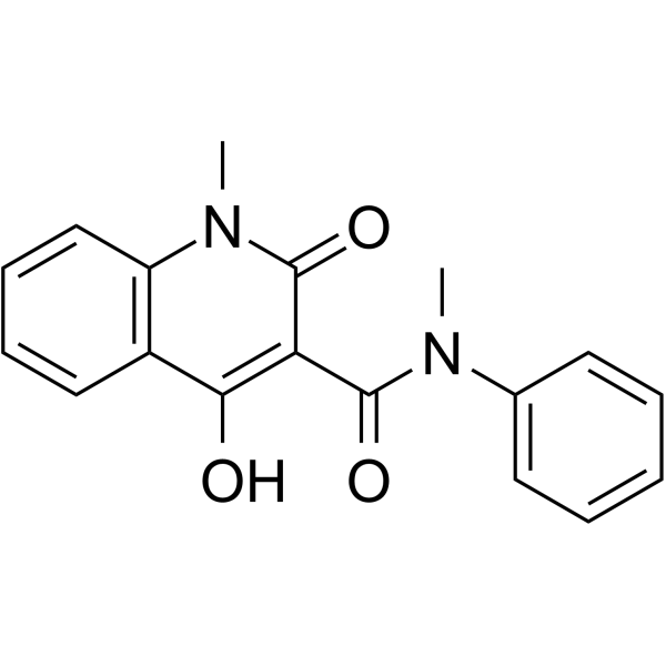
Roquinimex (Linomide) is an orally active immunomodulator with antineoplastic, anti-inflammatory, and antiangiogenic activity. Roquinimex suppresses TH1 lymphocyte cytokines (IL-2, IFN-γ), promotes TH2 lymphocyte cytokines (IL-4, IL-10), increases NK cell, activated monocyte, and T cell activity. Roquinimex blocks macrophage TNF-α production and suppresses IL-1/IL-6 secretion. Roquinimex exhibits in vivo antitumour activity, suppresses rodent autoimmune disease signs, and ameliorates murine colitis and psoriasis. Roquinimex can be used for the research of leukemia, inflammatory bowel disease, multiple sclerosis, and psoriasis.
For research use only. We do not sell to patients.
- Purity : 99.69%
- CAS No.: 84088-42-6
- Formula: C18H16N2O3
- Molecular Weight:308.33
-
Storage:Powder -20°C, 3 years , 4°C, 2 years ; In solvent -80°C, 2 years , -20°C, 1 year

To place orders, for customer services and technical support, please contact: MedChemExpress USA
Tel: 609-228-6898 E-mail: [email protected] [email protected]


-
 Biological Activity
-
Chemical Information
-
Solvent & Solubility
- Protocols
- Purity & Documentation
- References
-
 Help & FAQs


-
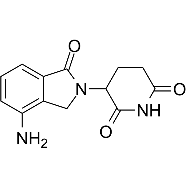
HY-A0003
LenalidomideImmunomodulator -
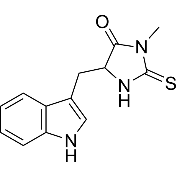
HY-15760
Necrostatin-1RIPK1 Inhibitor -

HY-13743R
Roquinimex (Standard)Roquinimex Standard -
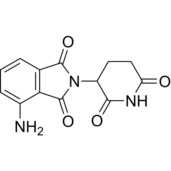
HY-10984
PomalidomideImmunomodulatory Agent -
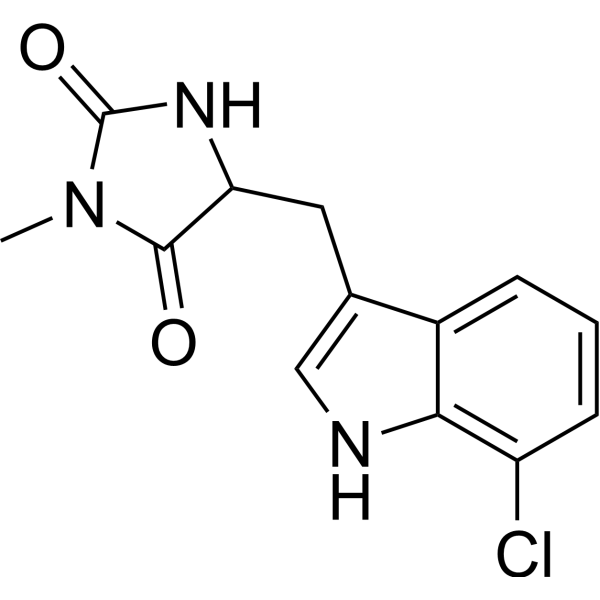
HY-14622A
Necrostatin 2 racemateRIPK1 Inhibitor -
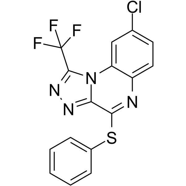
HY-110203
R-7050TNF Receptor Antagonist -
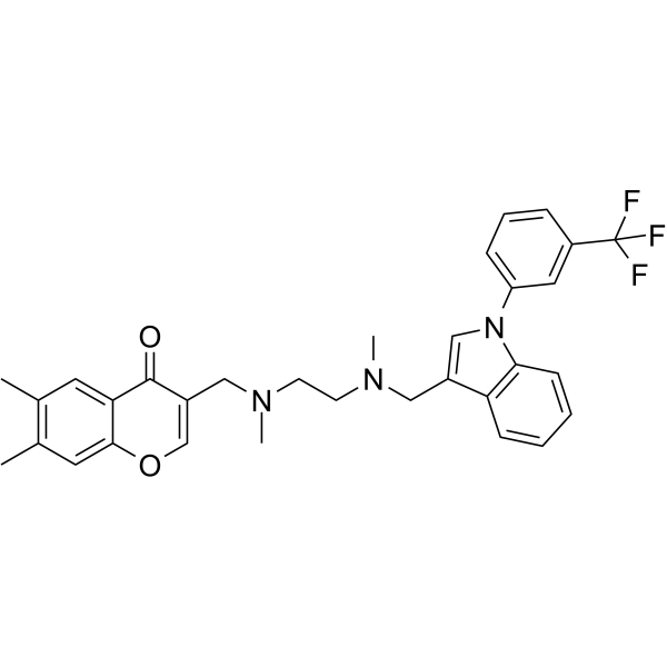
HY-111255
SPD304TNFα Inhibitor -

HY-108847
EtanerceptTNF Inhibitor -

HY-P9908
AdalimumabTNF-α Inhibitor -
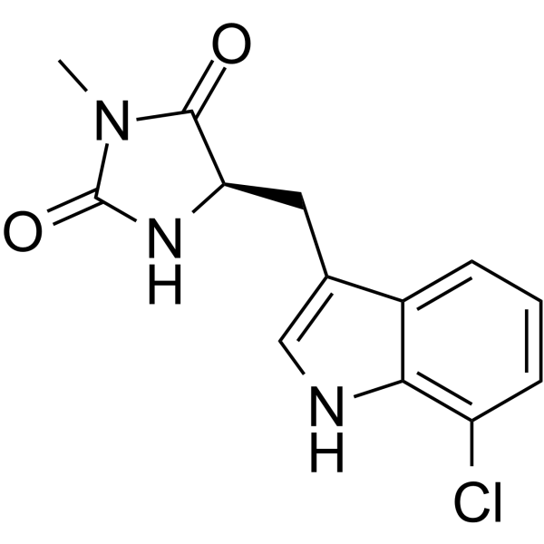
HY-14622
Necrostatin 2RIP kinase Inhibitor -

HY-A0003R
Lenalidomide (Standard)Reference Standard -
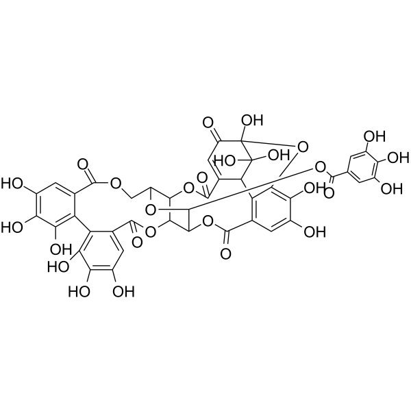
HY-N0472
GeraniinTNF Receptor Inhibitor


Biological Activity
Description
Cellular Effect
|
Cell Line

|
Type | Value | Description | References |
|---|---|---|---|---|
| HUVEC | IC50 |
13.95 μM
Compound: 1 (Linomide)
|
Inhibitory concentration against Human umbilical vein endothelial cell (HUVEC) proliferation
Inhibitory concentration against Human umbilical vein endothelial cell (HUVEC) proliferation
|
[PMID: 12643940] |
| PC-3 | IC50 |
>100 μM
Compound: 1 (Linomide)
|
Antiproliferative activity against PC-3 prostate cancer cells
Antiproliferative activity against PC-3 prostate cancer cells
|
[PMID: 12643940] |
In Vitro
Roquinimex (1-100 μg/mL; 48 h) significantly reduces the number of MBP-reactive IFN-γ secreting peripheral blood MNC, but does not affect PHA-reactive IFN-γ secreting cells[3].
Roquinimex (100 μg/mL; 48 h) reduces pro-inflammatory TNF-α mRNA expression across MBP-, PPD-, and PHA-reactive peripheral blood MNC, while increasing anti-inflammatory TGF-β and IL-10 mRNA expression in MBP- and PHA-reactive cells, with no effect on IFN-γ, LT, or IL-4 mRNA expression[3].
MedChemExpress (MCE) has not independently confirmed the accuracy of these methods. They are for reference only. Further protocols information, click here.
In Vivo
Roquinimex ameliorates manifestations of lupus-like disease in MRL/lpr mice[3].
Roquinimex (p.o.) provides complete protection from insulitis and maintains normal glucose tolerance for over 40 weeks in NOD mice[3].
Roquinimex (0.5-1% w/w; topical; twice daily; 8 days) exerts antipsoriatic effects in Imiquimod (HY-B0180)-induced mice[5].
MedChemExpress (MCE) has not independently confirmed the accuracy of these methods. They are for reference only.
-
Animal Model:Balb/c mice (male, 21-26 g) with colitis induced by 5% DDS in drinking water for 5 days[2]
-
Dosage:300 mg/kg/day
-
Administration:s.c.; daily; 8 days; strated from day 3 pre-DSS challenge
-
Result:Reduced disease activity index by 60% after 5 days of DSS exposure and by 72% over the full experimental period.
Reduced mucosal damage score.
Prevented DSS-induced crypt height reduction.
Reduced colonic myeloperoxidase activity by 53%.
All outcomes were statistically significant.
-
Animal Model:Swiss Albino mice (male, 8-12 weeks old, 24-30 g) with psoriasis induced by Imiquimod[5]
-
Dosage:0.5%; 1% w/w
-
Administration:topical; twice daily; 8 days
-
Result:Reduced mean Baker's histopathological score.
Increased skin tissue IL-10 levels.
Reduced skin tissue IL-17 levels.
Reduced skin tissue TNF-α levels.
Reduced skin tissue VEGF levels.
Improved histopathological features, including mild keratosis, no Munro's abscesses or parakeratosis, mild acanthosis, few or no rete ridges, mild papillary thinning, and mild or few dermal lymphocytic infiltrations.
Showed statistically significant improvements.


Chemical Information
-
CAS No. 84088-42-6
-
Appearance Solid
-
Molecular Weight 308.33
-
Formula C18H16N2O3
-
Color White to off-white
-
SMILES
O=C(C1=C(O)C2=C(N(C)C1=O)C=CC=C2)N(C)C3=CC=CC=C3
-
Synonyms
Linomide; FCF89; ABR212616
-
Shipping
Room temperature in continental US; may vary elsewhere.
-
Storage
Powder -20°C 3 years 4°C 2 years In solvent -80°C 2 years -20°C 1 year


Solvent & Solubility
In Vitro:
DMSO : ≥ 83.3 mg/mL (270.17 mM; Hygroscopic DMSO has a significant impact on the solubility of product, please use newly opened DMSO)
* "≥" means soluble, but saturation unknown.
Please refer to the solubility information to select the appropriate solvent. Once prepared, please aliquot and store the solution to prevent product inactivation from repeated freeze-thaw cycles.
Storage method and period of stock solution: -80°C, 2 years; -20°C, 1 year. When stored at -80°C, please use it within 2 years. When stored at -20°C, please use it within 1 year.
Please refer to the solubility information to select the appropriate solvent. Once prepared, please aliquot and store the solution to prevent product inactivation from repeated freeze-thaw cycles.
Storage method and period of stock solution: -80°C, 2 years; -20°C, 1 year. When stored at -80°C, please use it within 2 years. When stored at -20°C, please use it within 1 year.
Concentration (start) × Volume (start) = Concentration (final) × Volume (final)

Protocols
-
Naïve CD4+ T-cell subset differentiation/polarization
Naïve CD4+ T-cell subset differentiation/polarization is an in vitro assay in which purified naïve CD4+ T cells are activated through TCR and CD28 costimulation and cultured with defined cytokines and neutralizing antibodies to generate Th0, Th1, Th2, Th17, or induced Treg-like populations. Differentiation is detected by subset-associated cytokines and transcription factors: IFN-γ/T-bet for Th1, IL-4/GATA3 for Th2, IL-17A/RORγt for Th17, and Foxp3 for induced Treg cells. The assay readout is usually generated by intracellular cytokine staining after restimulation, transcription-factor staining by flow cytometry, ELISA of secreted cytokines, or gene-expression analysis. The result reflects cytokine-directed lineage commitment or polarization rather than antigen-specific immune protection by itself.
-
Imiquimod-Induced Psoriasiform Dermatitis
Imiquimod (IMQ)-induced psoriasiform dermatitis is a widely used murine model in which topical application of IMQ, a Toll-like receptor 7 (TLR7) agonist, triggers innate immune activation in the skin and induces a psoriasis-like inflammatory cascade characterized by epidermal hyperplasia, immune cell infiltration, and cytokine production dominated by the IL-23/IL-17 axis. This inflammatory response is mediated through activation of dendritic cells and downstream induction of IL-23, IL-17A, IL-22, and related pro-inflammatory mediators, recapitulating key features of human plaque psoriasis and enabling mechanistic and therapeutic studies. The model is commonly induced using Aldara (5% IMQ cream) applied topically to murine skin, resulting in rapid onset of erythema, scaling, and thickening that can be quantified as disease severity indices and validated histologically.
-
DSS-Induced Colitis
Dextran sulfate sodium (DSS)-induced colitis is generated by administering DSS in mouse drinking water, producing epithelial injury, barrier disruption, weight loss, diarrhea, fecal blood, colon shortening, histologic mucosal damage, and inflammatory mediator changes; the model is mainly used to study acute or chronic intestinal inflammation resembling selected features of ulcerative colitis. DSS injury is interpreted through clinical and tissue readouts rather than a single molecular endpoint: daily body weight, stool consistency, and bleeding are combined into a disease activity index, while colon length, histology, cytokines, myeloperoxidase activity, intestinal permeability, and tight-junction markers provide complementary measures of inflammation and barrier damage.
-
TNBS-Induced Colitis
TNBS-induced colitis is produced by intrarectal delivery of 2,4,6-trinitrobenzene sulfonic acid in ethanol, where ethanol disrupts the mucosal barrier and TNBS haptenates colonic proteins, generating immune-mediated colonic inflammation with weight loss, diarrhea, ulceration, transmural injury, inflammatory-cell infiltration, and cytokine responses. The model is used as an experimental intestinal inflammation model with Crohn’s disease–like features, especially when Th1-type responses, IL-12–dependent inflammation, chronic relapsing inflammation, or fibrosis-related endpoints are studied.
-
Research Protocol for Inflammation-related Diseases
The NLRP3 inflammasome is a cytosolic innate immune signaling platform that integrates priming signals and danger-signal activation to promote caspase-1 activation, maturation of IL-1β and IL-18, and gasdermin D-mediated pyroptotic cell death. The core experimental logic is to determine whether inflammatory disease phenotypes are driven by increased NLRP3 expression, ASC-containing inflammasome assembly, caspase-1 cleavage, GSDMD cleavage, and extracellular release of IL-1β/IL-18 rather than by nonspecific cell injury alone. The pathway is strongly linked to inflammation-related disease phenotypes because monosodium urate crystals activate NALP3/NLRP3 inflammasome signaling in gout-like crystal inflammation, cholesterol crystals activate NLRP3 inflammasomes in atherogenesis models, and DSS-induced intestinal inflammation has been reported to involve NLRP3 inflammasome activity. However, experimental colitis studies also show context-dependent protective effects of NLRP3 inflammasome co
-
Research Protocol for Cancer Immunology
Cancer immunology studies how the immune system recognizes, suppresses, edits, or fails to eliminate malignant cells through tumor antigen release, antigen presentation, T-cell priming, immune trafficking, tumor-cell killing, and feedback inhibition in the tumor microenvironment. The cancer-immunity cycle links tumor antigenicity, dendritic-cell priming, CD8+ T-cell infiltration, cytotoxic function, and immune-checkpoint regulation to tumor rejection or immune escape. Immune-checkpoint pathways such as PD-1/PD-L1 and CTLA-4 suppress antitumor T-cell activity and can be therapeutically blocked, but many tumors remain resistant because of poor antigen presentation, weak T-cell infiltration, suppressive myeloid cells, regulatory T cells, and tumor-intrinsic immune-exclusion programs. Unresolved questions include which immune-cell states predict response, how tumor-intrinsic pathways exclude immune cells, how myeloid suppression limits checkpoint blockade, and which combination strategies


Purity & Documentation
-
Data Sheet (280 KB)
-
SDS (479 KB)
- English - EN (479 KB)
- Français - FR (479 KB)
- Deutsch - DE (479 KB)
- Norwegian - NO (479 KB)
- Español - ES (479 KB)
- Swedish - SV (479 KB)
- Italian - IT (479 KB)
- Korean - KR (479 KB)
- Portuguese - PT (479 KB)
-
Handling Instructions (2659 KB)
References
[1]. Ohsuga Y, et al. Dermatologic changes associated with roquinimex immunotherapy after autologous bone marrow transplant. J Am Acad Dermatol. 2000;43(3):437-441. [Content Brief]
[2]. Liu Q, et al. Roquinimex inhibits dextran sodium sulfate-induced murine colitis. Inflamm Res. 2003;52(2):64-68. [Content Brief]
[3]. Tian WZ, et al. Linomide (roquinimex) affects the balance between pro- and anti-inflammatory cytokines in vitro in multiple sclerosis. Acta Neurol Scand. 1998;98(2):94-101. [Content Brief]
[4]. Mackean MJ, et al. A feasibility study of roquinimex (Linomide) and alpha interferon in patients with advanced malignant melanoma or renal carcinoma. Br J Cancer. 1998;78(12):1620-1623. [Content Brief]
[5]. Hasan AM, et al. Novel effect of topical Roquinimex and its combination with Clobetasol on an imiquimod-induced model of psoriasis in mice. Naunyn Schmiedebergs Arch Pharmacol. 2024;397(7):5219-5232. [Content Brief]


Complete Stock Solution Preparation Table
Please refer to the solubility information to select the appropriate solvent. Once prepared, please aliquot and store the solution to prevent product inactivation from repeated freeze-thaw cycles.
Storage method and period of stock solution: -80°C, 2 years; -20°C, 1 year. When stored at -80°C, please use it within 2 years. When stored at -20°C, please use it within 1 year.
| Optional Solvent | Concentration Solvent Mass | 1 mg | 5 mg | 10 mg | 25 mg |
|---|---|---|---|---|---|
| DMSO | 1 mM | 3.2433 mL | 16.2164 mL | 32.4328 mL | 81.0820 mL |
| 5 mM | 0.6487 mL | 3.2433 mL | 6.4866 mL | 16.2164 mL | |
| 10 mM | 0.3243 mL | 1.6216 mL | 3.2433 mL | 8.1082 mL | |
| 15 mM | 0.2162 mL | 1.0811 mL | 2.1622 mL | 5.4055 mL | |
| 20 mM | 0.1622 mL | 0.8108 mL | 1.6216 mL | 4.0541 mL | |
| 25 mM | 0.1297 mL | 0.6487 mL | 1.2973 mL | 3.2433 mL | |
| 30 mM | 0.1081 mL | 0.5405 mL | 1.0811 mL | 2.7027 mL | |
| 40 mM | 0.0811 mL | 0.4054 mL | 0.8108 mL | 2.0270 mL | |
| 50 mM | 0.0649 mL | 0.3243 mL | 0.6487 mL | 1.6216 mL | |
| 60 mM | 0.0541 mL | 0.2703 mL | 0.5405 mL | 1.3514 mL | |
| 80 mM | 0.0405 mL | 0.2027 mL | 0.4054 mL | 1.0135 mL | |
| 100 mM | 0.0324 mL | 0.1622 mL | 0.3243 mL | 0.8108 mL |


Roquinimex Related Classifications
HY-13743 Related Classifications
 Powered by Bioz
Keywords
- Roquinimex
- 84088-42-6
- Linomide
- FCF89
- ABR212616
- FCF89
- FCF 89
- FCF-89
- ABR212616
- ABR 212616
- ABR-212616
- TNF Receptor
- Interleukin Related
- IFNAR
- acute myelogenous leukemia
- inflammatory bowel disease
- NK cell
- chronic myelogenous leukemia
- TH1 lymphocyte cytokines
- multiple sclerosis
- advanced renal cell carcinoma
- advanced malignant melanoma
- eccrine sweat gland
- TH2 lymphocyte cytokines
- Inhibitor
- inhibitor
- inhibit