



Aaptamine
Based on 2 publication(s) in Google Scholar
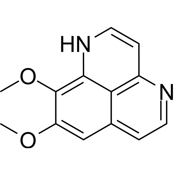
Aaptamine is an alkaloid that can be isolated from the sponge Aaptos suberitoides. Aaptamine is a competitive antagonist of the α-adrenergic receptor, as well as an inhibitor of the proteasome and cholinesterase. Aaptamine is cytotoxic to tumor cells and can induce apoptosis, cell cycle arrest of tumor cells, and the expression of p21 through a p53-independent pathway. Aaptamine has multiple activities such as anti-tumor, antioxidant, antibacterial, and analgesic effects.
Nur für Forschungszwecke. Wir verkaufen nicht an Patienten.
- Reinheit : 99.71%
- CAS. Nr.: 85547-22-4
- Formel: C13H12N2O2
- Molecular Weight:228.25
-
Speicherung:
4°C, protect from light
* In solvent : -80°C, 6 months; -20°C, 1 month (protect from light)

To place orders, for customer services and technical support, please contact: MedChemExpress USA
Tel: 609-228-6898 E-mail: [email protected] [email protected]


-
 Biologische Aktivität
-
Chemical Information
-
Lösungsmittel & Löslichkeit
- Protokoll
- Reinheit & Dokumentation
- Verweise
-
 Help & FAQs


Publications Citing Use of MedChemExpress (MCE) Aaptamine
MoreAlle Adrenergic Receptor Isoform-spezifische Produkte anzeigen
MoreAlle AP-1 Isoform-spezifische Produkte anzeigen
More

Biologische Aktivität
Beschreibung
IC50 & Target
[5]|
AChE 0.23 μg/mL (IC50) |
BChE 1.38 μg/mL (IC50) |
Cellular Effect
|
Cell Line

|
Type | Value | Description | References |
|---|---|---|---|---|
| 786-0 | EC50 |
>100 μM
Compound: 1
|
Inhibition of HIF2alpha in human 786-0 cells expressing truncated HIF1alpha assessed as reduction in luciferase activity after 24 hrs by reporter gene assay
Inhibition of HIF2alpha in human 786-0 cells expressing truncated HIF1alpha assessed as reduction in luciferase activity after 24 hrs by reporter gene assay
|
[PMID: 22928967] |
| A498 | ED50 |
3.2 μg/mL
Compound: 3
|
Antineoplastic activity against human A498 cells
Antineoplastic activity against human A498 cells
|
[PMID: 15043446] |
| A549 | IC50 |
2.8 μg/mL
Compound: 2
|
Cytotoxicity against human A549 cells after 6 days by MTT assay
Cytotoxicity against human A549 cells after 6 days by MTT assay
|
[PMID: 10514310] |
| HeLa | IC50 |
15 μg/mL
Compound: 1
|
Cytotoxicity against human HeLa cells after 3 days by MTT assay
Cytotoxicity against human HeLa cells after 3 days by MTT assay
|
[PMID: 20451377] |
| HeLa | IC50 |
19 μM
Compound: 1
|
Cytotoxicity against human HeLa cells assessed as reduction in cell viability after 48 hrs by CCK8 assay
Cytotoxicity against human HeLa cells assessed as reduction in cell viability after 48 hrs by CCK8 assay
|
[PMID: 33170001] |
| HL-60 | IC50 |
>55 μM
Compound: 7
|
Antioxidant activity against TPA-induced ROS production in human HL60 cells by 2',7'-dichlorodihydrofluorescein diacetate cellular-based assay
Antioxidant activity against TPA-induced ROS production in human HL60 cells by 2',7'-dichlorodihydrofluorescein diacetate cellular-based assay
|
[PMID: 12762791] |
| HT-29 | IC50 |
6.9 μg/mL
Compound: 2
|
Cytotoxicity against human HT-29 cells after 6 days by MTT assay
Cytotoxicity against human HT-29 cells after 6 days by MTT assay
|
[PMID: 10514310] |
| KM-20L2 | ED50 |
3.6 μg/mL
Compound: 3
|
Antineoplastic activity against human KM20L2 cells
Antineoplastic activity against human KM20L2 cells
|
[PMID: 15043446] |
| L5178Y | IC50 |
8.3 μM
Compound: 5
|
Cytotoxic activity against mouse L5178Y cells by MTT assay
Cytotoxic activity against mouse L5178Y cells by MTT assay
|
[PMID: 23282083] |
| MCF7 | IC50 |
>20 μM
Compound: 1
|
Cytotoxicity against human MCF7 cells assessed as reduction in cell viability after 48 hrs by CCK8 assay
Cytotoxicity against human MCF7 cells assessed as reduction in cell viability after 48 hrs by CCK8 assay
|
[PMID: 33170001] |
| NB-4 | IC50 |
>20 μM
Compound: 1
|
Cytotoxicity against human NB4 cells assessed as reduction in cell viability after 48 hrs by CCK8 assay
Cytotoxicity against human NB4 cells assessed as reduction in cell viability after 48 hrs by CCK8 assay
|
[PMID: 33170001] |
| NCI-H460 | ED50 |
3.2 μg/mL
Compound: 3
|
Antineoplastic activity against human NCI-H460 cells
Antineoplastic activity against human NCI-H460 cells
|
[PMID: 15043446] |
| OVCAR-3 | ED50 |
4.9 μg/mL
Compound: 3
|
Antineoplastic activity against human OVCAR-3 cells
Antineoplastic activity against human OVCAR-3 cells
|
[PMID: 15043446] |
| P388 | ED50 |
3.6 μg/mL
Compound: 3
|
Antineoplastic activity against mouse P388 cells
Antineoplastic activity against mouse P388 cells
|
[PMID: 15043446] |
| P388 | IC50 |
0.6 μg/mL
Compound: 2
|
Cytotoxicity against mouse P388 cells after 3 days by MTT assay
Cytotoxicity against mouse P388 cells after 3 days by MTT assay
|
[PMID: 10514310] |
| SF-295 | ED50 |
4.1 μg/mL
Compound: 3
|
Antineoplastic activity against human SF295 cells
Antineoplastic activity against human SF295 cells
|
[PMID: 15043446] |
| SK-MEL-5 | ED50 |
4.3 μg/mL
Compound: 3
|
Antineoplastic activity against human SK-MEL-5 cells
Antineoplastic activity against human SK-MEL-5 cells
|
[PMID: 15043446] |
In Vitro
Aaptamine (10 μM) can shift the dose-response curve of norepinephrine to the right in a parallel manner in isolated rabbit aorta and renal artery, without affecting the curves of histamine or potassium chloride[1].
Aaptamine (30 μg/mL; 0-48 h) can increase the expression of p21 and induce cell cycle arrest in MG63 cells[2].
Aaptamine (3 days) is cytotoxic to HeLa cells, with an IC50 of 15 μg/mL[3].
Aaptamine (50-100 μM; 6-24 h) can activate AP-1- and NF-κB-dependent transcriptional activity in JB6 Cl41 cells[4].
Aaptamine (5-55 µg/mL; 24 h) has an inhibitory effect on ESBL-producing Gram-negative uropathogenic bacteria[6].
MedChemExpress (MCE) has not independently confirmed the accuracy of these methods. They are for reference only.
-
Cell Line:MG63 cells
-
Concentration:30 μg/mL
-
Incubation Time:0, 3, 6, 12, 24 and 48 h
-
Result:Increased the level of p21.
In Vivo
MedChemExpress (MCE) has not independently confirmed the accuracy of these methods. They are for reference only.
-
Animal Model:Chronic constriction injury treated adult male Wistar rats (250-285 g)[7]
-
Dosage:5, 30 and 100 μg
-
Administration:Intrathecal injection; single dose
-
Result:Showed different durations of analgesic effects on CCI-induced nociception. The 5 μg, 30 μg, and 100 μg doses presented analgesic durations of 90, 150, and 150 minutes respectively. However, the variance in the group administered with 100 μg of aaptamine was the largest, and the rats presented some abnormal motor behavior.
- RNA extraction experimental
- Research Protocol for Infectious Diseases
- Detection of Apoptosis
- TUNEL staining for apoptotic DNA fragmentation
- Flow cytometric DNA-content cell-cycle staining
- Annexin V plus membrane-impermeant dye apoptosis staining
- BrdU Incorporation Assay
- Apoptosis Solutions
- Protocol for Cell Cycle
- Cell Viability Determination by MTT Colorimetric Assay


Chemical Information
-
CAS. Nr. 85547-22-4
-
Appearance Solid
-
Molecular Weight 228.25
-
Formel C13H12N2O2
-
Color White to yellow
-
SMILES
COC1=C2C3=C(N=CC=C3C=C1OC)C=CN2
-
Structure Classification
-
Initial Source
-
Versand
Room temperature in continental US; may vary elsewhere.
-
Speicherung
4°C, protect from light
* In solvent : -80°C, 6 months; -20°C, 1 month (protect from light)


Publications (2)
-
Journal Impact Factor
-
Most Recent
-
J Exp Pharmacol
Aaptamine Alters Vimentin Expression and Migration Capability of Triple-Negative Breast Cancer Cells. [Abstract]2025 May 22:17:239-247. PMID: 40420842 -


Lösungsmittel & Löslichkeit
In Vitro:
DMSO : 100 mg/mL (438.12 mM; Need ultrasonic; Hygroscopic DMSO has a significant impact on the solubility of product, please use newly opened DMSO)
Please refer to the solubility information to select the appropriate solvent. Once prepared, please aliquot and store the solution to prevent product inactivation from repeated freeze-thaw cycles.
Storage method and period of stock solution: -80°C, 6 months; -20°C, 1 month (protect from light). When stored at -80°C, please use it within 6 months. When stored at -20°C, please use it within 1 month.
Please refer to the solubility information to select the appropriate solvent. Once prepared, please aliquot and store the solution to prevent product inactivation from repeated freeze-thaw cycles.
Storage method and period of stock solution: -80°C, 6 months; -20°C, 1 month (protect from light). When stored at -80°C, please use it within 6 months. When stored at -20°C, please use it within 1 month.
Konzentration (Stammlösung) × Volumen (Stammlösung) = Konzentration (Ziellösung) × Volumen (Ziellösung)


Reinheit & Dokumentation
-
Data Sheet (283 KB)
-
SDS (252 KB)
- English - EN (252 KB)
- Français - FR (252 KB)
- Deutsch - DE (252 KB)
- Norwegian - NO (252 KB)
- Español - ES (252 KB)
- Swedish - SV (252 KB)
- Italian - IT (252 KB)
- Korean - KR (252 KB)
- Portuguese - PT (252 KB)
-
Handling Instructions (2659 KB)
Verweise
[1]. Ohizumi Y, et al. Alpha-adrenoceptor blocking action of aaptamine, a novel marine natural product, in vascular smooth muscle. J Pharm Pharmacol. 1984 Nov;36(11):785-6. [Content Brief]
[2]. Aoki S, et al. Aaptamine, a spongean alkaloid, activates p21 promoter in a p53-independent manner. Biochem Biophys Res Commun. 2006 Mar 31;342(1):101-6. [Content Brief]
[3]. Tsukamoto S, et al. Aaptamine, an alkaloid from the sponge Aaptos suberitoides, functions as a proteasome inhibitor. Bioorg Med Chem Lett. 2010 Jun 1;20(11):3341-3. [Content Brief]
[4]. Dyshlovoy SA, et al. Aaptamines from the marine sponge Aaptos sp. display anticancer activities in human cancer cell lines and modulate AP-1-, NF-κB-, and p53-dependent transcriptional activity in mouse JB6 Cl41 cells. Biomed Res Int. 2014;2014:469309. [Content Brief]
[7]. Sung CS, et al. Antinociceptive Effects of Aaptamine, a Sponge Component, on Peripheral Neuropathy in Rats. Mar Drugs. 2023 Feb 4;21(2):113. [Content Brief]


Complete Stock Solution Preparation Table
Please refer to the solubility information to select the appropriate solvent. Once prepared, please aliquot and store the solution to prevent product inactivation from repeated freeze-thaw cycles.
Storage method and period of stock solution: -80°C, 6 months; -20°C, 1 month (protect from light). When stored at -80°C, please use it within 6 months. When stored at -20°C, please use it within 1 month.
| Optional Solvent | Concentration Solvent Mass | 1 mg | 5 mg | 10 mg | 25 mg |
|---|---|---|---|---|---|
| DMSO | 1 mM | 4.3812 mL | 21.9058 mL | 43.8116 mL | 109.5290 mL |
| 5 mM | 0.8762 mL | 4.3812 mL | 8.7623 mL | 21.9058 mL | |
| 10 mM | 0.4381 mL | 2.1906 mL | 4.3812 mL | 10.9529 mL | |
| 15 mM | 0.2921 mL | 1.4604 mL | 2.9208 mL | 7.3019 mL | |
| 20 mM | 0.2191 mL | 1.0953 mL | 2.1906 mL | 5.4765 mL | |
| 25 mM | 0.1752 mL | 0.8762 mL | 1.7525 mL | 4.3812 mL | |
| 30 mM | 0.1460 mL | 0.7302 mL | 1.4604 mL | 3.6510 mL | |
| 40 mM | 0.1095 mL | 0.5476 mL | 1.0953 mL | 2.7382 mL | |
| 50 mM | 0.0876 mL | 0.4381 mL | 0.8762 mL | 2.1906 mL | |
| 60 mM | 0.0730 mL | 0.3651 mL | 0.7302 mL | 1.8255 mL | |
| 80 mM | 0.0548 mL | 0.2738 mL | 0.5476 mL | 1.3691 mL | |
| 100 mM | 0.0438 mL | 0.2191 mL | 0.4381 mL | 1.0953 mL |


Aaptamine Related Classifications
HY-N4225 Related Classifications
 Powered by Bioz