



Aplitabart

Based on 1 Customer Validation
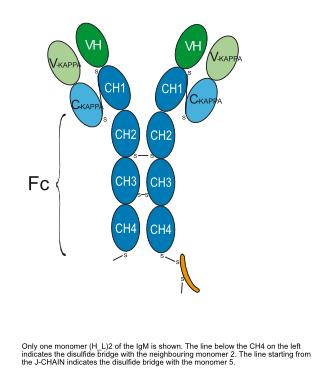
Aplitabart (IGM-8444) is a pentameric IgM death receptor 5 (DR5) agonist antibody with a KD of 85 nM and an apparent affinity of 11 pM. Aplitabart induces DR5 multimerization, thereby triggering cancer cell apoptosis via the extrinsic apoptotic pathway apoptosis. Aplitabart is applicable to cancer-related research such as colorectal cancer and gastric cancer.
For research use only. We do not sell to patients.
- Purity : 97.024%
- CAS No.: 2796349-94-3
- Molecular Weight:871.57 kDa
-
Storage:
Please store the product under the recommended conditions in the Certificate of Analysis.


To place orders, for customer services and technical support, please contact: MedChemExpress USA
Tel: 609-228-6898 E-mail: [email protected] [email protected]


-
 Biological Activity
-
Chemical Information
- Protocols
- Purity & Documentation
- References
-
 Help & FAQs




Biological Activity
Description
Isotype
Human IgM-kappa_Jchain
Recommend Isotype Controls
Species Reactivity
Human
IC50 & Target
|
DR5 |
In Vitro
IGM-8444 potently binds to Colo205 colorectal cancer cells (EC50 = 170 ng/mL) and H-EMC-SS chondrosarcoma cells (EC50 = 64 ng/mL)[1].
IGM-8444 (24 h) potently induces cytotoxicity in Colo205 colorectal cancer cells (IC50 = 1.1 ng/mL) and H-EMC-SS chondrosarcoma cells (IC50 = 0.32 ng/mL)[1].
IGM-8444 (1 μg/mL; 0.5-6 h) rapidly and potently activates caspase-3/7, caspase-8, and caspase-9 in Colo205 colorectal cancer cells and H-EMC-SS chondrosarcoma cells[1].
Treatment with IGM-8444 (1 μg/mL; 1-6 h) for 6 h induces early apoptosis in 23% of Colo205 colorectal cancer cells and 15% of H-EMC-SS chondrosarcoma cells[1].
IGM-8444 (72 h) exhibits synergistic cytotoxicity with chemotherapeutic agents and ABT-199 (HY-15531) in HCT15 colorectal cancer cells, NCI-H2228 non-small cell lung cancer cells, and MV-411 acute myeloid leukemia cells, with a maximum Bliss score ranging from 16.8 to 53.4[1].
MedChemExpress (MCE) has not independently confirmed the accuracy of these methods. They are for reference only. Further protocols information, click here.
-
Cell Line:Colo205 colorectal cancer cells, H-EMC-SS chondrosarcoma cells
-
Concentration:1 μg/mL
-
Incubation Time:1, 3, 6 h
-
Result:Induced early apoptosis in 23% of Colo205 cells and 15% of H-EMC-SS cells after 6 hours of treatment.
Showed significantly more early apoptosis induction than the corresponding anti-DR5 IgG (4% and 2% early apoptotic cells for Colo205 and H-EMC-SS, respectively), which was similar to isotype controls.
In Vivo
IGM-8444 (5 mg/kg; intravenous injection; once every 2 days; 11 doses total) induces 101% tumor growth inhibition (TGI) in NCI-H2122 non-small cell lung cancer xenografts and extends the median event-free survival by 40 days[1].
IGM-8444 (0.3-10 mg/kg; intravenous injection; once every other day) induces potent and sustained tumor regression in the GXF251 gastric cancer PDX model[1].
IGM-8444 (5 mg/kg; intravenous injection; once every other day; 7 doses total) , in combination with irinotecan (HY-16562), 5-FU (HY-90006) or Paclitaxel (HY-B0015), enhances the antitumor efficacy in the Colo205 colorectal cancer xenograft model[1].
IGM-8444 (5 mg/kg; intravenous injection; once every two days; 11 doses total) combined with ABT-199 induces 102% TGI and prolongs survival in mice bearing DOHH-2 non-Hodgkin's lymphoma xenografts[1].
MedChemExpress (MCE) has not independently confirmed the accuracy of these methods. They are for reference only.
-
Animal Model:Crl:NU (Ncr)-Foxn1nu (female, 8-12-weeks-old, subcutaneous implantation of 2×10⁶ Colo205 cells)[1]
-
Dosage:0.3, 1, 3 and 10 mg/kg (tumor growth inhibition); 0.3, 1, 3, 10 and 20 mg/kg (biomarker analysis)
-
Administration:i.v.; every other day; 11 doses (tumor growth inhibition); i.v.; single dose (biomarker analysis)
-
Result:Induced 23%, 29%, 75%, and 84% tumor growth inhibition (TGI) on day 22 at doses of 0.3, 1, 3, and 10 mg/kg, respectively.
Increased serum M30 up to 10-fold and M65 up to 5-fold in a dose-dependent manner 24 hours after single doses.
-
Animal Model:Crl:NU (Ncr)-Foxn1nu (female, 8-12-weeks-old, subcutaneous implantation of 107 NCI-H2122 cells)[1]
-
Dosage:5 mg/kg
-
Administration:i.v.; every other day; 11 doses (tumor growth and survival); i.v.; single dose (biomarker analysis)
-
Result:Induced 101% TGI on day 26, with 5/10 animals achieving a partial response (PR).
Extended median event-free survival by 40 days compared with vehicle.
Significantly elevated cleaved caspase-3 (CC3) staining in tumor tissue 24 hours after a single dose.
-
Animal Model:NMRI nude (female, 5-7-weeks-old, subcutaneous implantation of 3-4 mm GXF251 PDX tumor fragments)[1]
-
Dosage:0.3, 1, 3 and 10 mg/kg (dose-dependent study); 5 mg/kg (fixed dose, different tumor sizes)
-
Administration:i.v.; every other day; 7 doses (fixed dose); i.v.; every other day (dose-dependent study); i.v.; weekly (weekly dosing study)
-
Result:Induced 112% TGI on day 46 at 5 mg/kg every other day for 7 doses, with 8/10 complete responses (CR), 2/10 partial responses (PR), 7/10 event-free animals, and 6/10 tumor-free survivors (TFS) at day 99.
Elevated serum M30 10-fold and M65 6-fold 24 hours post-treatment.
Induced 30%, 55%, 101%, and 99% TGI on day 59 at doses of 0.3, 1, 3, and 10 mg/kg every other day, respectively, with 2/7 tumor-free animals in the 10 mg/kg group at day 100.
Induced 58% and 102% TGI on day 28 with weekly dosing of 1 and 10 mg/kg, respectively.
Induced 112%, 103%, and 101% TGI on day 32 when dosed into tumors with initial volumes of 100, 300, or 500 mm3, respectively.
-
Animal Model:Crl:NU (Ncr)-Foxn1nu (female, 8-12-weeks-old, subcutaneous implantation of 2×10⁶ Colo205 cells)[1]
-
Dosage:5 mg/kg (single agent); 5 mg/kg + 100 mg/kg irinotecan; 5 mg/kg + 100 mg/kg 5-FU; 5 mg/kg + 25 mg/kg paclitaxel
-
Administration:i.v.; every other day; 7 doses (IGM-8444); i.p.; weekly; 3 weeks (Irinotecan, 5-FU); i.v.; every other day; 5 doses (Paclitaxel)
-
Result:Induced 70%-76% TGI as a single agent at 5 mg/kg.
Combination with Irinotecan induced 103% TGI with 2/10 PRs and extended median event-free survival by 42 days compared with vehicle.
Combination with 5-FU induced 104% TGI with 3/10 PRs and 1/10 CR/TFS, without additional toxicity compared with 5-FU alone.
Combination with paclitaxel increased CR rate from 30% (Paclitaxel alone) to 100%, with 8/10 animals tumor-free at day 100 compared with 2/10 in the Paclitaxel-only group.
-
Animal Model:Fox Chase SCID, CB17/Icr-Prkdcscid/IcrIcoCrl (female, 9-weeks-old, subcutaneous implantation of 107 DOHH-2 cells)[1]
-
Dosage:5 mg/kg (single agent); 5 mg/kg + 100 mg/kg ABT-199
-
Administration:i.v.; every other day; 11 doses (IGM-8444); p.o.; daily; 21 days (ABT-199)
-
Result:Induced 45% TGI as a single agent at 5 mg/kg.
Combination with ABT-199 induced 102% TGI with 3/10 PRs and 2/10 CRs.
Significantly extended median event-free survival compared with vehicle or ABT-199 alone.
Clinical Trial
| NCT Number | Sponsor | Condition | Start Date |
Phase

|
|---|---|---|---|---|
| NCT01329991 | Plexxikon| | 2011-05 | PHASE1 |
Gene ID
Accession
Conjugated
Unconjugated
Reconsititution
The product can be reconstituted/diluted with sterile PBS or saline.
Format
-
IgM-kappa_Jchain
Application
ELISA, FACS, Functional assay
Verified Bioactivity
-
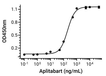 Immobilized TRAIL R2/TNFRSF10B Protein, Human (HY-P72779) can bind Aplitabart. The EC50 for this effect is 201.8 ng/mL.


Chemical Information
-
CAS No. 2796349-94-3
-
Appearance Liquid
-
Molecular Weight 871.57 kDa
-
Color Colorless to off-white
-
SMILES
[Aplitabart]
-
Synonyms
IGM-8444
-
Shipping
Shipping with dry ice.
-
Formulation
Please refer to the lot-specific COA for specific buffer information.
-
Storage
Please store the product under the recommended conditions in the Certificate of Analysis.

Protocols
-
Apoptosis
Apoptosis, also called programmed cell death, is generally characterized by distinct morphological characteristics.
-
TUNEL staining for apoptotic DNA fragmentation
TUNEL staining detects DNA strand breaks by using terminal deoxynucleotidyl transferase to add labeled nucleotides to exposed 3′-OH DNA termini, generating either microscopic staining in fixed cells or tissue sections, or fluorescence/cytometric signal in cell suspensions. TUNEL positivity reflects DNA fragmentation but should not be interpreted alone as definitive apoptosis, because TUNEL can also label necrotic, autolytic, mechanically damaged, or DNA-repair-associated DNA breaks.
-
Annexin V plus membrane-impermeant dye apoptosis staining
Annexin V-based apoptosis assays rely on the detection of phosphatidylserine (PS) externalization from the inner leaflet of the plasma membrane to the outer leaflet, an early biochemical hallmark of apoptosis. Fluorescently labeled Annexin V binds PS in a calcium-dependent manner, enabling identification of early apoptotic cells by flow cytometry or fluorescence microscopy. When combined with a membrane-impermeant DNA-binding dye (e. g. , propidium iodide), this approach allows discrimination between viable (Annexin V−/dye−), early apoptotic (Annexin V+/dye−), and late apoptotic or necrotic (Annexin V+/dye+) cell populations by assessing membrane integrity and PS exposure.
-
Apoptosis Solutions
Apoptosis is a regulated, generally non-lytic cell-death pathway that removes unwanted, damaged, infected, or abnormal cells through coordinated morphological changes, caspase activation, DNA fragmentation, and membrane remodeling. The intrinsic apoptosis pathway is controlled mainly by mitochondrial outer membrane permeabilization, BCL-2 family proteins, cytochrome c release, apoptosome formation, caspase-9 activation, and downstream executioner caspase-3/7 activation. The extrinsic apoptosis pathway is initiated by death receptors such as Fas, TNFR, and TRAIL receptors, which recruit adaptor proteins and activate caspase-8 before engaging executioner caspases or mitochondrial amplification through BID cleavage. Apoptosis is linked to many phenotypes, including cancer cell killing, tissue homeostasis, immune regulation, neurodegeneration, infection response, and treatment-induced cytotoxicity; unresolved questions include how apoptosis interacts with necroptosis, pyroptosis, ferroptos


Purity & Documentation
-
Data Sheet (268 KB)
-
SDS (251 KB)
- English - EN (251 KB)
- Français - FR (251 KB)
- Deutsch - DE (251 KB)
- Norwegian - NO (251 KB)
- Español - ES (251 KB)
- Swedish - SV (251 KB)
- Italian - IT (251 KB)
- Korean - KR (251 KB)
- Portuguese - PT (251 KB)
-
Inhibitory Antibodies User Guide (603 KB)
References

Calculators
Concentration (start) × Volume (start) = Concentration (final) × Volume (final)


Aplitabart Related Classifications
HY-P990901 Related Classifications
 Powered by Bioz
Keywords
- Aplitabart
- 2796349-94-3
- IGM-8444
- IGM8444
- IGM 8444
- TNF Receptor
- Apoptosis
- acute myeloid leukemia
- cancer cells
- Death receptor 5
- patient-derived tumor models
- Colo205 colorectal cancer cells
- DR5
- xenograft models
- primary human hepatocytes
- extrinsic apoptotic pathway
- H-EMC-SS chondrosarcoma cells
- Inhibitor
- inhibitor
- inhibit