



bpV(phen)
Based on 3 publication(s) in Google Scholar
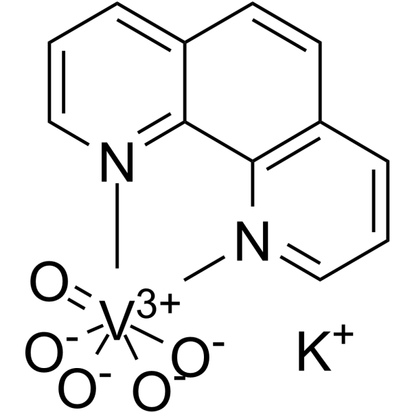
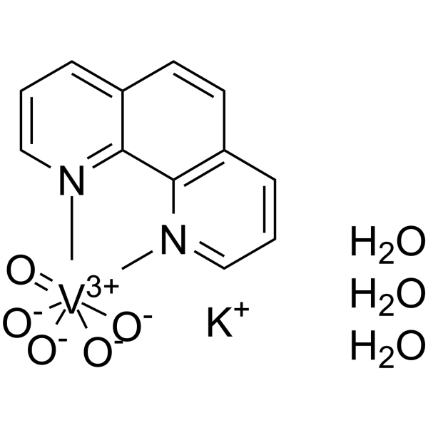
bpV(phen), a insulin-mimetic agent, is a potent protein tyrosine phosphatase (PTP) and PTEN inhibitor with IC50s of 38 nM, 343 nM and 920 nM for PTEN, PTP-β and PTP-1B, respectively. bpV(phen) inhibits proliferation of the protozoan parasite Leishmania in vitro. bpV(phen) strongly induces the secretion of a large number of chemokines and pro-inflammatory cytokines, and it activates a Th1-type pathway (IL-12, IFNγ). bpV(phen) can also induce cell apoptosis, and has anti-angiogenic and anti-tumor activity.
Nur für Forschungszwecke. Wir verkaufen nicht an Patienten.
- Reinheit : 98.0%
- CAS. Nr.: 42494-73-5
- Formel: C12H8KN2O5V
- Molecular Weight:350.24
-
Speicherung:
4°C, sealed storage, away from moisture
* In solvent : -80°C, 6 months; -20°C, 1 month (sealed storage, away from moisture)

To place orders, for customer services and technical support, please contact: MedChemExpress USA
Tel: 609-228-6898 E-mail: [email protected] [email protected]


-
 Biologische Aktivität
-
Chemical Information
- Protokoll
- Reinheit & Dokumentation
- Verweise
-
 Help & FAQs



Publications Citing Use of MedChemExpress (MCE) bpV(phen)
MoreAlle Parasite Isoform-spezifische Produkte anzeigen
More

Biologische Aktivität
Beschreibung
IC50 & Target
IC50: 38 nM (PTEN), 343 nM (PTP-β) and 920 nM (PTP-1B)[3]
Parasite Leishmania[2]
Apoptosis[1]
In Vitro
bpV(phen) (5 μM; 24.5 hours; H9c2 cells) treatment causes a further decrease of cell viability in H/R-injured H9c2 cells[1].
bpV(phen) (5 μM; 24.5 hours; H9c2 cells) treatment increases the apoptosis of H/R-injured H9c2 cells[1].
bpV(phen) (5 μM; 24.5 hours; H9c2 cells) treatment significantly promotes the accumulation of cytoplasmic Cytochrome C in H/R-injured H9c2 cells[1].
After stimulation of bpV(phen), PTEN-induced putative kinase protein 1 (PINK1)/Parkin-mediated mitophagy is inhibited[1].
bpV(phen) is an insulin-mimetic agent following insulin-receptor tyrosine kinase hyperphosphorylation and activation[4].
MedChemExpress (MCE) has not independently confirmed the accuracy of these methods. They are for reference only.
-
Cell Line:Hypoxia/reoxygenation (H/R)-injured H9c2 cells
-
Concentration:5 μM
-
Incubation Time:24.5 hours (hypoxia for 24 h; reoxygenation for 30 minutes)
-
Result:Caused a further decrease of cell viability.
-
Cell Line:Hypoxia/reoxygenation (H/R)-injured H9c2 cells
-
Concentration:5 μM
-
Incubation Time:24.5 hours (hypoxia for 24 h; reoxygenation for 30 minutes)
-
Result:Increased the apoptosis of H/R-injured H9c2 cells.
-
Cell Line:Hypoxia/reoxygenation (H/R)-injured H9c2 cells
-
Concentration:5 μM
-
Incubation Time:24.5 hours (hypoxia for 24 h; reoxygenation for 30 minutes)
-
Result:Showed an increased release of Cytochrome C.
In Vivo
MedChemExpress (MCE) has not independently confirmed the accuracy of these methods. They are for reference only.
-
Animal Model:Male BALB/c nude (nu/nu) athymic mice (6-7 weeks old) injected with PC-3 cells[2]
-
Dosage:5 mg/kg
-
Administration:Intraperitoneal injection; daily; for 38 days
-
Result:Caused a significant reduction in average tumor volume.
- Detection of Apoptosis
- TUNEL staining for apoptotic DNA fragmentation
- LPS-Induced Endotoxemia/Systemic Inflammation
- Naïve CD4+ T-cell subset differentiation/polarization
- Annexin V plus membrane-impermeant dye apoptosis staining
- Research Protocol for Endocrine Diseases
- Apoptosis Solutions
- Research Protocol for Inflammation-related Diseases
- Cell Viability Determination by MTT Colorimetric Assay


Chemical Information
-
CAS. Nr. 42494-73-5
-
Appearance Solid
-
Molecular Weight 350.24
-
Formel C12H8KN2O5V
-
Color Light yellow to yellow
-
SMILES
O=[V+3]12([N]3=CC=C4)([O-][O-]1)([O-][O-]2)[N]5=C6C3=C4C=CC6=CC=C5.[K+]
-
Versand
Room temperature in continental US; may vary elsewhere.
-
Speicherung
4°C, sealed storage, away from moisture
* In solvent : -80°C, 6 months; -20°C, 1 month (sealed storage, away from moisture)


Publications (3)
-
Journal Impact Factor
-
Most Recent
-
Nat Commun
The phosphatase PTEN links platelets with immune regulatory functions of mouse T follicular helper cells. [Abstract]2022 May 19;13(1):2762. PMID: 35589797 -
Biochem Biophys Res Commun
2020 Sep 3;529(4):1045-1052. PMID: 32819563 -
Am J Mens Health
The Guangsi Yulin Decoction Inhibits Oxidative Stress and Inflammation-Induced Spermatogenesis Dysfunction via the PTEN/ PI3K/ AKT/ FoxO1 Pathway. [Abstract]2026 Mar-Apr;20(2):15579883261427164. PMID: 41807111


Reinheit & Dokumentation
-
Data Sheet (298 KB)
-
SDS (393 KB)
- English - EN (393 KB)
- Français - FR (393 KB)
- Deutsch - DE (393 KB)
- Norwegian - NO (393 KB)
- Español - ES (393 KB)
- Swedish - SV (393 KB)
- Italian - IT (393 KB)
- Korean - KR (393 KB)
- Portuguese - PT (393 KB)
-
Handling Instructions (2659 KB)
Verweise
[1]. Tang W, et al. PTEN-mediated mitophagy and APE1 overexpression protects against cardiac hypoxia/reoxygenation injury. In Vitro Cell Dev Biol Anim. 2019 Oct;55(9):741-748. [Content Brief]
[2]. Caron D, et al. Protein tyrosine phosphatase inhibition induces anti-tumor activity: evidence of Cdk2/p27 kip1 and Cdk2/SHP-1 complex formation in human ovarian cancer cells. Cancer Lett. 2008 Apr 18;262(2):265-75. [Content Brief]
[3]. Schmid AC, et al. Bisperoxovanadium compounds are potent PTEN inhibitors. FEBS Lett. 2004 May 21;566(1-3):35-8. [Content Brief]
[4]. Band CJ, et al. Early signaling events triggered by peroxovanadium [bpV(phen)] are insulin receptor kinase (IRK)-dependent: specificity of inhibition of IRK-associated protein tyrosine phosphatase(s) by bpV(phen). Mol Endocrinol. 1997 Dec;11(13):1899-910 [Content Brief]
[5]. Chen Q, et al. Potassium Bisperoxo(1,10-phenanthroline)oxovanadate (bpV(phen)) Induces Apoptosis and Pyroptosis and Disrupts the P62-HDAC6 Protein Interaction to Suppress the Acetylated Microtubule-dependent Degradation of Autophagosomes. J Biol Chem. 201 [Content Brief]

Calculators
Konzentration (Stammlösung) × Volumen (Stammlösung) = Konzentration (Ziellösung) × Volumen (Ziellösung)


bpV(phen) Related Classifications
HY-136065 Related Classifications
 Powered by Bioz