



M190S

Based on 1 Customer Validation
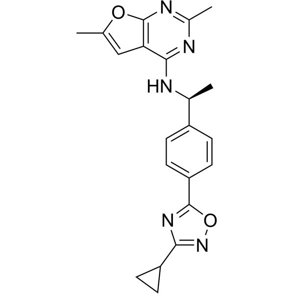
M109S is a novel small molecule protecting cells from mitochondria-dependent apoptosis both in vitro and in vivo. M109S has the potential to become a research tool for studying cell death mechanisms and to develop therapeutics targeting mitochondria-dependent cell death pathway. M109S has orally bioactivity with excellent brain permeability.
Nur für Forschungszwecke. Wir verkaufen nicht an Patienten.
- Reinheit : 99.65%
- CAS. Nr.: 2578300-07-7
- Formel: C21H21N5O2
- Molecular Weight:375.42
-
Speicherung:Powder -20°C, 3 years , 4°C, 2 years ; In solvent -80°C, 6 months , -20°C, 1 month

To place orders, for customer services and technical support, please contact: MedChemExpress USA
Tel: 609-228-6898 E-mail: [email protected] [email protected]


-
 Biologische Aktivität
-
Chemical Information
-
Lösungsmittel & Löslichkeit
- Protokoll
- Reinheit & Dokumentation
- Verweise
-
 Help & FAQs


Alle Caspase Isoform-spezifische Produkte anzeigen
More

Biologische Aktivität
Beschreibung
IC50 & Target
|
caspase-3 23.4 nM (EC50) |
Caspase 3 23.4 nM (EC50) |
In Vitro
M109S (0.1-10000 nM, 24-48 h) inhibits apoptosis induced by Bax as well as Bak[1].
M109S (0-10μM, 4 h) suppresses Staurosporine (HY-15141 STS)-induced apoptosis in MEFs [1].
M109S (0-10μM, 24 h) inhibits Etoposide (HY-13629)-induced apoptosis in Neuro2a cells[1].
M109S (500 nM, 24 h) inhibits Obatoclax (HY-10969A)-induced apoptosis in ARPE19 cells[1].
M109S (500 nM, 48 h) suppresses the conformation change (N-terminal exposure) and mitochondrial translocation of Bax[1].
M109S (1.0 μM, 4h) decreases mitochondrial oxygen consumption and reactive oxygen species, whereas M109S (0.1-1 nM, 4 h) increases glycolysis[1].
MedChemExpress (MCE) has not independently confirmed the accuracy of these methods. They are for reference only.
-
Cell Line:MEF(Wt, Bax only, Bak only)
-
Concentration:0.1 nM, 1 nM, 10 nM, 100 nM, 10000 nM
-
Incubation Time:24 h((WT and Bax-only), 48 h (Bak-only)
-
Result:Showed a dose-dependent suppression of caspase activation in all three types of MEFs.
-
Cell Line:MEF
-
Concentration:0 nM, 1.6 nM, 8 nM, 40 nM, 200 nM, 10000 nM
-
Incubation Time:4 h
-
Result:Suppressed STS-induced caspase activation in a dose-dependent manner.
-
Cell Line:Neuro2a
-
Concentration:0 nM, 40 nM, 200 nM, 10000 nM
-
Incubation Time:24 h
-
Result:Suppressed Etoposide-induced caspase activation in a dose-dependent manner.
-
Cell Line:ARPE19
-
Concentration:500 nM
-
Incubation Time:24 h
-
Result:Significantly inhibited Obatoclax-induced apoptosis in ARPE19 cells comparing to control.
-
Cell Line:iBax cells
-
Concentration:500 nM
-
Incubation Time:48 h
-
Result:Significantly suppressed the amount of immunoprecipitated Bax without a significant change in the total Bax expression.
-
Cell Line:iBax cells
-
Concentration:500 nM
-
Incubation Time:48 h
-
Result:Showed the frequency of the punctuated staining was significantly reduced.
In Vivo
M109S (1 mg/kg, i.p., i.v., 5 mg/kg, o.p. 10 mg/kg 1 time) is an orally bioactive cell death inhibitor penetrating blood-brain/retina-barrier[1].
MedChemExpress (MCE) has not independently confirmed the accuracy of these methods. They are for reference only.
-
Animal Model:Abca4-/-Rdh8-/- mice
-
Dosage:10 mg/kg
-
Administration:Oral Gavage (p.o.)
-
Result:Comparing to mice with M109S, the number of AF spots was similar to that detected in the dark-adapted mice.
-
Animal Model:Mice and Rat
-
Dosage:Intraperitoneal injection (i.p., 1 mg/kg), Intravenous injection (i.v., 5 mg/kg), or Oral gavage (p.o., 10 mg/kg)
-
Administration:Intraperitoneal injection (i.p.), Intravenous injection (i.v.), or Oral gavage (p.o.).
-
Result:Showed plasma concentration reached 1.0 mg/mL (2.6 mM) within 30 min from p.o. in mice, and it remained at 596± 134 ng/mL (1.6±0.36 mM) after 24 h, the same as in rat. Plasmic M109S was 565.3±188.3 nM in rats, and 171.0±52.0 nM in retina, 222.7±74.7 nM in brain, respectively.


Chemical Information
-
CAS. Nr. 2578300-07-7
-
Appearance Solid
-
Molecular Weight 375.42
-
Formel C21H21N5O2
-
Color White to off-white
-
SMILES
CC1=NC(OC(C)=C2)=C2C(N[C@@H](C)C3=CC=C(C4=NC(C5CC5)=NO4)C=C3)=N1
-
Versand
Room temperature in continental US; may vary elsewhere.
-
Speicherung
Powder -20°C 3 years 4°C 2 years In solvent -80°C 6 months -20°C 1 month


Lösungsmittel & Löslichkeit
In Vitro:
DMSO : 100 mg/mL (266.37 mM; Need ultrasonic; Hygroscopic DMSO has a significant impact on the solubility of product, please use newly opened DMSO)
Please refer to the solubility information to select the appropriate solvent. Once prepared, please aliquot and store the solution to prevent product inactivation from repeated freeze-thaw cycles.
Storage method and period of stock solution: -80°C, 6 months; -20°C, 1 month. When stored at -80°C, please use it within 6 months. When stored at -20°C, please use it within 1 month.
Please refer to the solubility information to select the appropriate solvent. Once prepared, please aliquot and store the solution to prevent product inactivation from repeated freeze-thaw cycles.
Storage method and period of stock solution: -80°C, 6 months; -20°C, 1 month. When stored at -80°C, please use it within 6 months. When stored at -20°C, please use it within 1 month.
Konzentration (Stammlösung) × Volumen (Stammlösung) = Konzentration (Ziellösung) × Volumen (Ziellösung)
In Vivo:
Select the appropriate dissolution method based on your experimental animal and administration route.
- For the following dissolution methods, please ensure to first prepare a clear stock solution using an In Vitro approach and then sequentially add co-solvents:
- To ensure reliable experimental results, the clarified stock solution can be appropriately stored based on storage conditions. As for the working solution for In Vivo experiments, it is recommended to prepare freshly and use it on the same day.
- The percentages shown for the solvents indicate their volumetric ratio in the final prepared solution. If precipitation or phase separation occurs during preparation, heat and/or sonication can be used to aid dissolution.
Add each solvent one by one: 10% DMSO 40% PEG300 5% Tween-80 45% Saline
Solubility: 2.5 mg/mL (6.66 mM); Clear solution; Need ultrasonic
This protocol yields a clear solution of 2.5 mg/mL.
Taking 1 mL working solution as an example, add 100 μL DMSO stock solution (25.0 mg/mL) to 400 μL PEG300, and mix evenly; then add 50 μL Tween-80 and mix evenly; then add 450 μL Saline to adjust the volume to 1 mL.
Preparation of Saline: Dissolve 0.9 g sodium chloride in ddH₂O and dilute to 100 mL to obtain a clear Saline solution.
Add each solvent one by one: 10% DMSO 90% (20% SBE-β-CD in Saline)
Solubility: 2.5 mg/mL (6.66 mM); Clear solution; Need ultrasonic
This protocol yields a clear solution of 2.5 mg/mL.
Taking 1 mL working solution as an example, add 100 μL DMSO stock solution (25.0 mg/mL) to 900 μL 20% SBE-β-CD in Saline, and mix evenly.
Preparation of 20% SBE-β-CD in Saline (4°C, storage for one week): 2 g SBE-β-CD powder is dissolved in 10 mL Saline, completely dissolve until clear.
In Vivo Dissolution Calculator
Please enter the basic information of animal experiments:
-
-
-
-
Recommended: Prepare an additional quantity of animals to account for potential losses during experiments.
Please enter your animal formula composition:
-
%DMSO +
Recommended: Keep the proportion of DMSO in working solution below 2% if your animal is weak.
-
%+
-
+%Tween-80 + +
-
%Saline +
The co-solvents required include: DMSO, . All of co-solvents are available by MedChemExpress (MCE). , Tween 80. All of co-solvents are available by MedChemExpress (MCE).
Working solution concentration: 0.22 mg/mL
Method for preparing stock solution: mg drug dissolved in μL DMSO. Stock solution concentration: mg/mL.
1. Take μL DMSO stock solution;
2. Add μL .
μL , mix evenly;
3. Then add μL Tween 80, mix evenly;
4. Then add μL
Please ensure that the stock solution in the first step is dissolved to a clear state, and add co-solvents in sequence. You can use ultrasonic heating (ultrasonic cleaner, recommended frequency 20-40 kHz), vortexing, etc. to assist dissolution.


Reinheit & Dokumentation
-
Data Sheet (278 KB)
-
SDS (254 KB)
- English - EN (254 KB)
- Français - FR (254 KB)
- Deutsch - DE (254 KB)
- Norwegian - NO (254 KB)
- Español - ES (254 KB)
- Swedish - SV (254 KB)
- Italian - IT (254 KB)
- Korean - KR (254 KB)
- Portuguese - PT (254 KB)
-
Handling Instructions (2659 KB)
Verweise


Complete Stock Solution Preparation Table
Please refer to the solubility information to select the appropriate solvent. Once prepared, please aliquot and store the solution to prevent product inactivation from repeated freeze-thaw cycles.
Storage method and period of stock solution: -80°C, 6 months; -20°C, 1 month. When stored at -80°C, please use it within 6 months. When stored at -20°C, please use it within 1 month.
| Optional Solvent | Concentration Solvent Mass | 1 mg | 5 mg | 10 mg | 25 mg |
|---|---|---|---|---|---|
| DMSO | 1 mM | 2.6637 mL | 13.3184 mL | 26.6368 mL | 66.5921 mL |
| 5 mM | 0.5327 mL | 2.6637 mL | 5.3274 mL | 13.3184 mL | |
| 10 mM | 0.2664 mL | 1.3318 mL | 2.6637 mL | 6.6592 mL | |
| 15 mM | 0.1776 mL | 0.8879 mL | 1.7758 mL | 4.4395 mL | |
| 20 mM | 0.1332 mL | 0.6659 mL | 1.3318 mL | 3.3296 mL | |
| 25 mM | 0.1065 mL | 0.5327 mL | 1.0655 mL | 2.6637 mL | |
| 30 mM | 0.0888 mL | 0.4439 mL | 0.8879 mL | 2.2197 mL | |
| 40 mM | 0.0666 mL | 0.3330 mL | 0.6659 mL | 1.6648 mL | |
| 50 mM | 0.0533 mL | 0.2664 mL | 0.5327 mL | 1.3318 mL | |
| 60 mM | 0.0444 mL | 0.2220 mL | 0.4439 mL | 1.1099 mL | |
| 80 mM | 0.0333 mL | 0.1665 mL | 0.3330 mL | 0.8324 mL | |
| 100 mM | 0.0266 mL | 0.1332 mL | 0.2664 mL | 0.6659 mL |


M190S Related Classifications
HY-156168 Related Classifications
 Powered by Bioz