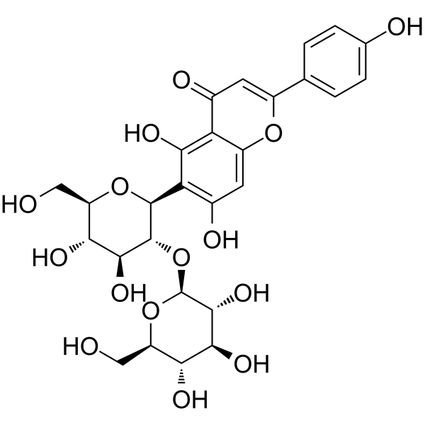
Meloside A
Based on 5 publication(s) in Google Scholar
Meloside A (Isovitexin 2''-O-glucoside) is a flavonoid with antioxidant activity. Meloside A can inhibit cell apoptosis and ROS production. Meloside A can inhibit androgen receptor (AR) nuclear translocation and AR protein expression. Meloside A can reduce IL-6, TGF-β1 and DKK-1 levels. Meloside A can be used for the researches of inflammation and endocrinology, such as hair loss.
Nur für Forschungszwecke. Wir verkaufen nicht an Patienten.
- Reinheit : 98.72%
- CAS. Nr.: 60767-80-8
- Formel: C27H30O15
- Molecular Weight:594.52
-
Speicherung:
4°C, sealed storage, away from moisture and light
* In solvent : -80°C, 6 months; -20°C, 1 month (sealed storage, away from moisture and light)
Publications Citing Use of MedChemExpress (MCE) Meloside A
More
Biologische Aktivität
Beschreibung
IC50 & Target
[1]|
IL-6 |
TGF-β1 |
In Vitro
Meloside A (5-100 μg/mL, 24 h) does not induce significant cytotoxicity in HDPCs[1].
Meloside A (50-100 μg/mL, 6-24 h) inhibits DHT-induced AR nuclear translocation and AR protein expression in HDPCs[1].
Meloside A (100 μg/mL, 24 h) reduces ROS levels in DHT-induced HDPCs[1].
Meloside A (50-100 μg/mL, 48 h) reduces the secretion of DKK-1, IL-6, and TGF-β1 in DHT-induced HDPCs[1].
MedChemExpress (MCE) has not independently confirmed the accuracy of these methods. They are for reference only. Further protocols information, click here.
-
Cell Line:DHT-induced HDPCs
-
Concentration:50 and 100 μg/mL
-
Incubation Time:24 h
-
Result:Decreased AR protein expression.
Chemical Information
-
CAS. Nr. 60767-80-8
-
Appearance Solid
-
Molecular Weight 594.52
-
Formel C27H30O15
-
Color Light yellow to yellow
-
SMILES
O=C1C=C(C2=CC=C(O)C=C2)OC3=CC(O)=C([C@H]4[C@@H]([C@H]([C@@H]([C@@H](CO)O4)O)O)O[C@H]5[C@@H]([C@H]([C@@H]([C@@H](CO)O5)O)O)O)C(O)=C13
-
Synonyms
Isovitexin 2''-O-glucoside; Isovitexin 2''-O-β-D-glucoside
-
Structure Classification
-
Versand
Room temperature in continental US; may vary elsewhere.
-
Speicherung
4°C, sealed storage, away from moisture and light
* In solvent : -80°C, 6 months; -20°C, 1 month (sealed storage, away from moisture and light)
Publications (5)
-
Journal Impact Factor
-
Most Recent
-
J Agric Food Chem
Quantification of Stress- and Resistance-Related Metabolites in Barley Leaves (Hordeum vulgare L.) Infected with Bipolaris sorokiniana via UHPLC-MS/MSMRM. [Abstract]2026 Jul 1;74(25):20051-20061. PMID: 42299507 -
J Agric Food Chem
UPLC-ESI-TOF-MS Profiling of Metabolome Alterations in Barley (Hordeum vulgare L.) Leaves Induced by Bipolaris sorokiniana. [Abstract]2025 Oct 1;73(39):24662-24687. PMID: 40963221 -
-
-
Lösungsmittel & Löslichkeit
In Vitro:
DMSO : 100 mg/mL (168.20 mM; Need ultrasonic; Hygroscopic DMSO has a significant impact on the solubility of product, please use newly opened DMSO)
Please refer to the solubility information to select the appropriate solvent. Once prepared, please aliquot and store the solution to prevent product inactivation from repeated freeze-thaw cycles.
Storage method and period of stock solution: -80°C, 6 months; -20°C, 1 month (sealed storage, away from moisture and light). When stored at -80°C, please use it within 6 months. When stored at -20°C, please use it within 1 month.
Please refer to the solubility information to select the appropriate solvent. Once prepared, please aliquot and store the solution to prevent product inactivation from repeated freeze-thaw cycles.
Storage method and period of stock solution: -80°C, 6 months; -20°C, 1 month (sealed storage, away from moisture and light). When stored at -80°C, please use it within 6 months. When stored at -20°C, please use it within 1 month.
Konzentration (Stammlösung) × Volumen (Stammlösung) = Konzentration (Ziellösung) × Volumen (Ziellösung)
Protokoll
-
Cytoplasmic-Nuclear Fractionated Protein Extraction
Cytoplasmic-nuclear fractionated protein extraction separates soluble cytoplasmic proteins from nuclear-enriched proteins by mild plasma-membrane permeabilization, differential centrifugation, washing of nuclei, and extraction of nuclear proteins for downstream immunoblotting or related molecular analysis. The readout is the relative abundance of a protein in cytoplasmic and nuclear fractions, commonly assessed by western blotting together with compartment markers such as tubulin or pyruvate kinase for cytoplasm and lamin, nucleoporin, hnRNP, H2AX, or Lamin B for nuclear fractions.
-
RNA extraction experimental
By lysing cells, releasing RNA, and removing impurities such as proteins and DNA, high-purity RNA products are finally obtained. The commonly used traditional method is the guanidine isothiocyanate/phenol/chloroform method (Trizol), which is suitable for a variety of animal materials including animal tissues, microorganisms, cultured cells, etc., and most plant materials.
-
Western Blot
Western blotting (WB) is a commonly used experimental method in molecular biology, biochemistry, and immunogenetics for identifying and quantifying target proteins. It combines gel electrophoresis with immunoassay, enabling researchers to analyze protein expression, post-translational modifications, and molecular weight.
-
Apoptosis
Apoptosis, also called programmed cell death, is generally characterized by distinct morphological characteristics.
-
TUNEL staining for apoptotic DNA fragmentation
TUNEL staining detects DNA strand breaks by using terminal deoxynucleotidyl transferase to add labeled nucleotides to exposed 3′-OH DNA termini, generating either microscopic staining in fixed cells or tissue sections, or fluorescence/cytometric signal in cell suspensions. TUNEL positivity reflects DNA fragmentation but should not be interpreted alone as definitive apoptosis, because TUNEL can also label necrotic, autolytic, mechanically damaged, or DNA-repair-associated DNA breaks.
-
Annexin V plus membrane-impermeant dye apoptosis staining
Annexin V-based apoptosis assays rely on the detection of phosphatidylserine (PS) externalization from the inner leaflet of the plasma membrane to the outer leaflet, an early biochemical hallmark of apoptosis. Fluorescently labeled Annexin V binds PS in a calcium-dependent manner, enabling identification of early apoptotic cells by flow cytometry or fluorescence microscopy. When combined with a membrane-impermeant DNA-binding dye (e. g. , propidium iodide), this approach allows discrimination between viable (Annexin V−/dye−), early apoptotic (Annexin V+/dye−), and late apoptotic or necrotic (Annexin V+/dye+) cell populations by assessing membrane integrity and PS exposure.
-
ROS/oxidative-stress fluorescent staining
ROS/oxidative-stress fluorescent staining uses cell-permeant fluorogenic probes that become fluorescent after oxidation inside cells or tissues; commonly used examples include DCFH-DA/DCFDA for broad cellular oxidant detection, DHE for superoxide-related signal detection, MitoSOX for mitochondrial superoxide-related signal detection, and CellROX probes for oxidative-stress-associated fluorescence readouts. The assay detects probe oxidation rather than a single ROS species unless the probe and analysis method have been chemically validated for that species. DCFH-DA enters cells, is deacetylated by intracellular esterases to DCFH, and produces fluorescent DCF after oxidation, so the readout is used as an operational measure of total cellular oxidative stress rather than a species-specific ROS measurement. DHE and MitoSOX can report superoxide-related oxidation, but red fluorescence alone can include non-specific ethidium-like oxidation products; HPLC or optimized spectral approaches are
-
Research Protocol for Endocrine Diseases
Endocrine diseases often arise from disrupted hormone production, hormone signaling, or target-tissue responsiveness; for diabetes-focused endocrine disease models, insulin signaling regulates glucose uptake, hepatic glucose output, lipid metabolism, and β-cell compensation. Type 2 diabetes develops through interacting defects in insulin resistance, β-cell dysfunction, adipose inflammation, hepatic glucose overproduction, altered incretin signaling, and ectopic lipid metabolism. A major unresolved question is whether endocrine dysfunction is driven primarily by target-tissue insulin resistance, intrinsic β-cell failure, immune/inflammatory stress, or combined multi-organ failure that differs by disease stage.
-
Apoptosis Solutions
Apoptosis is a regulated, generally non-lytic cell-death pathway that removes unwanted, damaged, infected, or abnormal cells through coordinated morphological changes, caspase activation, DNA fragmentation, and membrane remodeling. The intrinsic apoptosis pathway is controlled mainly by mitochondrial outer membrane permeabilization, BCL-2 family proteins, cytochrome c release, apoptosome formation, caspase-9 activation, and downstream executioner caspase-3/7 activation. The extrinsic apoptosis pathway is initiated by death receptors such as Fas, TNFR, and TRAIL receptors, which recruit adaptor proteins and activate caspase-8 before engaging executioner caspases or mitochondrial amplification through BID cleavage. Apoptosis is linked to many phenotypes, including cancer cell killing, tissue homeostasis, immune regulation, neurodegeneration, infection response, and treatment-induced cytotoxicity; unresolved questions include how apoptosis interacts with necroptosis, pyroptosis, ferroptos
-
Research Protocol for Inflammation-related Diseases
The NLRP3 inflammasome is a cytosolic innate immune signaling platform that integrates priming signals and danger-signal activation to promote caspase-1 activation, maturation of IL-1β and IL-18, and gasdermin D-mediated pyroptotic cell death. The core experimental logic is to determine whether inflammatory disease phenotypes are driven by increased NLRP3 expression, ASC-containing inflammasome assembly, caspase-1 cleavage, GSDMD cleavage, and extracellular release of IL-1β/IL-18 rather than by nonspecific cell injury alone. The pathway is strongly linked to inflammation-related disease phenotypes because monosodium urate crystals activate NALP3/NLRP3 inflammasome signaling in gout-like crystal inflammation, cholesterol crystals activate NLRP3 inflammasomes in atherogenesis models, and DSS-induced intestinal inflammation has been reported to involve NLRP3 inflammasome activity. However, experimental colitis studies also show context-dependent protective effects of NLRP3 inflammasome co
-
Protocol For Protein Expression And Purification
Recombinant protein expression in Escherichia coli followed by purification of a His-tagged soluble protein by immobilized metal affinity chromatography (IMAC), with optional MBP fusion and TEV tag removal when the construct includes these elements. The biological readout is production of the encoded target protein, detected as an inducible band at the expected molecular mass by SDS-PAGE and quantified by total protein assay or chromatographic absorbance; the purification readout is enrichment of the target protein in elution fractions after selective binding of polyhistidine residues to immobilized Ni2+/metal-chelate resin and elution by imidazole-containing buffer. Expression is driven by an inducible bacterial expression system, commonly T7/lac-based, in which IPTG or lactose/auto-induction activates transcription and translation of the cloned gene; lower induction temperature, lower inducer concentration, induction timing, and solubility-enhancing fusion tags can influence the frac
Reinheit & Dokumentation
-
Data Sheet (280 KB)
-
SDS (254 KB)
- English - EN (254 KB)
- Français - FR (254 KB)
- Deutsch - DE (254 KB)
- Norwegian - NO (254 KB)
- Español - ES (254 KB)
- Swedish - SV (254 KB)
- Italian - IT (254 KB)
- Korean - KR (254 KB)
- Portuguese - PT (254 KB)
-
Handling Instructions (2659 KB)
Verweise
Complete Stock Solution Preparation Table
Please refer to the solubility information to select the appropriate solvent. Once prepared, please aliquot and store the solution to prevent product inactivation from repeated freeze-thaw cycles.
Storage method and period of stock solution: -80°C, 6 months; -20°C, 1 month (sealed storage, away from moisture and light). When stored at -80°C, please use it within 6 months. When stored at -20°C, please use it within 1 month.
| Optional Solvent | Concentration Solvent Mass | 1 mg | 5 mg | 10 mg | 25 mg |
|---|---|---|---|---|---|
| DMSO | 1 mM | 1.6820 mL | 8.4101 mL | 16.8203 mL | 42.0507 mL |
| 5 mM | 0.3364 mL | 1.6820 mL | 3.3641 mL | 8.4101 mL | |
| 10 mM | 0.1682 mL | 0.8410 mL | 1.6820 mL | 4.2051 mL | |
| 15 mM | 0.1121 mL | 0.5607 mL | 1.1214 mL | 2.8034 mL | |
| 20 mM | 0.0841 mL | 0.4205 mL | 0.8410 mL | 2.1025 mL | |
| 25 mM | 0.0673 mL | 0.3364 mL | 0.6728 mL | 1.6820 mL | |
| 30 mM | 0.0561 mL | 0.2803 mL | 0.5607 mL | 1.4017 mL | |
| 40 mM | 0.0421 mL | 0.2103 mL | 0.4205 mL | 1.0513 mL | |
| 50 mM | 0.0336 mL | 0.1682 mL | 0.3364 mL | 0.8410 mL | |
| 60 mM | 0.0280 mL | 0.1402 mL | 0.2803 mL | 0.7008 mL | |
| 80 mM | 0.0210 mL | 0.1051 mL | 0.2103 mL | 0.5256 mL | |
| 100 mM | 0.0168 mL | 0.0841 mL | 0.1682 mL | 0.4205 mL |