



BMAP-27
BMAP-27 is a cationic amphipathic α-helical antimicrobial peptide with anticancer activity. BMAP-27 disrupts the integrity and permeability of cell membranes, leading to the leakage of intracellular DNA, proteins and alkaline phosphatase. BMAP-27 increases intracellular ROS levels and reduces plasma endotoxin and TNF-α concentrations. BMAP-27 can be used in studies related to Salmonella infection, breast cancer, lung cancer and obstructive jaundice.
For research use only. We do not sell to patients.
- CAS No.: 184870-30-2
- Formula: C158H263N45O27
- Molecular Weight:3225.06
-
Storage:
Please store the product under the recommended conditions in the Certificate of Analysis.

To place orders, for customer services and technical support, please contact: MedChemExpress USA
Tel: 609-228-6898 E-mail: [email protected] [email protected]


-
 Biological Activity
-
Chemical Information
- Protocols
- Purity & Documentation
- References
-
 Help & FAQs




Biological Activity
Description
In Vitro
BMAP-27 (2-5 μM; 20-60 min) TFA rapidly kills S. aureus and E. coli, achieving near-complete bacterial death within 20 min at its MIC of 2 μM[2].
BMAP-27 TFA efficiently depolarises the cytoplasmic membranes of S. aureus and E. coli at concentrations lower than its MIC of 2-4 μM[2].
BMAP-27 (5-32 μM; 2-5 min) TFA potently disrupts anionic POPC/POPG (2:1) membranes, shows moderate disruption of zwitterionic POPC/cholesterol (4:1) membranes at high concentrations, and exhibits Phosphatidylserine (PS) (HY-A0183)-dependent membrane disruption that correlates with its anticancer activity[2].
MedChemExpress (MCE) has not independently confirmed the accuracy of these methods. They are for reference only.
In Vivo
MedChemExpress (MCE) has not independently confirmed the accuracy of these methods. They are for reference only.
-
Animal Model:BALB/c (male, 25-35 g, bile duct ligation surgery for obstructive jaundice model)[3]
-
Dosage:1 mg/kg
-
Administration:i.p.; single dose
-
Result:Reduced plasma endotoxin levels to 496.3 EU/mL at 2 hours post-LPS, 438.5 EU/mL at 6 hours, and 0.097 EU/mL at 24 hours in sham-operated mice.
Reduced plasma endotoxin levels to 553.1 EU/mL at 2 hours post-LPS, 553.1 EU/mL at 6 hours, and 0.283 EU/mL at 24 hours in BDL mice.
Reduced plasma TNF-α levels to 0.45 ng/mL at 2 hours post-LPS in sham-operated mice.
Reduced plasma TNF-α levels to 0.80 ng/mL at 2 hours post-LPS, 0.24 ng/mL at 6 hours, and 0.08 ng/mL at 24-48 hours in BDL mice.
Reduced plasma IL-6 levels to 76.3 pg/mL at 2 hours post-LPS, 199.5 pg/mL at 6 hours, 187.3 pg/mL at 24 hours, and 88.1 pg/mL at 48 hours in BDL mice.
Reduced lethality to 15% over 48 hours in BDL mice.
Reduced bacterial colonization in peritoneal fluid to a mean of 5.5 × 101 CFU/mL in BDL mice.
Reduced blood culture positivity to 10% in BDL mice.
Showed no drug-related adverse effects or physiological parameter changes in non-LPS-exposed mice.


Chemical Information
-
CAS No. 184870-30-2
-
Molecular Weight 3225.06
-
Formula C158H263N45O27
-
Sequence
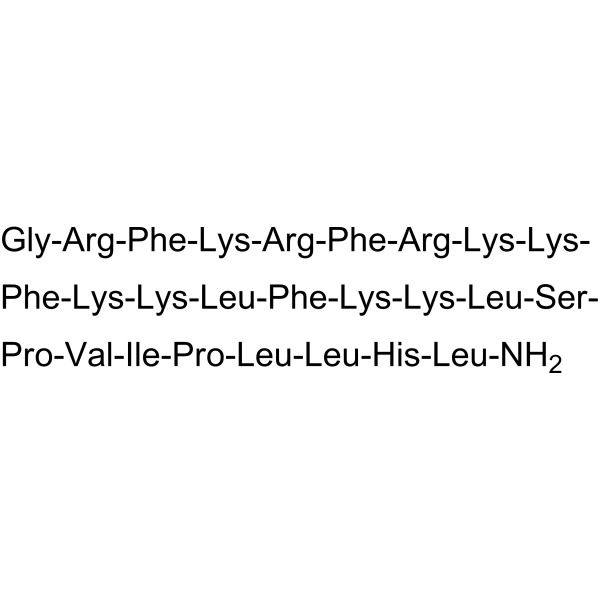
Gly-Arg-Phe-Lys-Arg-Phe-Arg-Lys-Lys-Phe-Lys-Lys-Leu-Phe-Lys-Lys-Leu-Ser-Pro-Val-Ile-Pro-Leu-Leu-His-Leu-NH2
-
Sequence Shortening
GRFKRFRKKFKKLFKKLSPVIPLLHL-NH2
-
Shipping
Room temperature in continental US; may vary elsewhere.
-
Storage
Please store the product under the recommended conditions in the Certificate of Analysis.


Purity & Documentation
References
[1]. Xia R, et al. Insight into the inhibitory activity and mechanism of bovine cathelicidin BMAP 27 against Salmonella Typhimurium. Microbial pathogenesis. 2024 Feb;187:106540. [Content Brief]
[2]. Yang S, et al. Structural analysis and mode of action of BMAP-27, a cathelicidin-derived antimicrobial peptide. Peptides. 2019 Aug;118:170106. [Content Brief]
[3]. Ghiselli R, et al. Effects of the antimicrobial peptide BMAP-27 in a mouse model of obstructive jaundice stimulated by lipopolysaccharide. Peptides. 2006 Nov;27(11):2592-9. [Content Brief]

Calculators
Concentration (start) × Volume (start) = Concentration (final) × Volume (final)


BMAP-27 Related Classifications
HY-P5286 Related Classifications
 Powered by Bioz