



Phosphatidylserine
Based on 3 publication(s) in Google Scholar
Phosphatidylserine (Phospholipids) is a well-conserved anti-inflammatory and immunosuppressive signal. Phosphatidylserine is involved in membrane translocation and the activation of protein kinase C, participating in Akt signaling through its interaction with PIP3. The local exposure of Phosphatidylserine can interact with complement and other proteins, promoting microglial phagocytosis during critical periods of synaptic refinement. Phosphatidylserine can promote blood coagulation in the extracellular environment and acts as a "eat me" signal to clear out apoptotic cells. Phosphatidylserine can suppress inflammation in tissues by inducing TGF-β secretion and inhibiting immune responses.
Nur für Forschungszwecke. Wir verkaufen nicht an Patienten.
- Reinheit : 98%
- CAS. Nr.: 1446756-47-3
- Molecular Weight:788.10
-
Speicherung:Powder -20°C, 3 years ; In solvent -80°C, 6 months , -20°C, 1 month

To place orders, for customer services and technical support, please contact: MedChemExpress USA
Tel: 609-228-6898 E-mail: [email protected] [email protected]


-
 Biologische Aktivität
-
Chemical Information
-
Lösungsmittel & Löslichkeit
- Protokoll
- Reinheit & Dokumentation
- Verweise
-
 Help & FAQs


-

HY-W250118
Cephalin form bovine brainBiochemical Assay Reagent -

HY-139533
Phosphatidylinositols, soya, sodium saltsPhosphatidylinositols Mixture -

HY-125853
Phosphatidylcholines,soyaPhosphatidylcholine -

HY-156041
Lysophosphatidylethanolamines, eggEndogenous Metabolite -

HY-18739
Phorbol 12-myristate 13-acetatePKC Activator -

HY-139414
LysophosphatidylcholinesHemolytic Agent -

HY-125854
Phosphatidylcholines, eggPhosphatidylcholine -

HY-W040255
1-Palmitoyl-2-glutaryl-sn-glycero-3-phosphocholineOxidized Phospholipid -
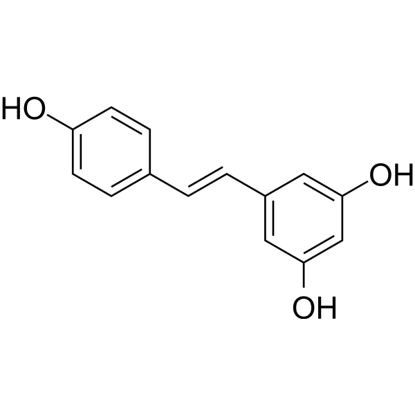
HY-16561
ResveratrolPolyphenolic Phytoalexin -
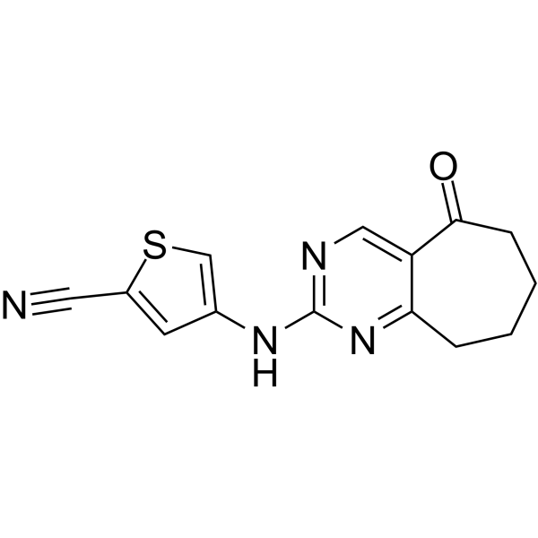
HY-W107464
G6PDi-1G6PD Inhibitor -
HY-W251428
Phosphatidylglycerols (egg) sodium saltBiochemical Assay Reagent -
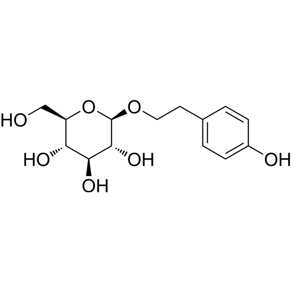
HY-N0109
SalidrosideProlyl Endopeptidase Inhibitor
Publications Citing Use of MedChemExpress (MCE) Phosphatidylserine
More

Biologische Aktivität
Beschreibung
In Vitro
Phosphatidylserine can inhibit inflammation in tissues by inducing TGF-β secretion, thereby avoiding activation of immune responses through the production of regulatory T cells[2]. Phosphatidylserine can promote neuronal survival, differentiation, and synaptic neurotransmission[3]. Phosphatidylserine mediates the pruning of microglia developmental synapses when undergoing local externalization[4].
MedChemExpress (MCE) has not independently confirmed the accuracy of these methods. They are for reference only.
In Vivo
MedChemExpress (MCE) has not independently confirmed the accuracy of these methods. They are for reference only.
-
Animal Model:BALB/cAnN and C57BL/6 male mice[2].
-
Dosage:Phosphatidylserine (PS) liposomes (0.5 mg of total lipid, PS-containing liposomes consisted of 30:30:40 molar ratio PS to PC to cholesterol)
-
Administration:Subcutaneous injection (s.c.); single dose
-
Result:Inhibited responses to Ags as determined by decreased draining lymph node tissue mass, with reduced numbers of total leukocytes and Ag-specific CD4+ T cells.
Clinical Trial
| NCT Number | Sponsor | Condition | Start Date |
Phase

|
|---|---|---|---|---|
| NCT01329991 | 2011-05 | PHASE1 |


Chemical Information
-
CAS. Nr. 1446756-47-3
-
Appearance Solid
-
Molecular Weight 788.10
-
Color Light yellow to yellow
-
SMILES
[Phosphatidylserine]
-
Synonyms
Phospholipids, phosphatidylserines; Serine glycerophosphatides
-
Initial Source
Most organisms
-
Versand
Room temperature in continental US; may vary elsewhere.
-
Speicherung
Powder -20°C 3 years In solvent -80°C 6 months -20°C 1 month


Publications (3)
-
Journal Impact Factor
-
Most Recent
-
-
Nat Commun
Transferrin-phosphatidylserine liposomes target TDP-43 and neuroinflammation in male mice with neuropathic pain. [Abstract]2025 Dec 12;16(1):11314. PMID: 41387431 -
Adv Sci (Weinh)
2026 Jul;13(39):e20095. PMID: 41972457


Lösungsmittel & Löslichkeit
In Vitro:
DMSO : < 1 mg/mL (insoluble or slightly soluble)


Reinheit & Dokumentation
-
Data Sheet (272 KB)
-
SDS (252 KB)
- English - EN (252 KB)
- Français - FR (252 KB)
- Deutsch - DE (252 KB)
- Norwegian - NO (252 KB)
- Español - ES (252 KB)
- Swedish - SV (252 KB)
- Italian - IT (252 KB)
- Korean - KR (252 KB)
- Portuguese - PT (252 KB)
-
Handling Instructions (2659 KB)
Verweise
[1]. Kay JG, et al. Phosphatidylserine-mediated cellular signaling. Adv Exp Med Biol. 2013;991:177-93. [Content Brief]
[2]. Hoffmann PR, et al. Interaction between phosphatidylserine and the phosphatidylserine receptor inhibits immune responses in vivo. J Immunol. 2005 Feb 1;174(3):1393-404. [Content Brief]
[3]. Kim HY, et al. Phosphatidylserine in the brain: metabolism and function. Prog Lipid Res. 2014 Oct;56:1-18. [Content Brief]
[4]. Scott-Hewitt N, et al. Local externalization of phosphatidylserine mediates developmental synaptic pruning by microglia. EMBO J. 2020 Aug 17;39(16):e105380. [Content Brief]

Calculators
Konzentration (Stammlösung) × Volumen (Stammlösung) = Konzentration (Ziellösung) × Volumen (Ziellösung)


Phosphatidylserine Related Classifications
HY-A0183 Related Classifications
 Powered by Bioz