



Ribostamycin
Based on 1 publication(s) in Google Scholar
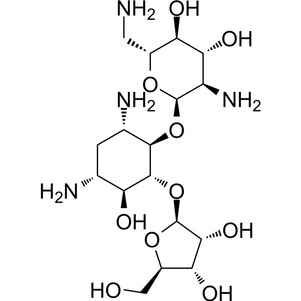
Ribostamycin (Vistamycin) is a broad-spectrum aminoglycoside antibiotic. Ribostamycin is effective against Gram-Negative and Gram-Positive bacterial infection. Ribostamycin also inhibits the chaperone activity of PDI.
Nos produits utilisent uniquement pour la recherche. Nous ne vendons pas aux patients.
- Pureté : 99.89%
- CAS No.: 25546-65-0
- Formule: C17H34N4O10
- Masse moléculaire:454.47
-
Stockage:Powder -20°C, 3 years , 4°C, 2 years ; In solvent -80°C, 6 months , -20°C, 1 month

To place orders, for customer services and technical support, please contact: MedChemExpress USA
Tel: 609-228-6898 E-mail: [email protected] [email protected]


-
 Activité biologique
-
Chemical Information
-
Solvant et solubilité
- Protocole
- Pureté et documentation
- Références
-
 Help & FAQs


Publications Citing Use of MedChemExpress (MCE) Ribostamycin
MoreVoir tous les produits spécifiques à Isoform Antibiotic
More

Activité biologique
Description
IC50 & Target
|
Aminoglycoside |
In Vitro
Ribostamycin inhibits Borrelia burgdorferi with a MIC90 of 32 mg/L[2].
Ribostamycin (1-100 μM, 0-15 min) inhibits the chaperone activity of PDI[3].
Ribostamycin inhibits Escherichia coli strains with a MIC of 0.9-7.2 μM[4].
MedChemExpress (MCE) has not independently confirmed the accuracy of these methods. They are for reference only. Further protocols information, click here.
-
Cell Line:Escherichia coli strains
-
Concentration:0-64 μg/mL
-
Incubation Time:14 h
-
Result:Inhibited Escherichia coli strains with a MIC of 0.9-7.2 μM.
In Vivo
MedChemExpress (MCE) has not independently confirmed the accuracy of these methods. They are for reference only.


Chemical Information
-
CAS No. 25546-65-0
-
Appearance Solid
-
Masse moléculaire 454.47
-
Formule C17H34N4O10
-
Color Off-white to light yellow
-
SMILES
NC[C@H]([C@H]([C@@H]([C@H]1N)O)O)O[C@@H]1O[C@H]([C@H](C[C@H]([C@@H]2O)N)N)[C@@H]2O[C@@H]3O[C@@H]([C@H]([C@H]3O)O)CO
-
Synonyms
Vistamycin; SF-733
-
Structure Classification
-
Initial Source
Streptomyces ribosidificus
-
Livraison
Room temperature in continental US; may vary elsewhere.
-
Stockage
Powder -20°C 3 years 4°C 2 years In solvent -80°C 6 months -20°C 1 month


Publications (1)
-
Journal Impact Factor
-
Most Recent
-
J Pharm Biomed Anal
Development and validation of a sensitive LC-MS/MS method without derivatization/ion-pairing agents for etimicin quantification in rat plasma, internal ear and kidney. [Abstract]2017 Nov 30:146:96-102. PMID: 28881316


Solvant et solubilité
In Vitro:
H2O : 50 mg/mL (110.02 mM; Need ultrasonic)
Please refer to the solubility information to select the appropriate solvent. Once prepared, please aliquot and store the solution to prevent product inactivation from repeated freeze-thaw cycles.
Storage method and period of stock solution: -80°C, 6 months; -20°C, 1 month. When stored at -80°C, please use it within 6 months. When stored at -20°C, please use it within 1 month.
* Note: If you choose water as the stock solution, please dilute it to the working solution, then filter and sterilize it with a 0.22 μm filter before use.
Please refer to the solubility information to select the appropriate solvent. Once prepared, please aliquot and store the solution to prevent product inactivation from repeated freeze-thaw cycles.
Storage method and period of stock solution: -80°C, 6 months; -20°C, 1 month. When stored at -80°C, please use it within 6 months. When stored at -20°C, please use it within 1 month.
* Note: If you choose water as the stock solution, please dilute it to the working solution, then filter and sterilize it with a 0.22 μm filter before use.
Concentration (start) × Volume (start) = Concentration (final) × Volume (final)

Protocole
-
Research Protocol for Infectious Diseases
Infectious-disease experiments test how pathogens interact with host barriers, innate immune receptors, inflammatory signaling, pathogen replication, and tissue injury; pattern-recognition receptors such as TLRs, RIG-I-like receptors, NOD-like receptors, and inflammasomes detect microbial molecules and activate NF-κB, interferon, and cytokine responses. The central hypothesis is that infection severity reflects the balance between pathogen burden and host response: protective inflammation restricts pathogen growth, whereas excessive or mislocalized inflammation contributes to tissue damage and disease phenotype. Unresolved questions include which host pathways are protective versus pathogenic, why some infection models fail to translate to human disease, and which combined readouts best predict clinically relevant infection outcomes.
-
Gram Staining of Tissue Sections
Gram staining of tissue sections is a histochemical technique used to differentiate Gram-positive and Gram-negative bacteria within histological specimens based on differences in bacterial cell wall structure and dye retention, adapted from classical bacteriological Gram staining into tissue-compatible “histological Gram stain” variants. In tissue applications, modifications of the Brown-Hopps and Brown-Brenn methods are commonly used to improve differentiation of microorganisms embedded within host connective tissue and to reduce overstaining or loss of Gram-negative signal, which are known limitations of earlier approaches. The principle relies on crystal violet-iodine complex retention in Gram-positive organisms and subsequent decolorization and counterstaining steps that allow contrast visualization of Gram-negative organisms against tissue background.


Pureté et documentation
-
Fiche technique (281 KB)
-
SDS (393 KB)
- English - EN (393 KB)
- Français - FR (393 KB)
- Deutsch - DE (393 KB)
- Norwegian - NO (393 KB)
- Español - ES (393 KB)
- Swedish - SV (393 KB)
- Italian - IT (393 KB)
- Korean - KR (393 KB)
- Portuguese - PT (393 KB)
-
Instruction de manipulation (2659 KB)
Références
[1]. Zheng T, et al. Linear self-assembly formation between gold nanoparticles and aminoglycoside antibiotics. Colloids Surf B Biointerfaces. 2018 Apr 1;164:185-191. [Content Brief]
[2]. Hunfeld KP, et al. In vitro activity of mezlocillin, meropenem, aztreonam, vancomycin, teicoplanin, ribostamycin and fusidic acid against Borrelia burgdorferi. Int J Antimicrob Agents. 2001 Mar;17(3):203-8. [Content Brief]
[3]. Horibe T, et al. Ribostamycin inhibits the chaperone activity of protein disulfide isomerase. Biochem Biophys Res Commun. 2001 Dec 21;289(5):967-72. [Content Brief]
[4]. Kong J, et al. Exploration of Antibiotic Activity of Aminoglycosides, in Particular Ribostamycin Alone and in Combination With Ethylenediaminetetraacetic Acid Against Pathogenic Bacteria. Front Microbiol. 2020 Jul 29;11:1718. [Content Brief]
[5]. Kitasato I, et al. Comparative nephrotoxicity of ribostamycin and gentamicin in rats evaluated by urinalysis. Drugs Exp Clin Res. 1989;15(6-7):273-89. [Content Brief]


Complete Stock Solution Preparation Table
Please refer to the solubility information to select the appropriate solvent. Once prepared, please aliquot and store the solution to prevent product inactivation from repeated freeze-thaw cycles.
Storage method and period of stock solution: -80°C, 6 months; -20°C, 1 month. When stored at -80°C, please use it within 6 months. When stored at -20°C, please use it within 1 month.
| Optional Solvent | Concentration Solvent Mass | 1 mg | 5 mg | 10 mg | 25 mg |
|---|---|---|---|---|---|
| H2O | 1 mM | 2.2004 mL | 11.0018 mL | 22.0037 mL | 55.0091 mL |
| 5 mM | 0.4401 mL | 2.2004 mL | 4.4007 mL | 11.0018 mL | |
| 10 mM | 0.2200 mL | 1.1002 mL | 2.2004 mL | 5.5009 mL | |
| 15 mM | 0.1467 mL | 0.7335 mL | 1.4669 mL | 3.6673 mL | |
| 20 mM | 0.1100 mL | 0.5501 mL | 1.1002 mL | 2.7505 mL | |
| 25 mM | 0.0880 mL | 0.4401 mL | 0.8801 mL | 2.2004 mL | |
| 30 mM | 0.0733 mL | 0.3667 mL | 0.7335 mL | 1.8336 mL | |
| 40 mM | 0.0550 mL | 0.2750 mL | 0.5501 mL | 1.3752 mL | |
| 50 mM | 0.0440 mL | 0.2200 mL | 0.4401 mL | 1.1002 mL | |
| 60 mM | 0.0367 mL | 0.1834 mL | 0.3667 mL | 0.9168 mL | |
| 80 mM | 0.0275 mL | 0.1375 mL | 0.2750 mL | 0.6876 mL | |
| 100 mM | 0.0220 mL | 0.1100 mL | 0.2200 mL | 0.5501 mL |
* Note: If you choose water as the stock solution, please dilute it to the working solution, then filter and sterilize it with a 0.22 μm filter before use.


Ribostamycin Related Classifications
HY-142127 Related Classifications
 Powered by Bioz