



SDH-IN-3
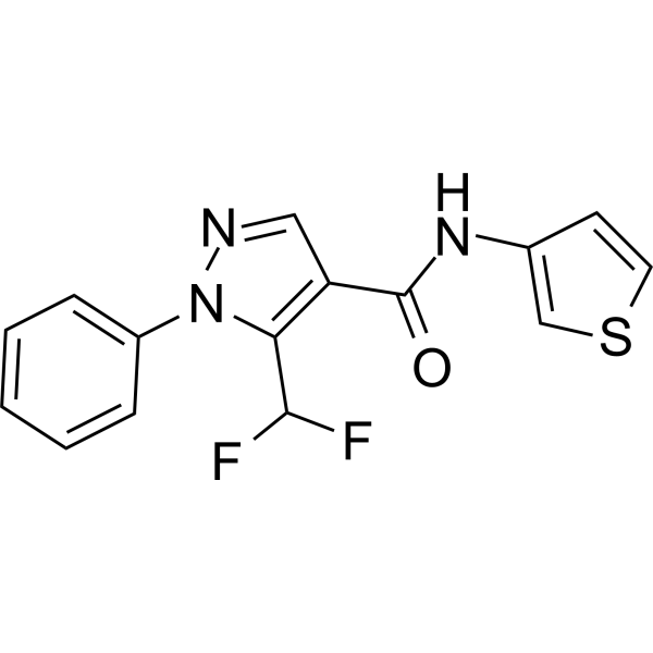
SDH-IN-3 is a succinate dehydrogenase (SDH) inhibitor with an IC50 of 7.2 μg/mL. SDH-IN-3 exhibits excellent antifungal activities against Nigrospora oryzae with an EC50 of 1.9 μg/mL. SDH-IN-3 can be used for anti-infection research.
Nos produits utilisent uniquement pour la recherche. Nous ne vendons pas aux patients.
- CAS No.: 2805297-11-2
- Formule: C15H11F2N3OS
- Masse moléculaire:319.33
-
Stockage:
Please store the product under the recommended conditions in the Certificate of Analysis.

To place orders, for customer services and technical support, please contact: MedChemExpress USA
Tel: 609-228-6898 E-mail: [email protected] [email protected]


-
 Activité biologique
-
Chemical Information
- Protocole
- Pureté et documentation
- Références
-
 Help & FAQs


Voir tous les produits spécifiques à Isoform Antibiotic
More

Activité biologique
Description
IC50 & Target
IC50: 7.2 mg/L succinate dehydrogenase (SDH)[1]
In Vitro
SDH-IN-3(Compound T6) (40 μg/mL; 6-7 days) has in vivo protective and curative activities against rice infected with N.oryzae mycelia 81.5% and 43.0%, respectively[1].
SDH-IN-3 (0-40 μg/mL; 14-80 hours) significantly inhibits the growth of N.oryzae mycelia and hinders spore germination and germ tube elongation[1].
SDH-IN-3(0-20 μg/mL; 12-36 hours) affects the mycelium membrane integrity of N.oryzae mycelia by increasing cell membrane permeability and causing peroxidation of cellular lipids[1].
MedChemExpress (MCE) has not independently confirmed the accuracy of these methods. They are for reference only.


Chemical Information
-
CAS No. 2805297-11-2
-
Masse moléculaire 319.33
-
Formule C15H11F2N3OS
-
SMILES
O=C(NC1=CSC=C1)C2=C(C(F)F)N(C3=CC=CC=C3)N=C2
-
Livraison
Room temperature in continental US; may vary elsewhere.
-
Stockage
Please store the product under the recommended conditions in the Certificate of Analysis.


Pureté et documentation
Références

Calculators
Concentration (start) × Volume (start) = Concentration (final) × Volume (final)


SDH-IN-3 Related Classifications
HY-155004 Related Classifications
 Powered by Bioz