



Daurisoline
Based on 5 publication(s) in Google Scholar
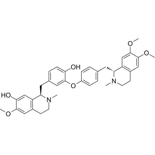
Daurisoline is a bis-benzylisoquinoline alkaloid that can be isolated from Menispermum dauricum and Rhizoma Menispermi. Daurisoline exerts a blocking effect on hERG and has antiarrhythmic effects. Daurisoline is a potent autophagy blocker that can be used for the research of cancer.
Nur für Forschungszwecke. Wir verkaufen nicht an Patienten.
- Reinheit : 99.90%
- CAS. Nr.: 70553-76-3
- Formel: C37H42N2O6
- Molecular Weight:610.74
-
Speicherung:Powder -20°C, 3 years , 4°C, 2 years ; In solvent -80°C, 2 years , -20°C, 1 year

To place orders, for customer services and technical support, please contact: MedChemExpress USA
Tel: 609-228-6898 E-mail: [email protected] [email protected]


-
 Biologische Aktivität
-
Chemical Information
-
Lösungsmittel & Löslichkeit
- Protokoll
- Reinheit & Dokumentation
- Verweise
-
 Help & FAQs


-
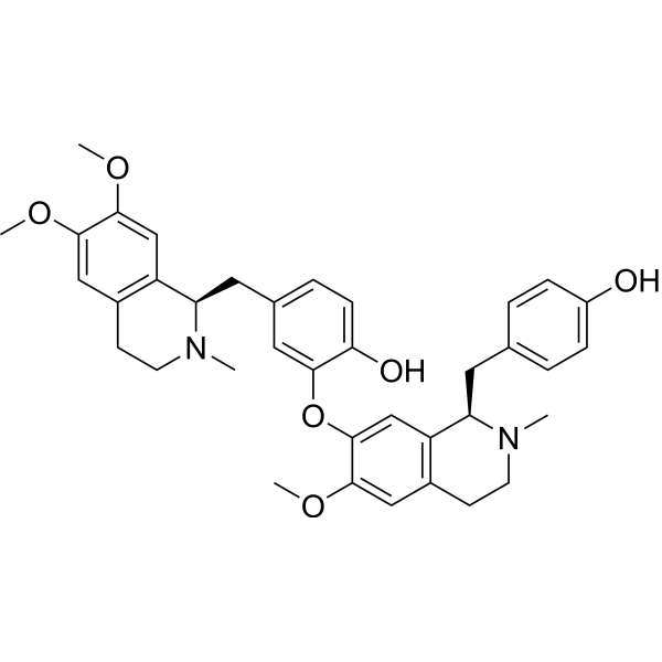
HY-N0484
LiensininePI3K/AKT/MAPK/JAK2/STAT3 Inhibitor -
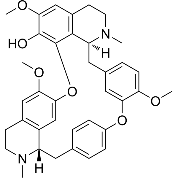
HY-N1372A
FangchinolineAutophagy Inducer -
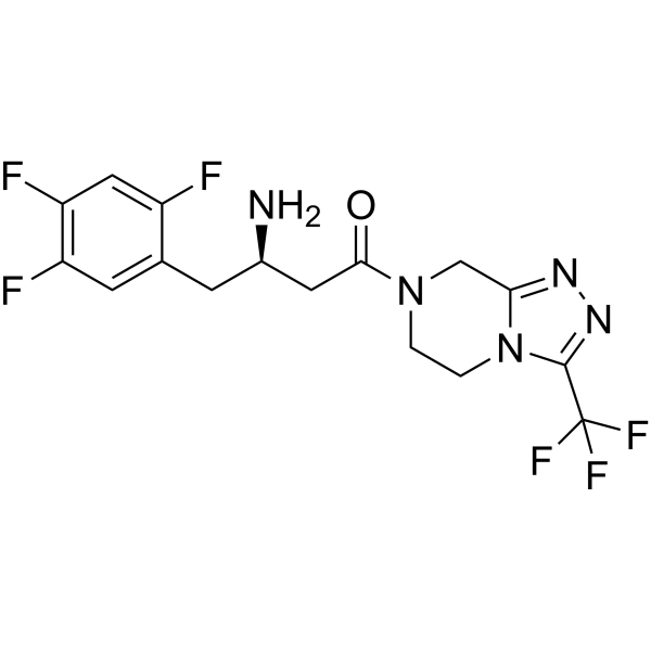
HY-13749
SitagliptinDPP4 Inhibitor -
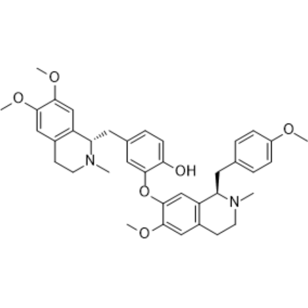
HY-N0441
NeferineAutophagy Modulator -
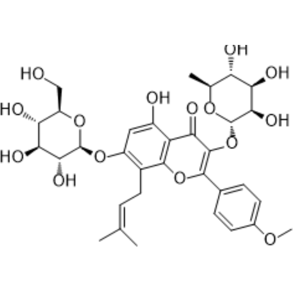
HY-N0014
IcariinPPAR Activator -
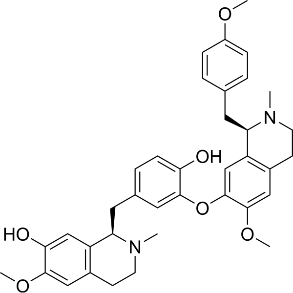
HY-N0770
IsoliensinineApoptosis Inducer -
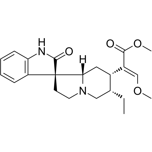
HY-N0901
CorynoxineAutophagy Enhancer -
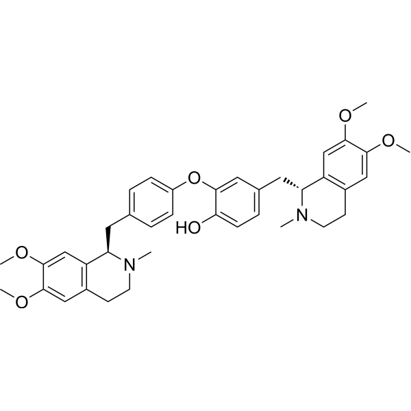
HY-N0220
DauricineApoptosis Inducer -
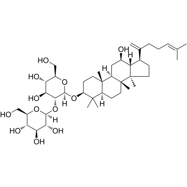
HY-N2515
Ginsenoside Rk1Apoptosis Inducer -
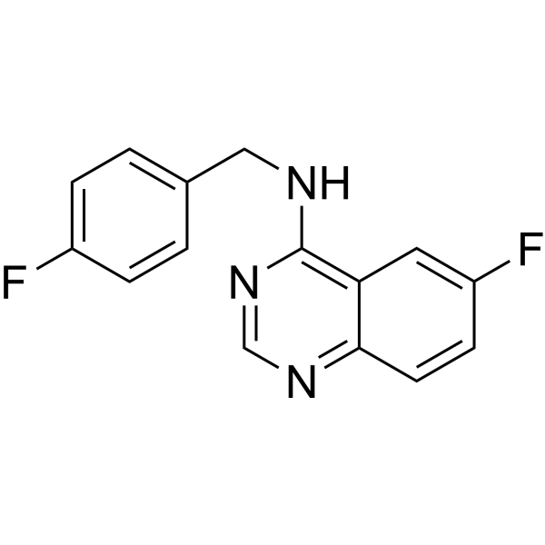
HY-12990
Spautin-1Autophagy Inhibitor -

HY-N0221R
Daurisoline (Standard)Autophagy Blocker -
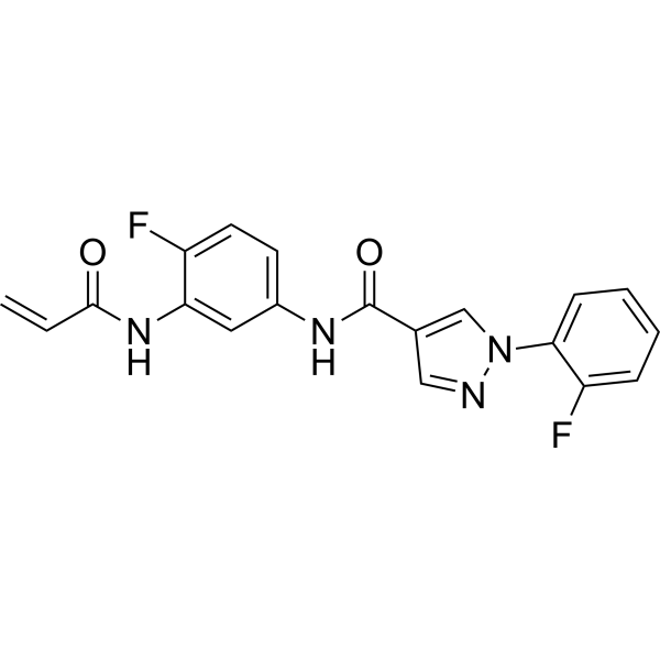
HY-128892
EN6Autophagy Activator
Publications Citing Use of MedChemExpress (MCE) Daurisoline
More Customer Validation & Images

-
RT-PCR
-
WB
-
ELISA
-
Flow Cytometry
-
Cell Proliferation/Viability Assay


Biologische Aktivität
Beschreibung
IC50 & Target
Cellular Effect
|
Cell Line

|
Type | Value | Description | References |
|---|---|---|---|---|
| A673 | GI50 |
9.2 μM
Compound: 17
|
Antiproliferative activity against human A673 assessed as cell growth inhibition after 48 hrs by SRB assay
Antiproliferative activity against human A673 assessed as cell growth inhibition after 48 hrs by SRB assay
|
[PMID: 33226219] |
| HCC1806 | GI50 |
17 μM
Compound: 17
|
Antiproliferative activity against human HCC1806 assessed as cell growth inhibition after 48 hrs by SRB assay
Antiproliferative activity against human HCC1806 assessed as cell growth inhibition after 48 hrs by SRB assay
|
[PMID: 33226219] |
| HCC1937 | GI50 |
6.9 μM
Compound: 17
|
Antiproliferative activity against human HCC1937 assessed as cell growth inhibition after 48 hrs by SRB assay
Antiproliferative activity against human HCC1937 assessed as cell growth inhibition after 48 hrs by SRB assay
|
[PMID: 33226219] |
| HCC70 | GI50 |
8.8 μM
Compound: 17
|
Antiproliferative activity against human HCC70 assessed as cell growth inhibition after 48 hrs by SRB assay
Antiproliferative activity against human HCC70 assessed as cell growth inhibition after 48 hrs by SRB assay
|
[PMID: 33226219] |
| HEK293 | IC50 |
9.1 μM
Compound: 1
|
Inhibition of human ERG stepped current expressed in HEK293 cells at + 20 mV holding potential by patch clamp assay
Inhibition of human ERG stepped current expressed in HEK293 cells at + 20 mV holding potential by patch clamp assay
|
[PMID: 22974355] |
| HEK293 | IC50 |
9.6 μM
Compound: 1
|
Inhibition of human ERG tail current expressed in HEK293 cells at + 60 mV holding potential by patch clamp assay
Inhibition of human ERG tail current expressed in HEK293 cells at + 60 mV holding potential by patch clamp assay
|
[PMID: 22974355] |
| MDA-MB-231 | GI50 |
13 μM
Compound: 17
|
Antiproliferative activity against human MDA-MB-231 assessed as cell growth inhibition after 48 hrs by SRB assay
Antiproliferative activity against human MDA-MB-231 assessed as cell growth inhibition after 48 hrs by SRB assay
|
[PMID: 33226219] |
| MDA-MB-453 | GI50 |
9 μM
Compound: 17
|
Antiproliferative activity against human MDA-MB-453 assessed as cell growth inhibition after 48 hrs by SRB assay
Antiproliferative activity against human MDA-MB-453 assessed as cell growth inhibition after 48 hrs by SRB assay
|
[PMID: 33226219] |
| SJRH30 | GI50 |
3.7 μM
Compound: 17
|
Antiproliferative activity against human SJRH30 assessed as cell growth inhibition after 48 hrs by SRB assay
Antiproliferative activity against human SJRH30 assessed as cell growth inhibition after 48 hrs by SRB assay
|
[PMID: 33226219] |
| Ventricular myocyte | IC50 |
19.9 μM
Compound: 1
|
Inhibition of Ikr tail current in rabbit ventricular myocytes
Inhibition of Ikr tail current in rabbit ventricular myocytes
|
[PMID: 22974355] |
| Ventricular myocyte | IC50 |
52.5 μM
Compound: 1
|
Inhibition of Iks tail current in rabbit ventricular myocytes
Inhibition of Iks tail current in rabbit ventricular myocytes
|
[PMID: 22974355] |
In Vitro
Daurisoline (compound 1) shows a maximal inhibitory effect on the end of depolarization (IhERG-step) at +20 mV and on the peak tail current (IhERG-tail) at +60 mV. At concentrations of 1, 3, 10, and 30 μM, the inhibition ratios for current amplitude at the end of depolarization (IhERG-step) are 32.2±4.2%, 41.6±2.6%, 62.1±5.9%, and 74.8±6.8%, respectively; the IC50 is 9.1 μM. In turn, the inhibition ratios for IhERG-tail are 16.7±5.8%, 31.1±4.5%, 55.1±7.2%, and 81.2±7.0%, respectively; the IC50 is 9.6 μM[1]. Daurisoline (DAS) inhibits the CPT-induced autophagy in different cancer cell lines, with IC50s of 74.75±1.03, 50.54±1.02 and 80.81±1.10 μM in HeLa, A549 and HCT-116 cells, respectively. DAC and Daurisoline also impair lysosomal function and lysosomal acidification, via inhibiting the lysosome V-type ATPase acitivity in DAC and Daurisoline treated cells[2].
MedChemExpress (MCE) has not independently confirmed the accuracy of these methods. They are for reference only.
In Vivo
MedChemExpress (MCE) has not independently confirmed the accuracy of these methods. They are for reference only.


Chemical Information
-
CAS. Nr. 70553-76-3
-
Appearance Solid
-
Molecular Weight 610.74
-
Formel C37H42N2O6
-
Color White to off-white
-
SMILES
OC1=CC2=C(C=C1OC)CCN(C)[C@@H]2CC3=CC=C(O)C(OC4=CC=C(C[C@H]5N(C)CCC6=C5C=C(OC)C(OC)=C6)C=C4)=C3
-
Synonyms
(R,R)-Daurisoline
-
Structure Classification
-
Initial Source
-
Versand
Room temperature in continental US; may vary elsewhere.
-
Speicherung
Powder -20°C 3 years 4°C 2 years In solvent -80°C 2 years -20°C 1 year


Publications (5)
-
Journal Impact Factor
-
Most Recent
-
Research (Wash D C)
Daurisoline Modulates the TBK1-Dependent Type I Interferon Pathway to Boost Anti-tumor Immunity via Targeting of LRP1. [Abstract]2025 Jul 4:8:0764. PMID: 40625660
Daurisoline purchased from MedChemExpress. Usage Cited in: Research (Wash D C). 2025 Jul 4:8:0764. [Abstract]
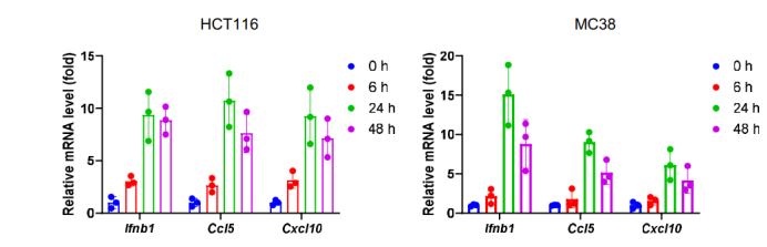
RNA (mRNA) expressions of Ifnb1, Ccl5, and Cxcl10 were measured by quantitative real-time PCR (qRT-PCR) treated with Daurisoline (DS).
Daurisoline purchased from MedChemExpress. Usage Cited in: Research (Wash D C). 2025 Jul 4:8:0764. [Abstract]
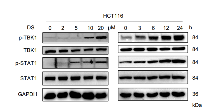
HCT116 cells were treated with the indicated concentrations of Daurisoline (DS) for 24 h or treated with 20 μM DS for the indicated time points; the phosphorylation levels of TANK-binding kinase 1 (TBK1) and STAT1 were determined by immunoblotting.
Daurisoline purchased from MedChemExpress. Usage Cited in: Research (Wash D C). 2025 Jul 4:8:0764. [Abstract]
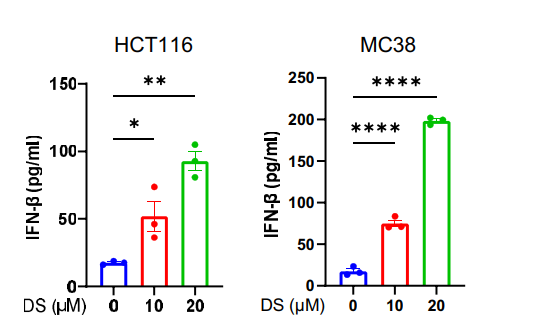
Enzyme-linked immunosorbent assay (ELISA) analysis of the IFN-β levels in the supernatant of HCT116 and MC38 cells after treatment with 20 μM Daurisoline (DS) for 48 h.
Daurisoline purchased from MedChemExpress. Usage Cited in: Research (Wash D C). 2025 Jul 4:8:0764. [Abstract]
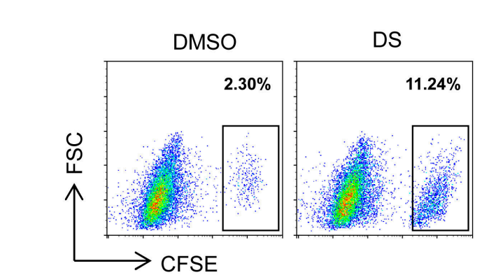
Carboxyfluorescein succinimidyl ester (CFSE)-stained MC38 cells were treated with DMSO or Daurisoline (DS) (20 μM) for 24 h, followed by coculturing with bone-marrow-derived macrophages (BMDMs) for an additional 24 h; the proportion of CFSE+ cells in CD11b+ F4/80+ macrophages was monitored by flow cytometry analysis.
-
Cell Commun Signal
Machine learning-aided discovery of T790M-mutant EGFR inhibitor CDDO-Me effectively suppresses non-small cell lung cancer growth. [Abstract]2024 Dec 5;22(1):585. PMID: 39639305 -
Cancer Biol Ther
2025 Dec;26(1):2442556. PMID: 39699276
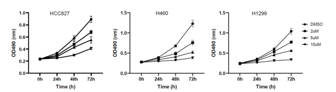
Daurisoline purchased from MedChemExpress. Usage Cited in: Cancer Biol Ther. 2025 Dec;26(1):2442556. [Abstract]
The CCK8 assay found that Daurisoline (2, 5, 10 μM) could decrease the proliferation of HCC827, H460 and H1299, and this function was dose and time-dependent.
-
Biochem Biophys Res Commun
Daurisoline inhibits hepatocellular carcinoma progression by restraining autophagy and promoting cispaltin-induced cell death. [Abstract]2021 Jan 1:534:1083-1090. PMID: 33213840 -
Oxid Med Cell Longev
Daurisoline Inhibits ESCC by Inducing G1 Cell Cycle Arrest and Activating ER Stress to Trigger Noxa-Dependent Intrinsic and CHOP-DR5-Dependent Extrinsic Apoptosis via p-eIF2 α-ATF4 Axis. [Abstract]2022 Aug 4;2022:5382263. PMID: 35965681


Lösungsmittel & Löslichkeit
In Vitro:
DMSO : 100 mg/mL (163.74 mM; Need ultrasonic; Hygroscopic DMSO has a significant impact on the solubility of product, please use newly opened DMSO)
Please refer to the solubility information to select the appropriate solvent. Once prepared, please aliquot and store the solution to prevent product inactivation from repeated freeze-thaw cycles.
Storage method and period of stock solution: -80°C, 2 years; -20°C, 1 year. When stored at -80°C, please use it within 2 years. When stored at -20°C, please use it within 1 year.
Please refer to the solubility information to select the appropriate solvent. Once prepared, please aliquot and store the solution to prevent product inactivation from repeated freeze-thaw cycles.
Storage method and period of stock solution: -80°C, 2 years; -20°C, 1 year. When stored at -80°C, please use it within 2 years. When stored at -20°C, please use it within 1 year.
Konzentration (Stammlösung) × Volumen (Stammlösung) = Konzentration (Ziellösung) × Volumen (Ziellösung)
In Vivo:
Select the appropriate dissolution method based on your experimental animal and administration route.
- For the following dissolution methods, please ensure to first prepare a clear stock solution using an In Vitro approach and then sequentially add co-solvents:
- To ensure reliable experimental results, the clarified stock solution can be appropriately stored based on storage conditions. As for the working solution for In Vivo experiments, it is recommended to prepare freshly and use it on the same day.
- The percentages shown for the solvents indicate their volumetric ratio in the final prepared solution. If precipitation or phase separation occurs during preparation, heat and/or sonication can be used to aid dissolution.
Add each solvent one by one: 10% DMSO 40% PEG300 5% Tween-80 45% Saline
Solubility: ≥ 2.5 mg/mL (4.09 mM); Clear solution
This protocol yields a clear solution of ≥ 2.5 mg/mL (saturation unknown).
Taking 1 mL working solution as an example, add 100 μL DMSO stock solution (25.0 mg/mL) to 400 μL PEG300, and mix evenly; then add 50 μL Tween-80 and mix evenly; then add 450 μL Saline to adjust the volume to 1 mL.
Preparation of Saline: Dissolve 0.9 g sodium chloride in ddH₂O and dilute to 100 mL to obtain a clear Saline solution.
Add each solvent one by one: 10% DMSO 90% (20% SBE-β-CD in Saline)
Solubility: ≥ 2.5 mg/mL (4.09 mM); Clear solution
This protocol yields a clear solution of ≥ 2.5 mg/mL (saturation unknown).
Taking 1 mL working solution as an example, add 100 μL DMSO stock solution (25.0 mg/mL) to 900 μL 20% SBE-β-CD in Saline, and mix evenly.
Preparation of 20% SBE-β-CD in Saline (4°C, storage for one week): 2 g SBE-β-CD powder is dissolved in 10 mL Saline, completely dissolve until clear.
For the following dissolution methods, please prepare the working solution directly:
It is recommended to prepare fresh solutions and use them promptly within a short period of time.
The percentages shown for the solvents indicate their volumetric ratio in the final prepared solution. If precipitation or phase separation occurs during preparation, heat and/or sonication can be used to aid dissolution.
Add each solvent one by one: 50% PEG300 50% Saline
Solubility: 20 mg/mL (32.75 mM); Clear solution; Need ultrasonic
In Vivo Dissolution Calculator
Please enter the basic information of animal experiments:
-
-
-
-
Recommended: Prepare an additional quantity of animals to account for potential losses during experiments.
Please enter your animal formula composition:
-
%DMSO +
Recommended: Keep the proportion of DMSO in working solution below 2% if your animal is weak.
-
%+
-
+%Tween-80 + +
-
%Saline +
The co-solvents required include: DMSO, . All of co-solvents are available by MedChemExpress (MCE). , Tween 80. All of co-solvents are available by MedChemExpress (MCE).
Working solution concentration: 0.22 mg/mL
Method for preparing stock solution: mg drug dissolved in μL DMSO. Stock solution concentration: mg/mL.
1. Take μL DMSO stock solution;
2. Add μL .
μL , mix evenly;
3. Then add μL Tween 80, mix evenly;
4. Then add μL
Please ensure that the stock solution in the first step is dissolved to a clear state, and add co-solvents in sequence. You can use ultrasonic heating (ultrasonic cleaner, recommended frequency 20-40 kHz), vortexing, etc. to assist dissolution.


Reinheit & Dokumentation
-
Data Sheet (279 KB)
-
SDS (392 KB)
- English - EN (392 KB)
- Français - FR (392 KB)
- Deutsch - DE (392 KB)
- Norwegian - NO (392 KB)
- Español - ES (392 KB)
- Swedish - SV (392 KB)
- Italian - IT (392 KB)
- Korean - KR (392 KB)
- Portuguese - PT (392 KB)
-
Handling Instructions (2659 KB)
Verweise
[1]. Liu Q, et al. Effect of daurisoline on HERG channel electrophysiological function and protein expression. J Nat Prod. 2012 Sep 28;75(9):1539-45. [Content Brief]
[2]. Wu MY, et al. Natural autophagy blockers, dauricine (DAC) and daurisoline (DAS), sensitize cancer cells tocamptothecin-induced toxicity. Oncotarget. 2017 Sep 8;8(44):77673-77684. [Content Brief]
[3]. Shi SJ, et al. Pharmacokinetic-pharmacodynamic modeling of daurisoline and dauricine in beagle dogs. Acta Pharmacol Sin. 2003 Oct;24(10):1011-5. [Content Brief]


Complete Stock Solution Preparation Table
Please refer to the solubility information to select the appropriate solvent. Once prepared, please aliquot and store the solution to prevent product inactivation from repeated freeze-thaw cycles.
Storage method and period of stock solution: -80°C, 2 years; -20°C, 1 year. When stored at -80°C, please use it within 2 years. When stored at -20°C, please use it within 1 year.
| Optional Solvent | Concentration Solvent Mass | 1 mg | 5 mg | 10 mg | 25 mg |
|---|---|---|---|---|---|
| DMSO | 1 mM | 1.6374 mL | 8.1868 mL | 16.3736 mL | 40.9339 mL |
| 5 mM | 0.3275 mL | 1.6374 mL | 3.2747 mL | 8.1868 mL | |
| 10 mM | 0.1637 mL | 0.8187 mL | 1.6374 mL | 4.0934 mL | |
| 15 mM | 0.1092 mL | 0.5458 mL | 1.0916 mL | 2.7289 mL | |
| 20 mM | 0.0819 mL | 0.4093 mL | 0.8187 mL | 2.0467 mL | |
| 25 mM | 0.0655 mL | 0.3275 mL | 0.6549 mL | 1.6374 mL | |
| 30 mM | 0.0546 mL | 0.2729 mL | 0.5458 mL | 1.3645 mL | |
| 40 mM | 0.0409 mL | 0.2047 mL | 0.4093 mL | 1.0233 mL | |
| 50 mM | 0.0327 mL | 0.1637 mL | 0.3275 mL | 0.8187 mL | |
| 60 mM | 0.0273 mL | 0.1364 mL | 0.2729 mL | 0.6822 mL | |
| 80 mM | 0.0205 mL | 0.1023 mL | 0.2047 mL | 0.5117 mL | |
| 100 mM | 0.0164 mL | 0.0819 mL | 0.1637 mL | 0.4093 mL |


Daurisoline Related Classifications
HY-N0221 Related Classifications
 Powered by Bioz