



MitoPBN

Based on 1 Customer Validation
MitoPBN is a AMPK/SIRT3/PGC-1α axis modulator, reactive oxygen species scavenger and mitochondrial function enhancer. MitoPBN increases the phosphorylation level of AMPK, restores SIRT3 expression and reverses the down-regulation of PGC-1α, thereby promoting mitochondrial biogenesis. MitoPBN regulates glucose metabolism, reduces blood glucose by inhibiting hepatic gluconeogenesis and increasing hepatic glucose uptake, while scavenging mitochondrial superoxide anion/hydrogen peroxide, maintaining membrane potential and increasing ATP production. MitoPBN also reduces cell apoptosis, improves sperm motility, survival rate and membrane integrity, but may induce reductive stress in cryopreserved sperm at high concentrations. MitoPBN is widely applicable to research related to diabetes and type 2 diabetes.
Nur für Forschungszwecke. Wir verkaufen nicht an Patienten.
- Reinheit : 95.00%
- CAS. Nr.: 652968-37-1
- Formel: C33H37BrNO2P
- Molecular Weight:590.53
-
Speicherung:
-20°C, sealed storage, away from moisture
* In solvent : -80°C, 6 months; -20°C, 1 month (sealed storage, away from moisture)


To place orders, for customer services and technical support, please contact: MedChemExpress USA
Tel: 609-228-6898 E-mail: [email protected] [email protected]


-
 Biologische Aktivität
-
Chemical Information
-
Lösungsmittel & Löslichkeit
- Protokoll
- Reinheit & Dokumentation
- Verweise
-
 Help & FAQs


-
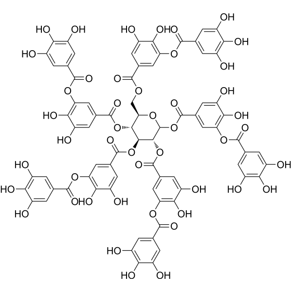
HY-B2136
Tannic acidhERG Channel Blocker -
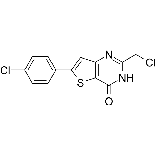
HY-162065
N6F11Ferroptosis Inducer -
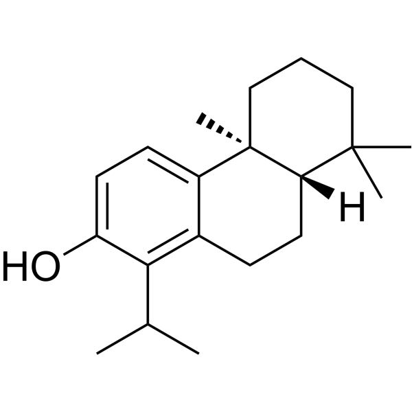
HY-N1136
(+)-TotarolDiterpenoid -
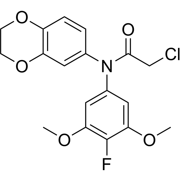
HY-155663
GPX4-IN-5GPX4 Inhibitor -
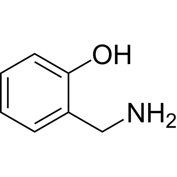
HY-34350
2-(Aminomethyl)phenolAntioxidant -
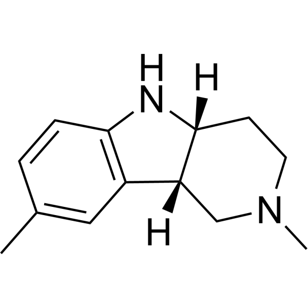
HY-W067572
StobadineAntioxidant -
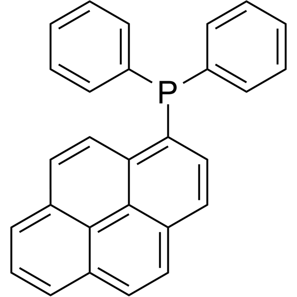
HY-118159
Diphenyl-1-pyrenylphosphinefluorescent probe -

HY-130743
Dieugenol -

HY-W587743
N1-Acetyl-5-methoxykynuramine hydrochlorideMelatonin Metabolites
Alle AMPK Isoform-spezifische Produkte anzeigen
More

Biologische Aktivität
Beschreibung
IC50 & Target
|
SIRT3 |
In Vitro
MitoPBN (40 μmol/l; 24 h) upregulates the AMPK/SIRT3/PGC-1α axis in STZ-treated primary mouse hepatocytes by increasing phosphorylated PGC-1α, phosphorylated AMPK, total AMPK, SIRT3, and NRF1 protein levels[1].
MitoPBN (40 μmol/l; 24 h) restores mitochondrial number in H2O2-stressed primary mouse hepatocytes, which was reduced by oxidative stress[1].
MitoPBN (100-150 μmol/L; ≥7 days) supplementation of isolated Ghezel ram sperm cryopreservation extender significantly improves post-thaw sperm motility, membrane integrity, mitochondrial activity, viability, antioxidant capacity, and ATP content while reducing ROS levels, abnormal sperm, and apoptosis, with 150 μmol/L being the optimal concentration[2].
MitoPBN (1-100 μmol/l; 3 h) potently scavenges peroxyl and carbon-centered free radicals in a cell-free in vitro system, as demonstrated by H2O2 consumption and adduct formation[3].
MitoPBN (5-20 μmol/l; 24 h) protects STZ-stressed L02 liver cells from cytotoxicity, with greater efficiency than PBN, and liposomal encapsulation as Nano-MitoPBN significantly reduces the effective protective concentration[3].
MitoPBN (40 μmol/l) inhibits hypoxia-induced mitophagy in L02 liver cells by reducing protein expression of LC3B and ATG5[3].
MedChemExpress (MCE) has not independently confirmed the accuracy of these methods. They are for reference only.
-
Cell Line:Streptozotocin (HY-13753) -stressed L02 human liver cells
-
Concentration:5, 20 μm
-
Incubation Time:24 h
-
Result:Protected L02 cells from Streptozotocin (STZ)-induced cytotoxicity.
Showed lower protective efficiency than Nano-MitoPBN, with 5 μmol/l Nano-MitoPBN achieving a comparable effect to 40 μmol/l free MitoPBN.
Exhibited greater protective efficiency than PBN, requiring a lower concentration to rescue cell survival.
In Vivo
Free MitoPBN (2.5 mg/kg; i.p.; daily; 8 weeks) reduces random blood glucose levels and improves oral glucose tolerance in high-fat diet-induced type 2 diabetic C57BL/6J mice[3].
MedChemExpress (MCE) has not independently confirmed the accuracy of these methods. They are for reference only.
-
Animal Model:Sperm cryopreservation induced Ghezel rams sheep model (sexually mature)[2]
-
Dosage:100 µmol/L; 150 µmol/L; 200 µmol/L; 250 µmol/L
-
Administration:added to tris extender for semen dilution prior to cryopreservation
-
Result:Effects at 100 µM
Increased total motility to 52.36%, progressive motility to 24.82%, membrane integrity to 50.01%, mitochondrial activity to 46.16%, and sperm viability to 48.99%.
Elevated total antioxidant capacity (TAC) to 1.85 mmol/L and glutathione peroxidase (GPx) activity to 61.16 U/mg.
Decreased ROS levels to 2.95 ×10³ cpm/106 sperm, abnormal sperm to 19.23%, and apoptotic sperm to 20.10%.
Increased straight-line velocity (VSL) to 55.24 µm/s and ATP content to 90 pmol/106 sperm.
Effects at 150 µmol/L (Optimal Dose)
Achieved highest total motility (54.16%), progressive motility (26.77%), membrane integrity (52.54%), mitochondrial activity (50.26%), and sperm viability (52.20%).
Maximized TAC to 1.93 mmol/L and GPx activity to 63.36 U/mg.
Reduced ROS levels to their lowest point at 2.80 ×10³ cpm/106 sperm, with abnormal sperm at 18.82% and apoptotic sperm at 18.70%.
Reached peak average path velocity (VAP) of 68.82 µm/s, VSL of 55.24 µm/s, and maximum ATP content of 116.29 pmol/106 sperm.
Effects at Higher Concentrations (200 & 250 µmol/L)
200 µmol/L (Diminishing Returns):
Showed increased sperm viability (44.12%) and ATP content, with reduced apoptotic sperm (~25%), but offered no significant improvements in motility, membrane integrity, or antioxidant markers compared to the 100 and 150 µmol/L doses.
250 µmol/L (Ineffective):
Did not provide significant beneficial effects on measured sperm parameters relative to the control group.
- Western Blot
- RNA extraction experimental
- Mitochondrial membrane-potential fluorescent assay
- Annexin V plus membrane-impermeant dye apoptosis staining
- ROS/oxidative-stress fluorescent staining
- Sperm Thawing for Functional Recovery
- Research Protocol for Metabolic Diseases
- Kinase activity and phosphorylation assays
- Detection of Apoptosis
- TUNEL staining for apoptotic DNA fragmentation
- Fluorescent plasma-membrane potential dye assay
- Mitochondrial membrane-potential and mitochondrial mass staining
- Apoptosis Solutions
- Protocol for Kinase activity and phosphorylation assays


Chemical Information
-
CAS. Nr. 652968-37-1
-
Appearance Solid
-
Molecular Weight 590.53
-
Formel C33H37BrNO2P
-
Color White to off-white
-
SMILES
[O-]/[N+](C(C)(C)C)=C\C1=CC=C(OCCCC[P+](C2=CC=CC=C2)(C3=CC=CC=C3)C4=CC=CC=C4)C=C1.[Br-]
-
Versand
Room temperature in continental US; may vary elsewhere.
-
Speicherung
-20°C, sealed storage, away from moisture
* In solvent : -80°C, 6 months; -20°C, 1 month (sealed storage, away from moisture)


Lösungsmittel & Löslichkeit
In Vitro:
DMF : 25 mg/mL (42.33 mM; Need ultrasonic and warming)
Please refer to the solubility information to select the appropriate solvent. Once prepared, please aliquot and store the solution to prevent product inactivation from repeated freeze-thaw cycles.
Storage method and period of stock solution: -80°C, 6 months; -20°C, 1 month (sealed storage, away from moisture). When stored at -80°C, please use it within 6 months. When stored at -20°C, please use it within 1 month.
Please refer to the solubility information to select the appropriate solvent. Once prepared, please aliquot and store the solution to prevent product inactivation from repeated freeze-thaw cycles.
Storage method and period of stock solution: -80°C, 6 months; -20°C, 1 month (sealed storage, away from moisture). When stored at -80°C, please use it within 6 months. When stored at -20°C, please use it within 1 month.
Konzentration (Stammlösung) × Volumen (Stammlösung) = Konzentration (Ziellösung) × Volumen (Ziellösung)


Reinheit & Dokumentation
-
Data Sheet (280 KB)
-
SDS (252 KB)
- English - EN (252 KB)
- Français - FR (252 KB)
- Deutsch - DE (252 KB)
- Norwegian - NO (252 KB)
- Español - ES (252 KB)
- Swedish - SV (252 KB)
- Italian - IT (252 KB)
- Korean - KR (252 KB)
- Portuguese - PT (252 KB)
-
Handling Instructions (2659 KB)
Verweise
[1]. Wu M, et al. Compartmentally scavenging hepatic oxidants through AMPK/SIRT3-PGC1α axis improves mitochondrial biogenesis and glucose catabolism. Free Radic Biol Med. 2021;168:117-128. [Content Brief]
[2]. Mehdipour M, et al. Mitochondrial specific antioxidant MitoPBN mitigates oxidative stress and improves mitochondrial function in cryopreserved ram sperm. Sci Rep. 2025;15(1):10526. Published 2025 Mar 27. [Content Brief]
[3]. Wu M, et al. Liver-targeted Nano-MitoPBN normalizes glucose metabolism by improving mitochondrial redox balance. Biomaterials. 2019;222:119457. [Content Brief]


Complete Stock Solution Preparation Table
Please refer to the solubility information to select the appropriate solvent. Once prepared, please aliquot and store the solution to prevent product inactivation from repeated freeze-thaw cycles.
Storage method and period of stock solution: -80°C, 6 months; -20°C, 1 month (sealed storage, away from moisture). When stored at -80°C, please use it within 6 months. When stored at -20°C, please use it within 1 month.
| Optional Solvent | Concentration Solvent Mass | 1 mg | 5 mg | 10 mg | 25 mg |
|---|---|---|---|---|---|
| DMF | 1 mM | 1.6934 mL | 8.4670 mL | 16.9339 mL | 42.3348 mL |
| 5 mM | 0.3387 mL | 1.6934 mL | 3.3868 mL | 8.4670 mL | |
| 10 mM | 0.1693 mL | 0.8467 mL | 1.6934 mL | 4.2335 mL | |
| 15 mM | 0.1129 mL | 0.5645 mL | 1.1289 mL | 2.8223 mL | |
| 20 mM | 0.0847 mL | 0.4233 mL | 0.8467 mL | 2.1167 mL | |
| 25 mM | 0.0677 mL | 0.3387 mL | 0.6774 mL | 1.6934 mL | |
| 30 mM | 0.0564 mL | 0.2822 mL | 0.5645 mL | 1.4112 mL | |
| 40 mM | 0.0423 mL | 0.2117 mL | 0.4233 mL | 1.0584 mL |


MitoPBN Related Classifications
HY-136855 Related Classifications
 Powered by Bioz