



VO-OHPic
Based on 42 publication(s) in Google Scholar
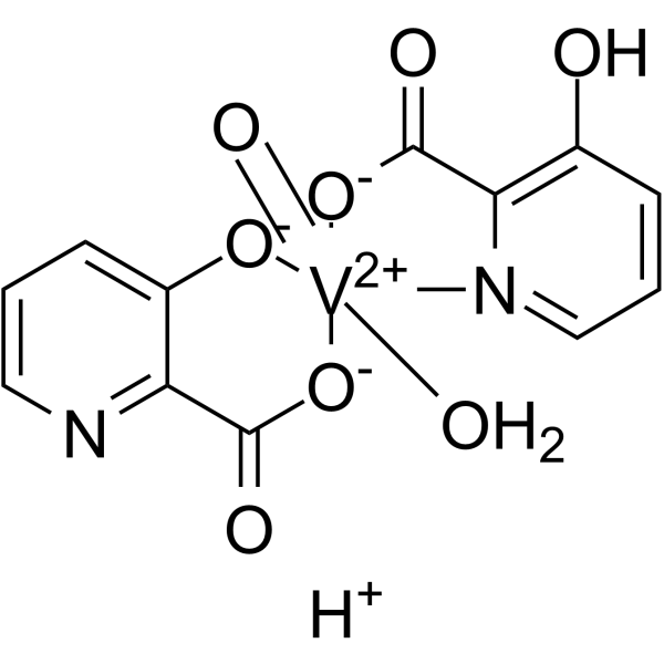
VO-OHPic is a reversible, noncompetitive PTEN inhibitor with an human IC50 value of 46 nM. VO-OHPic inhibits PTEN signaling, activates Akt-GSK3β and Nrf-2/HO-1 pathways, induces apoptosis resistance and elevates IL-10 levels. VO-OHPic inhibits autophagy, ferroptosis and oxidative stress. VO-OHPic can be used for the research of acute myocardial infarction, intervertebral disc degeneration, cardiomyopathy and cancer.
Nur für Forschungszwecke. Wir verkaufen nicht an Patienten.
- CAS. Nr.: 675848-25-6
- Formel: C12H10N2O8V
- Molecular Weight:361.16
-
Speicherung:
Please store the product under the recommended conditions in the Certificate of Analysis.

To place orders, for customer services and technical support, please contact: MedChemExpress USA
Tel: 609-228-6898 E-mail: [email protected] [email protected]


-
 Biologische Aktivität
-
Chemical Information
- Protokoll
- Reinheit & Dokumentation
- Verweise
-
 Help & FAQs



Publications Citing Use of MedChemExpress (MCE) VO-OHPic
More- Cell Stem Cell. 2022 Apr 7;29(4):545-558.e13. [Abstract]
- Bone Res. 2018 Nov 10:6:32. [Abstract]
- J Adv Res. 2025 Aug:74:589-607. [Abstract]
- Redox Biol. 2024 Jul:73:103200. [Abstract]
- Redox Biol. 2019 Jan:20:390-401. [Abstract]
- J Nanobiotechnology. 2025 May 25;23(1):376. [Abstract]
- Theranostics. 2019 Jul 9;9(18):5200-5213. [Abstract]
- Theranostics. 2019 Jul 9;9(16):4764-4778. [Abstract]
- Cell Death Dis. 2026 Jan 9;17(1):17. [Abstract]
- Cell Death Dis. 2025 Aug 7;16(1):595. [Abstract]
- Environ Sci Technol. 2020 Sep 1;54(17):10783-10796. [Abstract]
- Cell Death Dis. 2019 Apr 15;10(5):329. [Abstract]
- Phytomedicine. 2023 Dec:121:155117. [Abstract]
- Int J Mol Med. 2021 Jun;47(6):109. [Abstract]
- Clin Transl Med. 2022 Sep;12(9):e1061. [Abstract]
- Clin Transl Med. 2020 Dec;10(8):e240. [Abstract]
- Stem Cell Res Ther. 2019 Jul 29;10(1):217. [Abstract]
- Transl Psychiatry. 2021 Mar 26;11(1):185. [Abstract]
- Biochem Pharmacol. 2020 Jan:171:113715. [Abstract]
- Biochem Pharmacol. 2019 Oct:168:82-90. [Abstract]
- Commun Biol. 2026 Jan 23;9(1):311. [Abstract]
- Cell Biol Toxicol. 2025 Feb 27;41(1):53. [Abstract]
- Cell Regen. 2026 Aug 11;15(1):27.
- Int J Mol Sci. 2023 Jun 9;24(12):9954. [Abstract]
- Int Immunopharmacol. 2022 Feb:103:107840. [Abstract]
- Channels. 2026 Jun 22;20(1).
- Mol Med Rep. 2022 Jul;26(1):246. [Abstract]
- Biochim Biophys Acta Mol Basis Dis. 2022 Jan 1;1868(1):166292. [Abstract]
- Mol Med Rep. 2018 Dec;18(6):5489-5501. [Abstract]
- Mol Cell Biochem. 2026 May;481(5):2119-2137. [Abstract]
- J Cell Mol Med. 2019 Nov;23(11):7535-7544. [Abstract]
- iScience. 2024 Jun 19;27(7):110306. [Abstract]
- Mol Nutr Food Res. 2019 Dec;63(24):e1900418. [Abstract]
- BMC Cancer. 2019 Apr 25;19(1):391. [Abstract]
- Funct Integr Genomics. 2024 Apr 3;24(2):71. [Abstract]
- Cancer Med. 2019 Aug;8(9):4265-4277. [Abstract]
- Mol Vis. 2018 Jul 23:24:485-494. [Abstract]
- Biomed Pharmacother. 2026 Sep:202:119803.
- McGill University. 2026.
- Res Sq. 2026 Apr 30.
- SSRN. 2023 Jul 17.
- Eur Rev Med Pharmacol Sci. 2019 May;23(10):4406-4413. [Abstract]
 Customer Validation & Images

-
WB
-
WB
-
WB
-
WB
-
WB


Biologische Aktivität
Beschreibung
|
Akt |
IL-10 |
In Vitro
VO-OHpic (0.05-2.0 μg/mL; 2 h) dose-dependently improves viability of H/R-stressed isolated adult rat cardiac myocytes[1].
VO-OHpic (1 µM; 24-72 h) suppresses the proliferation of TSC2-/- MEFs in a time-dependent manner[2].
VO-OHpic (1 µM; 24-72 h) excessively inhibits autophagy in TSC2-/- MEFs[2].
VO-OHpic (1 µM; 24-72 h) modulates the PTEN/PRAS40 pathway in TSC2-/- MEFs[2].
VO-OHPic (1 μM; 24 h) inhibits TBHP-induced ferroptosis by restoring GPX4 and SLC7A11 expression in primary mouse CEP chondrocytes[3].
VO-OHPic (1 μM; 24 h) inhibits TBHP-induced ROS production in primary mouse CEP chondrocytes[3].
VO-OHPic (30 μM) alleviates IL-1β-induced degeneration in human nucleus pulposus cells by inhibiting PTEN and activating the PI3K/Akt pathway, thereby restoring collagen II and aggrecan levels[4].
VO-OHPic (30 μM) promotes proliferation in IL-1β-induced degenerated human nucleus pulposus cells by mediating cell cycle progression through the G1 to S phase[4].
VO-OHPic (30 μM) reduces oxidative stress in H2O2-induced degenerated human nucleus pulposus cells by lowering ROS levels and upregulating antioxidant enzymes SOD1, SOD2, CAT, and GSH[4].
VO-OHPic (1 μM; 48 h) activates the Nrf2 signaling pathway in Methylprednisolone (HY-B0260) -treated rat endothelial progenitor cells by increasing expression of Nrf2 and its downstream antioxidant proteins, and promoting Nrf2 nuclear translocation[5].
VO-OHPic (1 μM; 48 h) restores normal mitochondrial morphology in Methylprednisolone-treated rat endothelial progenitor cells[5].
VO-OHPic (1 μM; 48 h) suppresses Methylprednisolone-induced excessive reactive oxygen species generation in rat endothelial progenitor cells[5].
VO-OHPic (1 μM; 48 h) requires activation of Nrf2 to exert anti-apoptotic, antioxidant, and pro-angiogenic effects in Methylprednisolone-treated rat endothelial progenitor cells[5].
VO-OHpic (15-300 nM; 10 min) reversibly inhibits purified recombinant PTEN in a noncompetitive manner with an IC50 of 46 nM[6].
MedChemExpress (MCE) has not independently confirmed the accuracy of these methods. They are for reference only.
-
Cell Line:TSC2-/- murine embryonic fibroblasts (MEFs)
-
Concentration:1 µM
-
Incubation Time:24 h; 48 h; 72 h
-
Result:Reduced relative cell viability significantly at 48 h (P=0.005) and 72 h (P<0.001).
Showed no significant difference in relative cell viability at 24 h (P=0.333).
-
Cell Line:TSC2-/- murine embryonic fibroblasts (MEFs)
-
Concentration:1 µM
-
Incubation Time:24 h; 48 h; 72 h
-
Result:Reduced relative expression of LC3-I significantly at 24 h (P<0.001), 48 h (P=0.015), and 72 h (P=0.001).
Reduced relative expression of LC3-II significantly at 24 h (P=0.020), 48 h (P<0.001), and 72 h (P=0.007).\nReduced relative expression ratio of p-PTEN/PTEN significantly at 24 h (P=0.001), 48 h (P=0.005), and 72 h (P=0.011).
Reduced relative expression ratio of p-PRAS40/PRAS40 significantly at 48 h (P<0.001) and 72 h (P<0.001).
Showed no significant difference in relative expression ratio of p-PRAS40/PRAS40 at 24 h (P=0.137).
-
Cell Line:human nucleus pulposus (NP) cells
-
Concentration:10 μM; 30 μM; 50 μM; 100 μM
-
Incubation Time:48 h
-
Result:Achieves the highest cell viability at 30 μM compared to all other tested concentrations.
-
Cell Line:rat endothelial progenitor cells (EPCs)
-
Concentration:1 μM
-
Incubation Time:48 h
-
Result:Reversed MPS-induced increases in cleaved caspase-3, cleaved caspase-9, cytochrome C, and Bax protein expression.
Restored MPS-reduced Bcl-xL expression and the p-Bad/Bad ratio.
Suppressed MPS-promoted formation of the Bad/Bcl-xL pro-apoptotic complex.
Attenuated MPS-induced Bax translocation to mitochondria and cytochrome C release from mitochondria to cytoplasm.\nReversed MPS-induced reductions in SIRT1, Nrf2, HO-1, NQO-1, and Trx protein expression.
Increased nuclear Nrf2 localization, which was suppressed by MPS.
-
Cell Line:rat endothelial progenitor cells (EPCs)
-
Concentration:1 μM
-
Incubation Time:48 h
-
Result:Significantly increased VEGF concentration in the culture medium relative to the MPS-only group, reversing MPS-suppressed EPC VEGF secretion.
In Vivo
VO-OHpic (10 mg/kg; i.p.; every 2 days) attenuates LPS (HY-D1056)- and Methylprednisolone-induced osteonecrosis of the femoral head in rats, increases bone volume parameters, reduces empty bone lacunae, and promotes angiogenesis in the femoral head[5].
MedChemExpress (MCE) has not independently confirmed the accuracy of these methods. They are for reference only.
-
Animal Model:C57BL/6 (8-week-old male; surgically induced intervertebral disc instability)[3]
-
Dosage:10 mg/kg
-
Administration:i.p.; every other day; 12 weeks
-
Result:Reduced intervertebral disc degeneration histological score significantly compared to the IDD-only group.
Improved intervertebral disc height significantly compared to the IDD-only group.
Reduced bone volume/tissue volume (BV/TV) of the cartilage endplate significantly compared to the IDD-only group.
Increased COL2-positive cell levels in the cartilage endplate compared to the IDD-only group.
Decreased MMP3-positive cell levels in the cartilage endplate compared to the IDD-only group.
Increased Nrf-2-positive cell levels in the cartilage endplate compared to the IDD-only group.
Reduced COL10 and OCN-positive cell levels in the cartilage endplate compared to the IDD-only group.
-
Animal Model:Sprague-Dawley (male, 7 weeks old, body weight 250-300 g, osteonecrosis of the femoral head model induced by LPS + Methylprednisolone)[5]
-
Dosage:10 mg/kg
-
Administration:i.p.; every 2 days
-
Result:Significantly increased bone volume and bone volume/total volume compared to the methylprednisolone-only group.
Decreased bone surface/trabecular bone volume compared to the methylprednisolone-only group.
Reduced the number of empty bone lacunae in the femoral head compared to the methylprednisolone-only group.
Increased the relative staining intensity of CD31, VEGF, and VEGFR2 in subchondral bone trabeculae compared to the methylprednisolone-only group.
Increased the number of CD31-positive and VEGFR2-positive blood vessels per field compared to the methylprednisolone-only group.
- Detection of Apoptosis
- TUNEL staining for apoptotic DNA fragmentation
- Annexin V plus membrane-impermeant dye apoptosis staining
- ROS/oxidative-stress fluorescent staining
- Autophagy
- Apoptosis Solutions
- Research Protocol for Cardiovascular Diseases
- Lysosome and acidic-vesicle live-cell staining
- Ferroptosis Solutions
- Macroautophagy Solutions


Chemical Information
-
CAS. Nr. 675848-25-6
-
Molecular Weight 361.16
-
Formel C12H10N2O8V
-
SMILES
[OH2][V+2]([N]1=CC=CC(O)=C1C2=O)([O-]2)([O-]C3=CC=CN=C3C4=O)([O-]4)=O.[H+]
-
Versand
Room temperature in continental US; may vary elsewhere.
-
Speicherung
Please store the product under the recommended conditions in the Certificate of Analysis.


Publications (42)
-
Journal Impact Factor
-
Most Recent
-
Cell Stem Cell
A small-molecule cocktail promotes mammalian cardiomyocyte proliferation and heart regeneration. [Abstract]2022 Apr 7;29(4):545-558.e13. PMID: 35395187 -
Bone Res
NUMB maintains bone mass by promoting degradation of PTEN and GLI1 via ubiquitination in osteoblasts. [Abstract]2018 Nov 10:6:32. PMID: 30455992 -
J Adv Res
Salidroside sensitizes Triple-negative breast cancer to ferroptosis by SCD1-mediated lipogenesis and NCOA4-mediated ferritinophagy. [Abstract]2025 Aug:74:589-607. PMID: 39353532 -
Redox Biol
ARID3A enhances chemoresistance of pancreatic cancer via inhibiting PTEN-induced ferroptosis. [Abstract]2024 Jul:73:103200. PMID: 38781729 -
Redox Biol
Resveratrol as a new inhibitor of immunoproteasome prevents PTEN degradation and attenuates cardiac hypertrophy after pressure overload. [Abstract]2019 Jan:20:390-401. PMID: 30412827
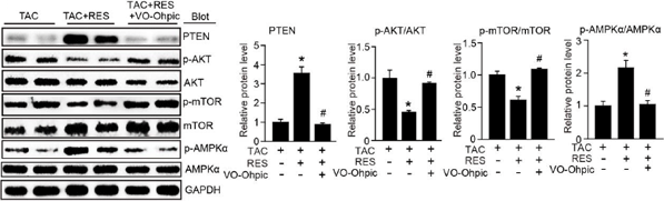
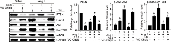
VO-OHPic purchased from MedChemExpress. Usage Cited in: Redox Biol. 2019 Jan:20:390-401. [Abstract]
Western analysis of related genes expression in mice with the treatment of TAC, TAC+RES, and TAC+RES+VO-Ophic.
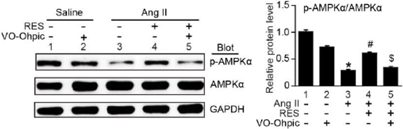
VO-OHPic purchased from MedChemExpress. Usage Cited in: Redox Biol. 2019 Jan:20:390-401. [Abstract]
Immunoblotting analysis show that RES treatment markedly inhibited Ang II-induced degradation of PTEN, activation of AKT and mTOR and inactivation of AMPK, but this effect is reversed by VO-OHpic.
VO-OHPic purchased from MedChemExpress. Usage Cited in: Redox Biol. 2019 Jan:20:390-401. [Abstract]
Immunoblotting analysis show that RES treatment markedly inhibited Ang II-induced degradation of PTEN, activation of AKT and mTOR and inactivation of AMPK, but this effect is reversed by VO-OHpic.
-
J Nanobiotechnology
An orally-administered nanotherapeutics with gold nanospheres supplying for rheumatoid arthritis therapy by re-shaping gut microbial tryptophan metabolism. [Abstract]2025 May 25;23(1):376. PMID: 40414887 -
Theranostics
Spontaneous evolution of human skin fibroblasts into wound-healing keratinocyte-like cells. [Abstract]2019 Jul 9;9(18):5200-5213. PMID: 31410210 -
Theranostics
Embryonic Stem Cells Modulate the Cancer-Permissive Microenvironment of Human Uveal Melanoma. [Abstract]2019 Jul 9;9(16):4764-4778. PMID: 31367256 -
Cell Death Dis
ZC3H15 regulates the ubiquitination of PTEN via recruitment of TRIM56 and promotes malignant progression of non-small cell lung cancer. [Abstract]2026 Jan 9;17(1):17. PMID: 41513632 -
Cell Death Dis
Activation of AKT via a dual mechanism enhances the susceptibility of melanoma cells to glucose deprivation. [Abstract]2025 Aug 7;16(1):595. PMID: 40774947 -
Environ Sci Technol
Tris(1,3-dichloro-2-propyl)phosphate Reduces the Lifespan via Activation of an Unconventional Insulin/Insulin-Like Growth Factor-1 Signaling Pathway. [Abstract]2020 Sep 1;54(17):10783-10796. PMID: 32786597 -
Cell Death Dis
S-nitrosylation of the Peroxiredoxin-2 promotes S-nitrosoglutathione-mediated lung cancer cells apoptosis via AMPK-SIRT1 pathway. [Abstract]2019 Apr 15;10(5):329. PMID: 30988280 -
Phytomedicine
Isovitexin alleviates hepatic fibrosis by regulating miR-21-mediated PI3K/Akt signaling and glutathione metabolic pathway: based on transcriptomics and metabolomics. [Abstract]2023 Dec:121:155117. PMID: 37820467 -
Int J Mol Med
Upregulation of NFKBIZ affects bladder cancer progression via the PTEN/PI3K/Akt signaling pathway. [Abstract]2021 Jun;47(6):109. PMID: 33907827 -
Clin Transl Med
PARK7 deficiency inhibits fatty acid β-oxidation via PTEN to delay liver regeneration after hepatectomy. [Abstract]2022 Sep;12(9):e1061. PMID: 36149763 -
Clin Transl Med
A selected small molecule prevents inflammatory osteolysis through restraining osteoclastogenesis by modulating PTEN activity. [Abstract]2020 Dec;10(8):e240. PMID: 33377656 -
Stem Cell Res Ther
Mouse embryonic palatal mesenchymal cells maintain stemness through the PTEN-Akt-mTOR autophagic pathway. [Abstract]2019 Jul 29;10(1):217. PMID: 31358051
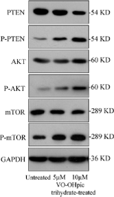
VO-OHPic purchased from MedChemExpress. Usage Cited in: Stem Cell Res Ther. 2019 Jul 29;10(1):217. [Abstract]
Western blot showing the levels of phosphorylated and non-phosphorylated PTEN, AKT, and mTOR proteins before and after VO-OHpic trihydrate treatment.
-
Transl Psychiatry
2021 Mar 26;11(1):185. PMID: 33771972 -
Biochem Pharmacol
Catalpol suppresses osteoclastogenesis and attenuates osteoclast-derived bone resorption by modulating PTEN activity. [Abstract]2020 Jan:171:113715. PMID: 31751538 -
Biochem Pharmacol
2,5-Dimethylcelecoxib prevents isoprenaline-induced cardiomyocyte hypertrophy and cardiac fibroblast activation by inhibiting Akt-mediated GSK-3 phosphorylation. [Abstract]2019 Oct:168:82-90. PMID: 31229551 -
Commun Biol
DTX1-mediated degradation of TUBB3 in Kupffer cells mitigates hepatocellular carcinoma progression by regulating M1/M2 polarization. [Abstract]2026 Jan 23;9(1):311. PMID: 41577987 -
Cell Biol Toxicol
Glycolysis regulates palatal mesenchyme proliferation through Pten-Glut1 axis via Pten classical and non-classical pathways. [Abstract]2025 Feb 27;41(1):53. PMID: 40014184 -
-
Int J Mol Sci
PTEN Inhibitor Treatment Lowers Muscle Plasma Membrane Damage and Enhances Muscle ECM Homeostasis after High-Intensity Eccentric Exercise in Mice. [Abstract]2023 Jun 9;24(12):9954. PMID: 37373102 -
Int Immunopharmacol
USP20 mitigates ischemic stroke in mice by suppressing neuroinflammation and neuron death via regulating PTEN signal. [Abstract]2022 Feb:103:107840. PMID: 34953448 -
-
Mol Med Rep
LncRNA LINC00961 regulates endothelial‑mesenchymal transition via the PTEN‑PI3K‑AKT pathway. [Abstract]2022 Jul;26(1):246. PMID: 35656895 -
Biochim Biophys Acta Mol Basis Dis
Mycobacterium tuberculosis ESAT6 modulates host innate immunity by downregulating miR-222-3p target PTEN. [Abstract]2022 Jan 1;1868(1):166292. PMID: 34710568 -
Mol Med Rep
miR‑371b‑5p inhibits endothelial cell apoptosis in monocrotaline‑induced pulmonary arterial hypertension via PTEN/PI3K/Akt signaling pathways. [Abstract]2018 Dec;18(6):5489-5501. PMID: 30387816 -
Mol Cell Biochem
S-equol promotes ferroptosis in triple negative breast cancer by coordinating NCOA4-mediated ferritinophagy and PPARγ-mediated lipid metabolism. [Abstract]2026 May;481(5):2119-2137. PMID: 41925800 -
J Cell Mol Med
Aspirin inhibits adipogenesis of tendon stem cells and lipids accumulation in rat injury tendon through regulating PTEN/PI3K/AKT signalling. [Abstract]2019 Nov;23(11):7535-7544. PMID: 31557405 -
iScience
Epidermal growth factor augments the self-renewal capacity of aged hematopoietic stem cells. [Abstract]2024 Jun 19;27(7):110306. PMID: 39055915 -
Mol Nutr Food Res
Resveratrol Attenuates Pressure Overload-Induced Cardiac Fibrosis and Diastolic Dysfunction via PTEN/AKT/Smad2/3 and NF-κB Signaling Pathways. [Abstract]2019 Dec;63(24):e1900418. PMID: 31655498 -
BMC Cancer
Numb inhibits epithelial-mesenchymal transition via RBP-Jκ-dependent Notch1/PTEN/FAK signaling pathway in tongue cancer. [Abstract]2019 Apr 25;19(1):391. PMID: 31023264 -
Funct Integr Genomics
MATN2 overexpression suppresses tumor growth in ovarian cancer via PTEN/PI3K/AKT pathway. [Abstract]2024 Apr 3;24(2):71. PMID: 38568332 -
Cancer Med
Embryonic stem cell microenvironment suppresses the malignancy of cutaneous melanoma cells by down-regulating PI3K/AKT pathway. [Abstract]2019 Aug;8(9):4265-4277. PMID: 31173492 -
Mol Vis
2018 Jul 23:24:485-494. PMID: 30967746
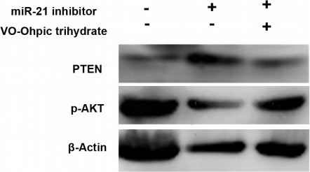
VO-OHPic purchased from MedChemExpress. Usage Cited in: Mol Vis. 2018 Jul 23:24:485-494. [Abstract]
The miR-21 inhibitor promotes the expression of the PTEN protein and inhibits the expression of p-AKT, and VO-Ohpic trihydrate reverses the effect.
-
-
-
-
-
Eur Rev Med Pharmacol Sci
2019 May;23(10):4406-4413. PMID: 31173315


Reinheit & Dokumentation
Verweise
[1]. Li Z, et al. PTEN signaling inhibitor VO-OHpic improves cardiac myocyte survival by mediating apoptosis resistance in vitro. Biomed Pharmacother. 2018;103:1217-1222. [Content Brief]
[2]. Wang W, et al. PTEN inhibitor VO-OHpic suppresses TSC2-/- MEFs proliferation by excessively inhibiting autophagy via the PTEN/PRAS40 pathway. Exp Ther Med. 2020;19(6):3565-3570. [Content Brief]
[3]. Cui X, et al. PTEN inhibitor VO-OHpic protects endplate chondrocytes against apoptosis and calcification via activating Nrf-2 signaling pathway. Aging (Albany NY). 2023;15(6):2275-2292. [Content Brief]
[4]. Lin Y, et al. VO-OHpic attenuates intervertebral disc degeneration via PTEN/Akt pathway. Eur Rev Med Pharmacol Sci. 2020;24(6):2811-2819. [Content Brief]
[5]. Yao X, et al. PTEN inhibitor VO-OHpic attenuates GC-associated endothelial progenitor cell dysfunction and osteonecrosis of the femoral head via activating Nrf2 signaling and inhibiting mitochondrial apoptosis pathway. Stem Cell Res Ther. 2020;11(1):140. Published 2020 Mar 30. [Content Brief]
[6]. Mak LH, et al. Characterisation of the PTEN inhibitor VO-OHpic. J Chem Biol. 2010;3(4):157-163. [Content Brief]

Calculators
Konzentration (Stammlösung) × Volumen (Stammlösung) = Konzentration (Ziellösung) × Volumen (Ziellösung)


VO-OHPic Related Classifications
HY-110067 Related Classifications
 Powered by Bioz
Keywords
- VO-OHPic
- 675848-25-6
- PTEN
- Apoptosis
- Autophagy
- Ferroptosis
- Reactive Oxygen Species (ROS)
- Interleukin Related
- Akt
- Keap1-Nrf2
- Akt-GSK3β
- Nrf-2/HO-1
- primary mouse CEP chondrocytes
- human nucleus pulposus cells
- rat endothelial progenitor cells
- acute myocardial infarction
- intervertebral disc degeneration
- adult rat cardiac myocytes
- TSC2-/- MEFs
- Inhibitor
- inhibitor
- inhibit