



10-OAHSA

Based on 1 Customer Validation
10-OAHSA is one of fatty acid esters of hydroxy fatty acids (FAHFAs). 10-POHSA increases glucose-stimulated insulin secretion (GSIS) at a high glucose concentration. 10-OAHSA reduces LPS (HY-D1056)-induced Tnf-α secretion in bone marrow-derived dendritic cells (BMDCs).
Nos produits utilisent uniquement pour la recherche. Nous ne vendons pas aux patients.
- Pureté : 99.0%
- CAS No.: 2005487-34-1
- Formule: C36H68O4
- Masse moléculaire:564.92
-
Stockage:
Solution, -20°C, 2 years


To place orders, for customer services and technical support, please contact: MedChemExpress USA
Tel: 609-228-6898 E-mail: [email protected] [email protected]


-
 Activité biologique
-
Chemical Information
- Protocole
- Pureté et documentation
- Références
-
 Help & FAQs


-
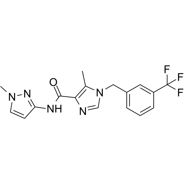
HY-141525
SCD1 inhibitor-4SCD1 Inhibitor -

HY-158651
10-SAHSAEndogenous Lipid -

HY-158625
10(R)-PAHSAEndogenous Lipid -

HY-158626
10(S)-PAHSAEndogenous Lipid -
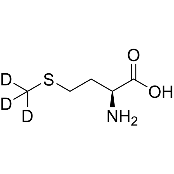
HY-N0326S7
L-Methionine-d3Stable Isotope -
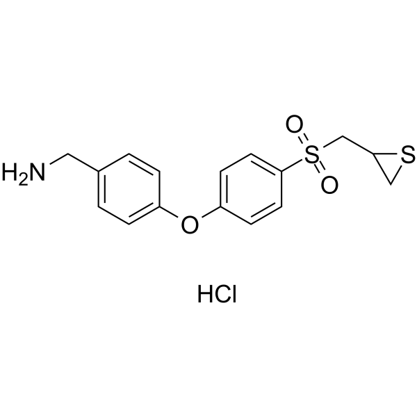
HY-124373
ND-336MMP Inhibitor -

HY-136796
12-PAHSAFatty Ester -

HY-Y1624
1,1'-(Azodicarbonyl)-dipiperidineBiochemical Assay Reagents -
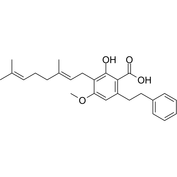
HY-113631
Amorfrutin BPPARγ agonist -
HY-N6181
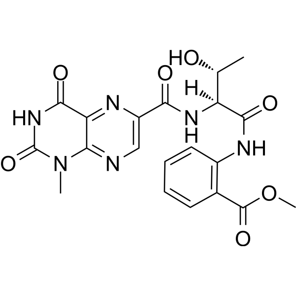
Terrelumamide A -

HY-145530
(S)-Hydroxychloroquine sulfateIsomer of Hydroxychloroquine -

HY-B1370E
(R)-Hydroxychloroquine phosphate(R)-isomer of Hydroxychloroquine
Voir tous les produits spécifiques à Isoform Endogenous Metabolite
MoreVoir tous les produits spécifiques à Isoform TNF Receptor
More

Activité biologique
Description


Chemical Information
-
CAS No. 2005487-34-1
-
Appearance Liquid
-
Masse moléculaire 564.92
-
Formule C36H68O4
-
Color Colorless to light yellow
-
SMILES
CCCCCCCC/C=C\CCCCCCCC(OC(CCCCCCCC)CCCCCCCCC(O)=O)=O
-
Livraison
Room temperature in continental US; may vary elsewhere.
-
Stockage
Solution, -20°C, 2 years


Pureté et documentation
-
Fiche technique (263 KB)
-
SDS (251 KB)
- English - EN (251 KB)
- Français - FR (251 KB)
- Deutsch - DE (251 KB)
- Norwegian - NO (251 KB)
- Español - ES (251 KB)
- Swedish - SV (251 KB)
- Italian - IT (251 KB)
- Korean - KR (251 KB)
- Portuguese - PT (251 KB)
-
Instruction de manipulation (2659 KB)
Références

Calculators
Concentration (start) × Volume (start) = Concentration (final) × Volume (final)


10-OAHSA Related Classifications
HY-158650 Related Classifications
 Powered by Bioz