



Y-320
Based on 7 publication(s) in Google Scholar
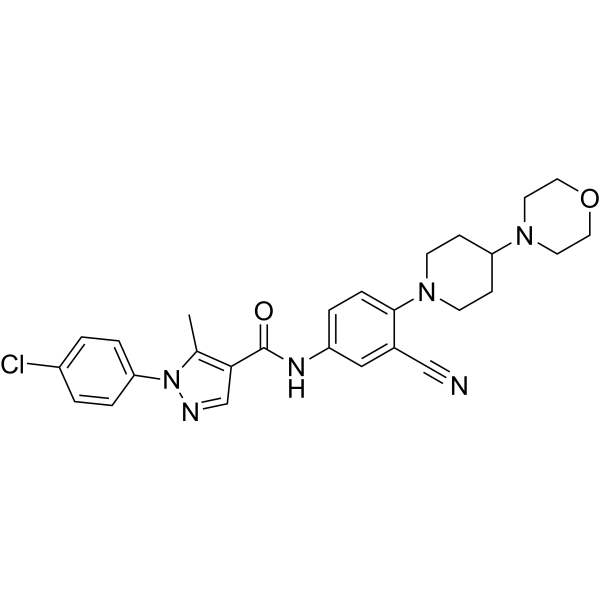
Y-320 is a potent, orally active phenylpyrazoleanilide immunomodulator. Y-320 inhibits IL-17 production by CD4 T cells stimulated with IL-15 with IC50 values of 20 to 60 nM. Y-320 enhances TP53, DMD, and COL17A1 PTC readthrough by G418 and increases cellular protein levels and protein synthesis. Y-320 concomitants use of with a low dose of Paclitaxel (HY-B0015) significantly sensitized multidrug resistance (MDR) tumors by inducing G2/M phase arrest and apoptosis. Y-320 can be used for research of rheumatoid arthritis (RA) and cancer.
Nos produits utilisent uniquement pour la recherche. Nous ne vendons pas aux patients.
- Pureté : 99.03%
- CAS No.: 288250-47-5
- Formule: C27H29ClN6O2
- Masse moléculaire:505.01
-
Stockage:Powder -20°C, 3 years , 4°C, 2 years ; In solvent -80°C, 2 years , -20°C, 1 year

To place orders, for customer services and technical support, please contact: MedChemExpress USA
Tel: 609-228-6898 E-mail: [email protected] [email protected]


-
 Activité biologique
-
Chemical Information
-
Solvant et solubilité
- Protocole
- Pureté et documentation
- Références
-
 Help & FAQs


-
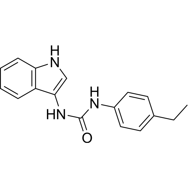
HY-112693
H-151STING Antagonist -
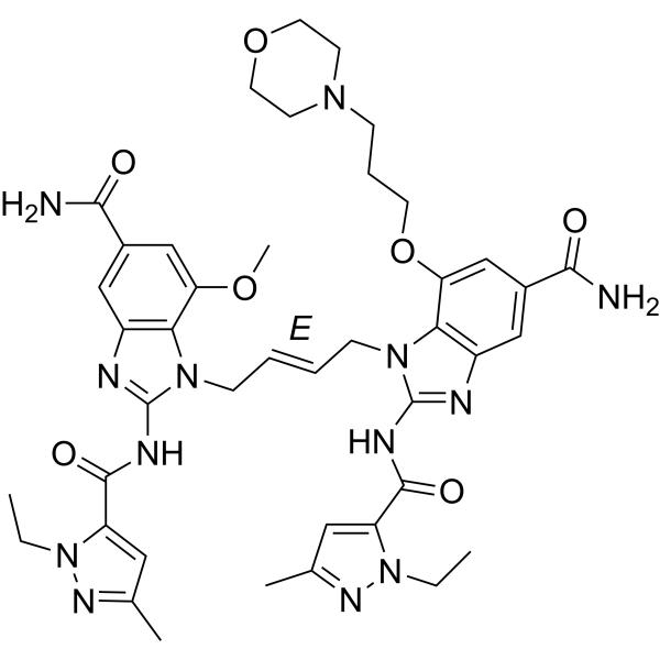
HY-112921A
diABZI STING agonist-1STING Agonist -
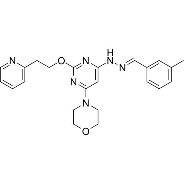
HY-14644
ApilimodIL-12/IL-23 Inhibitor -
HY-16668
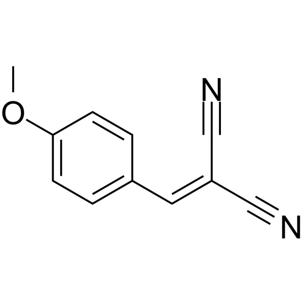
Tyrphostin A1Interleukin Related Inhibitor -
HY-14644A
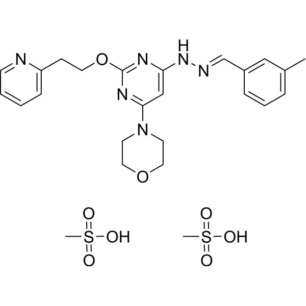
Apilimod mesylateIL-12/IL-23 Inhibitor -
HY-15614
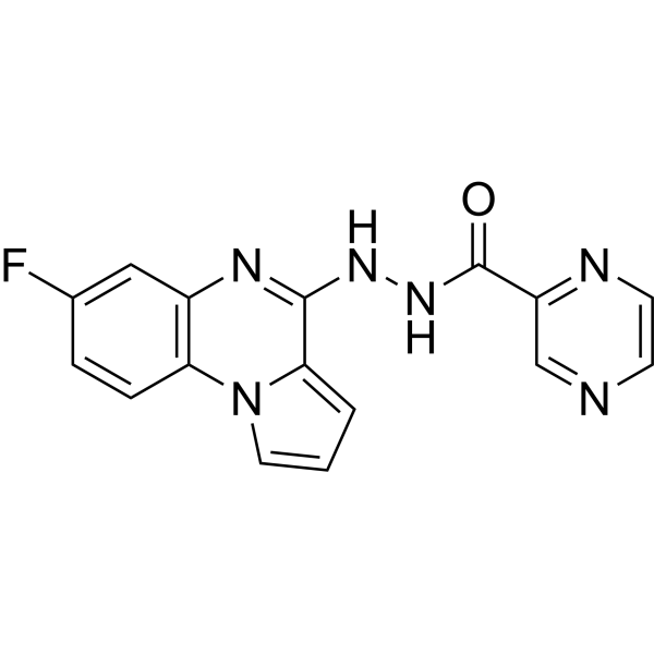
SC144GP130 Inhibitor -
HY-17002
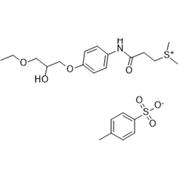
Suplatast (Tosilate)Th2 Cytokine Inhibitor -
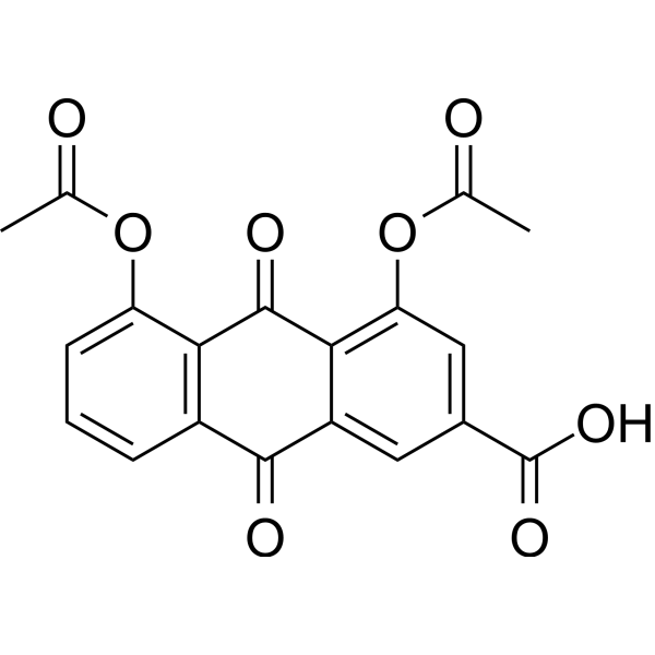
HY-N0283
DiacereinIL-1 Inhibitor -

HY-16668R
Tyrphostin A1 (Standard)Interleukin Related Inhibitor -
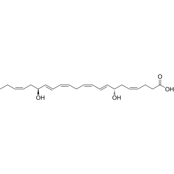
HY-145491
Resolvin D5Inflammation Inhibitor -
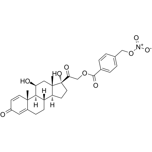
HY-101757
NO-prednisolonePrednisolone Derivative -
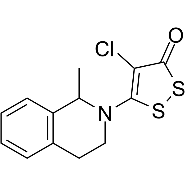
HY-101716
RP-54745
Publications Citing Use of MedChemExpress (MCE) Y-320
More- Cell Chem Biol. 2026 Apr 27:S2451-9456(26)00110-8. [Abstract]
- Int J Biol Macromol. 2024 May;266(Pt 1):130939. [Abstract]
- Ecotoxicol Environ Saf. 2025 Sep 15:303:118796. [Abstract]
- PLoS Genet. 2025 Oct 13;21(10):e1011907. [Abstract]
- Am J Transl Res. 2020 Feb 15;12(2):551-562. [Abstract]
- bioRxiv. 2026 Jun 26.
- bioRxiv. 2020 Jun.


Activité biologique
Description
IC50 & Target
[1]|
IL-15 |
IL-17 |
In Vitro
Y-320 (0-100 nM; 48 h) inhibits IL-17 production by murine and human CD4 T Cells stimulated with IL-15 with IC50 values of 25.7, 52.4 and 57.4 nM for murine CD4 T cells, murine Th17 cells and human CD4 T cells, respectively[1].
Y-320 (0-100 nM; 48 h) inhibits phosphorylation of JAK1/JAK3 in murine CD4 T cells stimulated with IL-15/CXCL12/anti-CD3 mAb[1].
Y-320 (0.25-2 μM; 48 h) enhances PTC readthrough by G418 in different cell lines[2].
Y-320 (0-2 μM; 48 h; HDQ-P1 cells) increases cellular protein levels and ribosome biogenesis in a concentration-dependent manner[2].
Y-320 (0-2 μM; 48 h; Tsc2-/- cells) causes a small decrease in phospho-S6K combination with G418 (100 μM)[2].
Y-320 (1 μM; 48 h; HDQ-P1 cells) up-regulates CXC chemokine expression including CXCL10, CXCL8, and CXCL2[2].
Y-320 (500 nM; 72 h) reverses the resistance to paclitaxel in MDR cancer cells. Y-320 has the reversal index (RI) combined with Paclitaxel (0-1000 nM) are 5.5 (Bads-200), 9.4 (Bats-72) and 1.7 (Huh7-TS-48)[3].
Y-320 (500 nM; 72 h; Bads-200 cells) enhances Paclitaxel-induced G2/M arrest and enhances Paclitaxel-induced (500 nM) tumor cell apoptosis[3].
Y-320 (0-20 μM; 72 h; Bads-200 cells) is a substrate of P-gp reverses MDR by inhibiting P-gp function[3].
MedChemExpress (MCE) has not independently confirmed the accuracy of these methods. They are for reference only.
-
Cell Line:Bads-200 cells
-
Concentration:500 nM
-
Incubation Time:72 hours
-
Result:Increased the percentage of cells at G2/M phase, from 6.3% to 42.5%.
-
Cell Line:Bads-200 cells
-
Concentration:500 nM
-
Incubation Time:72 hours
-
Result:Increased the ratio of apoptotic Bads-200 cells (30.8% versus 2.2%).
In Vivo
Y-320 (5 mg/kg; i.v.; every three days, for 18 d; Homozygous nude athymic mice with Bats-72 xenograft) sensitizes MDR xenograft tumor to Paclitaxel in vivo[3].
MedChemExpress (MCE) has not independently confirmed the accuracy of these methods. They are for reference only.
-
Animal Model:Type II collagen-induced arthritis (CIA) in DBA/1J mice[1]
-
Dosage:0, 0.1, 0.3, 1, and 3 mg/kg
-
Administration:Oral administration; daily, for 42 days
-
Result:Inhibited the development of CIA and the increase in paw thickness in a dose-dependent manner.
Inhibited joint destructions in a dose-dependent manner.
Improved inflammation and damage in the arthritic ankle joints in CIA mice.
-
Animal Model:Homozygous nude athymic mice with Bats-72 xenograft (female, 4-5 weeks old)[3]
-
Dosage:5 mg/kg; Paclitaxel (5 mg/kg)
-
Administration:Intravenous injection; every three days, for 18 days
-
Result:Inhibited tumor growth in Bats-72 xenografts without severe adverse effects.


Chemical Information
-
CAS No. 288250-47-5
-
Appearance Solid
-
Masse moléculaire 505.01
-
Formule C27H29ClN6O2
-
Color Off-white to pink
-
SMILES
O=C(C1=C(C)N(C2=CC=C(Cl)C=C2)N=C1)NC3=CC=C(N4CCC(N5CCOCC5)CC4)C(C#N)=C3
-
Livraison
Room temperature in continental US; may vary elsewhere.
-
Stockage
Powder -20°C 3 years 4°C 2 years In solvent -80°C 2 years -20°C 1 year


Publications (7)
-
Journal Impact Factor
-
Most Recent
-
Cell Chem Biol
2026 Apr 27:S2451-9456(26)00110-8. PMID: 42049033 -
Int J Biol Macromol
2024 May;266(Pt 1):130939. PMID: 38493816 -
Ecotoxicol Environ Saf
Vitamin C alleviates PM2.5-induced thyroid injury and ferroptosis by regulating the IL-17A/IL-17RA/ACT1 signaling pathway. [Abstract]2025 Sep 15:303:118796. PMID: 40753780 -
PLoS Genet
Genotype-phenotype characterization and functional reconstitution of pathogenic β-catenin variants from CTNNB1 syndrome patients. [Abstract]2025 Oct 13;21(10):e1011907. PMID: 41082542 -
Am J Transl Res
2020 Feb 15;12(2):551-562. PMID: 32194903 -
-


Solvant et solubilité
In Vitro:
DMSO : 5.5 mg/mL (10.89 mM; Need ultrasonic; Hygroscopic DMSO has a significant impact on the solubility of product, please use newly opened DMSO)
Please refer to the solubility information to select the appropriate solvent. Once prepared, please aliquot and store the solution to prevent product inactivation from repeated freeze-thaw cycles.
Storage method and period of stock solution: -80°C, 2 years; -20°C, 1 year. When stored at -80°C, please use it within 2 years. When stored at -20°C, please use it within 1 year.
Please refer to the solubility information to select the appropriate solvent. Once prepared, please aliquot and store the solution to prevent product inactivation from repeated freeze-thaw cycles.
Storage method and period of stock solution: -80°C, 2 years; -20°C, 1 year. When stored at -80°C, please use it within 2 years. When stored at -20°C, please use it within 1 year.
Concentration (start) × Volume (start) = Concentration (final) × Volume (final)
In Vivo:
Select the appropriate dissolution method based on your experimental animal and administration route.
- For the following dissolution methods, please ensure to first prepare a clear stock solution using an In Vitro approach and then sequentially add co-solvents:
- To ensure reliable experimental results, the clarified stock solution can be appropriately stored based on storage conditions. As for the working solution for In Vivo experiments, it is recommended to prepare freshly and use it on the same day.
- The percentages shown for the solvents indicate their volumetric ratio in the final prepared solution. If precipitation or phase separation occurs during preparation, heat and/or sonication can be used to aid dissolution.
Add each solvent one by one: 10% DMSO 40% PEG300 5% Tween-80 45% Saline
Solubility: ≥ 0.5 mg/mL (0.99 mM); Clear solution
This protocol yields a clear solution of ≥ 0.5 mg/mL (saturation unknown).
Taking 1 mL working solution as an example, add 100 μL DMSO stock solution (5.0 mg/mL) to 400 μL PEG300, and mix evenly; then add 50 μL Tween-80 and mix evenly; then add 450 μL Saline to adjust the volume to 1 mL.
Preparation of Saline: Dissolve 0.9 g sodium chloride in ddH₂O and dilute to 100 mL to obtain a clear Saline solution.
Add each solvent one by one: 10% DMSO 90% (20% SBE-β-CD in Saline)
Solubility: ≥ 0.5 mg/mL (0.99 mM); Clear solution
This protocol yields a clear solution of ≥ 0.5 mg/mL (saturation unknown).
Taking 1 mL working solution as an example, add 100 μL DMSO stock solution (5.0 mg/mL) to 900 μL 20% SBE-β-CD in Saline, and mix evenly.
Preparation of 20% SBE-β-CD in Saline (4°C, storage for one week): 2 g SBE-β-CD powder is dissolved in 10 mL Saline, completely dissolve until clear.
In Vivo Dissolution Calculator
Please enter the basic information of animal experiments:
-
-
-
-
Recommended: Prepare an additional quantity of animals to account for potential losses during experiments.
Please enter your animal formula composition:
-
%DMSO +
Recommended: Keep the proportion of DMSO in working solution below 2% if your animal is weak.
-
%+
-
+%Tween-80 + +
-
%Saline +
The co-solvents required include: DMSO, . All of co-solvents are available by MedChemExpress (MCE). , Tween 80. All of co-solvents are available by MedChemExpress (MCE).
Working solution concentration: 0.22 mg/mL
Method for preparing stock solution: mg drug dissolved in μL DMSO. Stock solution concentration: mg/mL.
1. Take μL DMSO stock solution;
2. Add μL .
μL , mix evenly;
3. Then add μL Tween 80, mix evenly;
4. Then add μL
Please ensure that the stock solution in the first step is dissolved to a clear state, and add co-solvents in sequence. You can use ultrasonic heating (ultrasonic cleaner, recommended frequency 20-40 kHz), vortexing, etc. to assist dissolution.


Pureté et documentation
-
Fiche technique (278 KB)
-
SDS (393 KB)
- English - EN (393 KB)
- Français - FR (393 KB)
- Deutsch - DE (393 KB)
- Norwegian - NO (393 KB)
- Español - ES (393 KB)
- Swedish - SV (393 KB)
- Italian - IT (393 KB)
- Korean - KR (393 KB)
- Portuguese - PT (393 KB)
-
Instruction de manipulation (2659 KB)
Références
[1]. Ushio H, et, al. A new phenylpyrazoleanilide, y-320, inhibits interleukin 17 production and ameliorates collagen-induced arthritis in mice and cynomolgus monkeys. Pharmaceuticals (Basel). 2013 Dec 23;7(1):1-17. [Content Brief]
[2]. Hosseini-Farahabadi S, et, al. Small molecule Y-320 stimulates ribosome biogenesis, protein synthesis, and aminoglycoside-induced premature termination codon readthrough. PLoS Biol. 2021 May 3;19(5):e3001221. [Content Brief]
[3]. Hong J, et, al. Y-320, a novel immune-modulator, sensitizes multidrug-resistant tumors to chemotherapy. Am J Transl Res. 2020 Feb 15;12(2):551-562. [Content Brief]


Complete Stock Solution Preparation Table
Please refer to the solubility information to select the appropriate solvent. Once prepared, please aliquot and store the solution to prevent product inactivation from repeated freeze-thaw cycles.
Storage method and period of stock solution: -80°C, 2 years; -20°C, 1 year. When stored at -80°C, please use it within 2 years. When stored at -20°C, please use it within 1 year.
| Optional Solvent | Concentration Solvent Mass | 1 mg | 5 mg | 10 mg | 25 mg |
|---|---|---|---|---|---|
| DMSO | 1 mM | 1.9802 mL | 9.9008 mL | 19.8016 mL | 49.5040 mL |
| 5 mM | 0.3960 mL | 1.9802 mL | 3.9603 mL | 9.9008 mL | |
| 10 mM | 0.1980 mL | 0.9901 mL | 1.9802 mL | 4.9504 mL |


Y-320 Related Classifications
HY-15898 Related Classifications
 Powered by Bioz