



Crocetin
Based on 3 publication(s) in Google Scholar
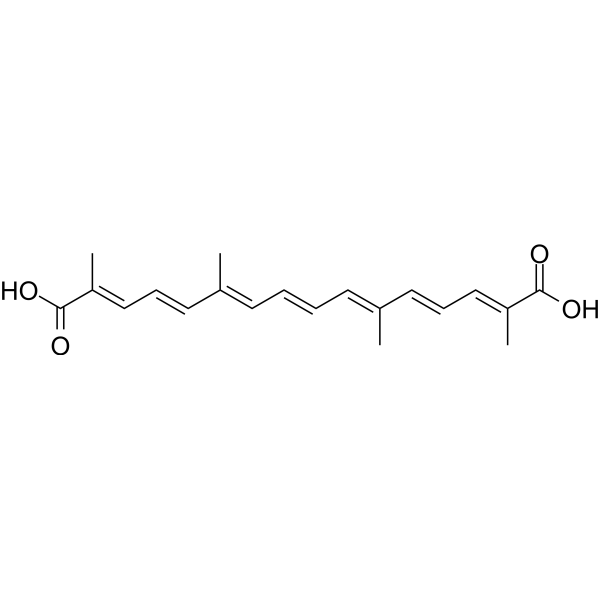
Crocetin (Transcrocetin) is an aglycone of crocin. Crocetin is an orally active and brain-penetrant. Crocetin shows strong NMDA receptor affinity and channel opening activity. Crocetin can downregulate the proinflammatory cytokines and COX-2 exoression. Crocetin can inhibit apoptosis and activation of MAPK. Crocetin can delay delays brain and body aging. Crocetin can be used for the researches of cancer, neurological disease and inflammation, such as cervical cancer and ischemia.
For research use only. We do not sell to patients.
- Purity : 98.07%
- CAS No.: 27876-94-4
- Formula: C20H24O4
- Molecular Weight:328.40
-
Storage:Powder -20°C, 3 years , 4°C, 2 years
* The compound is unstable in solutions, freshly prepared is recommended.

To place orders, for customer services and technical support, please contact: MedChemExpress USA
Tel: 609-228-6898 E-mail: [email protected] [email protected]


-
 Biological Activity
-
Chemical Information
-
Solvent & Solubility
- Protocols
- Purity & Documentation
- References
-
 Help & FAQs


-
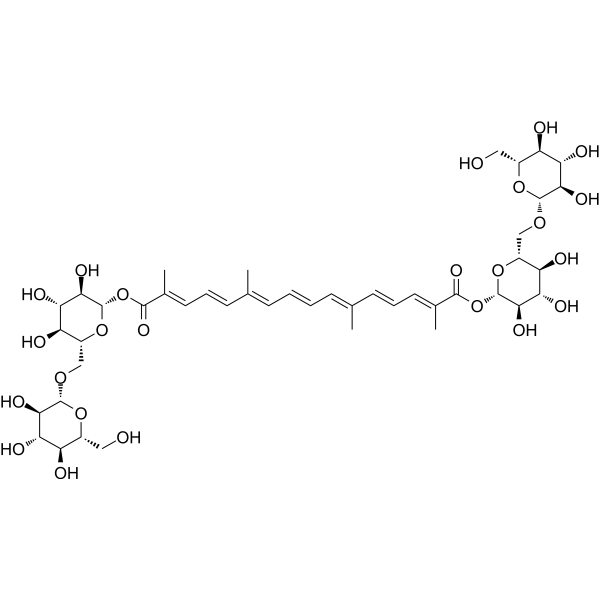
HY-N0697
CrocinEndogenous Metabolite -
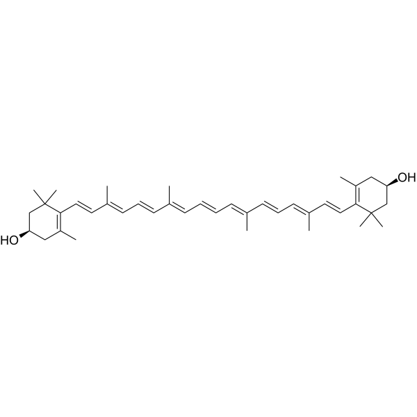
HY-120318
ZeaxanthinDiet-obtained Carotenoid -
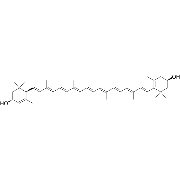
HY-N6947
LuteinAnti-inflammatory Agent -
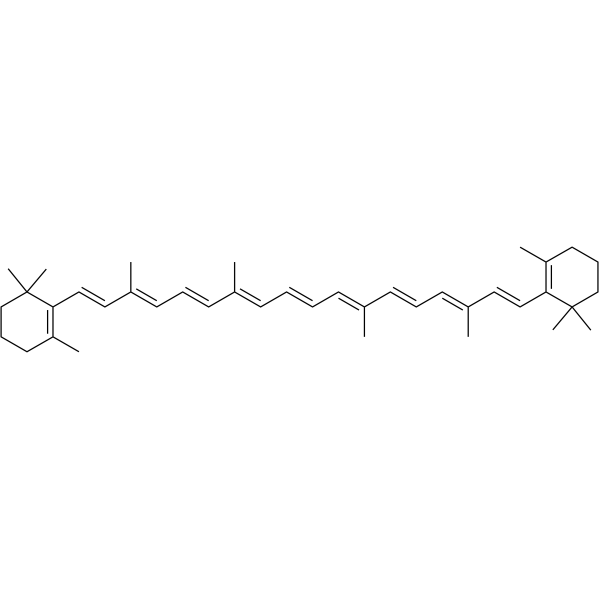
HY-N0411
β-CaroteneCarotenoid compound -
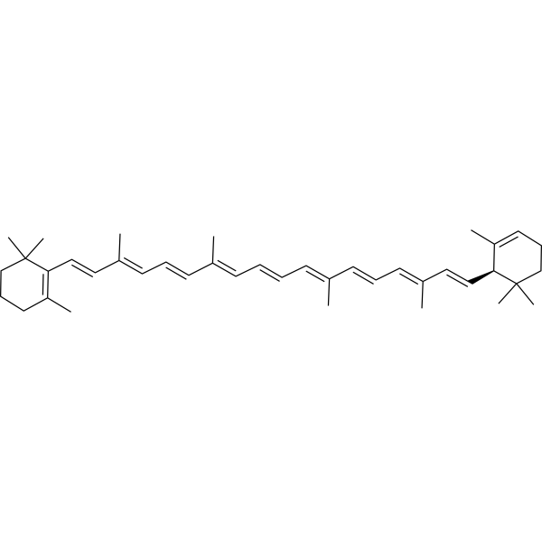
HY-113462
α-CaroteneVitamin A Precursor -
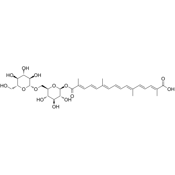
HY-N6644
Crocin IIICrocin -
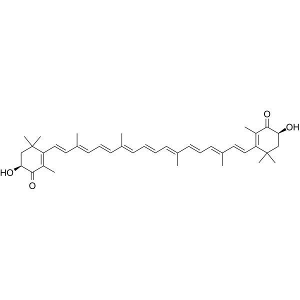
HY-B2163
AstaxanthinAntioxidant -

HY-N2072R
Crocetin (Standard)Anticancer/Neuroprotective Agent -
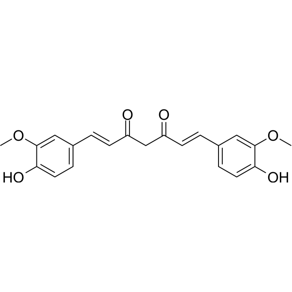
HY-N0005
Curcuminp300 Histone Acetylatransferase Inhibitor -
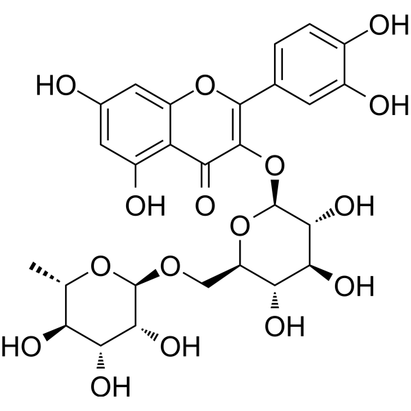
HY-N0148
RutinCBR1 Inhibitor -
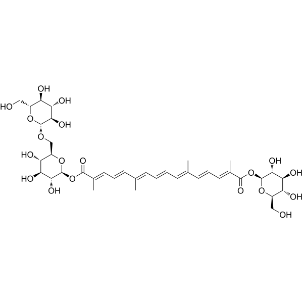
HY-N0698
Crocin IIα-synuclein Inhibitor -
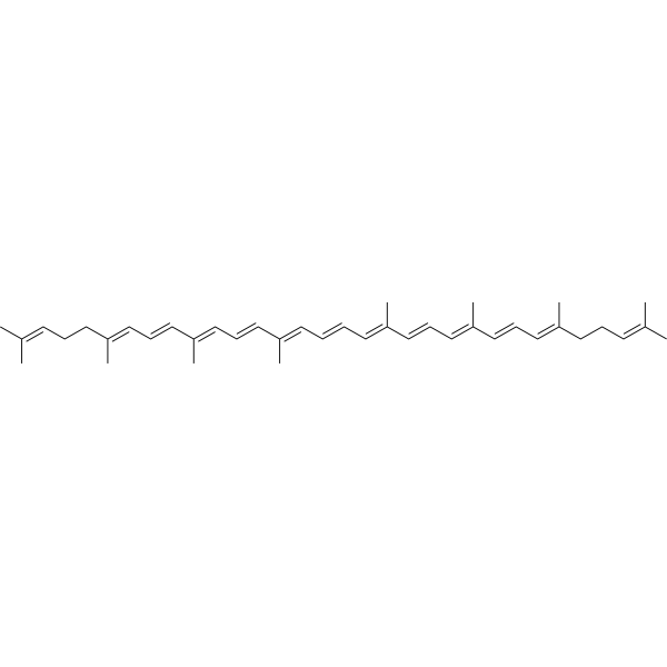
HY-N0287
LycopeneAntioxidant
Publications Citing Use of MedChemExpress (MCE) Crocetin
More Customer Validation & Images

-
Cell Proliferation/Viability Assay

-
IF

-
Histological Imaging/Staining

-
Flow Cytometry

All iGluR Isoforms
More

Biological Activity
Description
IC50 & Target
[1]|
NMDA Receptor |
COX-2 |
Cellular Effect
|
Cell Line

|
Type | Value | Description | References |
|---|---|---|---|---|
| RAW264.7 | IC50 |
5.99 μM
Compound: 18
|
Inhibition of nitric oxide production in lipopolysaccharide-activated mouse RAW264.7 cells by Griess reaction based method
Inhibition of nitric oxide production in lipopolysaccharide-activated mouse RAW264.7 cells by Griess reaction based method
|
[PMID: 23305920] |
In Vitro
Crocetin (1-100 μM, 24 h) inhibits COX-2 expression in HeLa cells[1].
MedChemExpress (MCE) has not independently confirmed the accuracy of these methods. They are for reference only. Further protocols information, click here.
-
Cell Line:HeLa cells
-
Concentration:1, 10, 50 and 100 μM
-
Incubation Time:24 h
-
Result:Reduced COX-2 mRNA levels.
In Vivo
Crocetin (20 mg/kg, p.o., 1 h before ischemia) inhibits retinal ischemic damage in ischemia/reperfusion-induced mice models[2].
Crocetin (25 mg/mL, p.o., daily) delays brain and body aging in aged C57BL/6J mice[3].
MedChemExpress (MCE) has not independently confirmed the accuracy of these methods. They are for reference only.
-
Animal Model:Methylcholanthrene- (MCA)-induced uterine cervix tumor mice models[1]
-
Dosage:10, 20 and 40 mg/kg
-
Administration:Orally administration, twice a day for 35 days
-
Result:Decreased MDA levels.
Decreased PMNs activity.
Reduced IL-1β, TNF-α and iNOS levels.
-
Animal Model:Ischemia/reperfusion-induced mice models[2]
-
Dosage:20 mg/kg
-
Administration:Orally administration, 1 h before ischemia
-
Result:Prevented the reduction in IPL thickness.
Prevented the induction of cell apoptosis.
Inhibited functional damage and DNA oxidation in the retina.
Prevented the activation of MAPK and redox-sensitive transcription factors in the retina.
-
Animal Model:Aged C57BL/6J mice[3]
-
Dosage:25 mg/mL
-
Administration:Orally administration, daily
-
Result:Increased ATP and NAD+ levels.
Upregulated the electron transport chain to yield more energy.
Restored the ETC function of mitochondria.
Attenuated the basal level of oxidative stress.


Chemical Information
-
CAS No. 27876-94-4
-
Appearance Solid
-
Molecular Weight 328.40
-
Formula C20H24O4
-
Color Yellow to orange
-
SMILES
O=C(O)/C(C)=C/C=C/C(C)=C/C=C/C=C(C)/C=C/C=C(C)/C(O)=O
-
Synonyms
Transcrocetin; trans-Crocetin
-
Structure Classification
-
Initial Source
-
Shipping
Room temperature in continental US; may vary elsewhere.
-
Storage
Powder -20°C 3 years 4°C 2 years * The compound is unstable in solutions, freshly prepared is recommended.


Publications (3)
-
Journal Impact Factor
-
Most Recent
-
Int J Pharm X
RGD peptide-functionalized micelles loaded with crocetin ameliorate doxorubicin-induced cardiotoxicity. [Abstract]2025 Feb 26:9:100326. PMID: 40103672
Crocetin purchased from MedChemExpress. Usage Cited in: Int J Pharm X. 2025 Feb 26:9:100326. [Abstract]
The protective analysis of RGD@M(Cro) nanoparticles containing different Crocetin (Cro) centrations (0, 2.5, 5, 10, 20, 40 μM) in HL-1 cells via CCK8 assay.
Crocetin purchased from MedChemExpress. Usage Cited in: Int J Pharm X. 2025 Feb 26:9:100326. [Abstract]
TUNEL analysis of cell apoptosis treated with Cro, M(Cro), or RGD@ M(Cro) containing 40 μM Crocetin (Cro).
Crocetin purchased from MedChemExpress. Usage Cited in: Int J Pharm X. 2025 Feb 26:9:100326. [Abstract]
The mice were administered Crocetin (Cro), M(Cro), or RGD@M(Cro) (20 mg/kg) for one week, followed by a single intraperitoneal injection of Dox (15 mg/kg) on the seventh day. H&E staining and Masson staining of cardiac tissue in mice.
Crocetin purchased from MedChemExpress. Usage Cited in: Int J Pharm X. 2025 Feb 26:9:100326. [Abstract]
Flow cytometric analysis of HL-1 cells treated with M(Cro) or RGD@M(Cro) nanomicelles containing 20 μM Crocetin (Cro) for 2 h at 37 °C.
-
Int Immunopharmacol
Rutin ameliorates hepatic ischemia-reperfusion injury by targeting CD36 to suppress hepatocyte ferroptosis. [Abstract]2026 Mar 1:172:116231. PMID: 41576568 -
J Phys Chem B
2026 Jan 8;130(1):205-214. PMID: 41404815


Solvent & Solubility
In Vitro:
DMSO : 20 mg/mL (60.90 mM; Need ultrasonic; Hygroscopic DMSO has a significant impact on the solubility of product, please use newly opened DMSO)
Please refer to the solubility information to select the appropriate solvent. The compound is unstable in solutions, freshly prepared is recommended.
Please refer to the solubility information to select the appropriate solvent. The compound is unstable in solutions, freshly prepared is recommended.
Concentration (start) × Volume (start) = Concentration (final) × Volume (final)

Protocols
-
Apoptosis
Apoptosis, also called programmed cell death, is generally characterized by distinct morphological characteristics.
-
TUNEL staining for apoptotic DNA fragmentation
TUNEL staining detects DNA strand breaks by using terminal deoxynucleotidyl transferase to add labeled nucleotides to exposed 3′-OH DNA termini, generating either microscopic staining in fixed cells or tissue sections, or fluorescence/cytometric signal in cell suspensions. TUNEL positivity reflects DNA fragmentation but should not be interpreted alone as definitive apoptosis, because TUNEL can also label necrotic, autolytic, mechanically damaged, or DNA-repair-associated DNA breaks.
-
Annexin V plus membrane-impermeant dye apoptosis staining
Annexin V-based apoptosis assays rely on the detection of phosphatidylserine (PS) externalization from the inner leaflet of the plasma membrane to the outer leaflet, an early biochemical hallmark of apoptosis. Fluorescently labeled Annexin V binds PS in a calcium-dependent manner, enabling identification of early apoptotic cells by flow cytometry or fluorescence microscopy. When combined with a membrane-impermeant DNA-binding dye (e. g. , propidium iodide), this approach allows discrimination between viable (Annexin V−/dye−), early apoptotic (Annexin V+/dye−), and late apoptotic or necrotic (Annexin V+/dye+) cell populations by assessing membrane integrity and PS exposure.
-
Apoptosis Solutions
Apoptosis is a regulated, generally non-lytic cell-death pathway that removes unwanted, damaged, infected, or abnormal cells through coordinated morphological changes, caspase activation, DNA fragmentation, and membrane remodeling. The intrinsic apoptosis pathway is controlled mainly by mitochondrial outer membrane permeabilization, BCL-2 family proteins, cytochrome c release, apoptosome formation, caspase-9 activation, and downstream executioner caspase-3/7 activation. The extrinsic apoptosis pathway is initiated by death receptors such as Fas, TNFR, and TRAIL receptors, which recruit adaptor proteins and activate caspase-8 before engaging executioner caspases or mitochondrial amplification through BID cleavage. Apoptosis is linked to many phenotypes, including cancer cell killing, tissue homeostasis, immune regulation, neurodegeneration, infection response, and treatment-induced cytotoxicity; unresolved questions include how apoptosis interacts with necroptosis, pyroptosis, ferroptos
-
Research Protocol for Inflammation-related Diseases
The NLRP3 inflammasome is a cytosolic innate immune signaling platform that integrates priming signals and danger-signal activation to promote caspase-1 activation, maturation of IL-1β and IL-18, and gasdermin D-mediated pyroptotic cell death. The core experimental logic is to determine whether inflammatory disease phenotypes are driven by increased NLRP3 expression, ASC-containing inflammasome assembly, caspase-1 cleavage, GSDMD cleavage, and extracellular release of IL-1β/IL-18 rather than by nonspecific cell injury alone. The pathway is strongly linked to inflammation-related disease phenotypes because monosodium urate crystals activate NALP3/NLRP3 inflammasome signaling in gout-like crystal inflammation, cholesterol crystals activate NLRP3 inflammasomes in atherogenesis models, and DSS-induced intestinal inflammation has been reported to involve NLRP3 inflammasome activity. However, experimental colitis studies also show context-dependent protective effects of NLRP3 inflammasome co
-
Research Protocol for Neurological Diseases
PINK1/Parkin-mediated mitophagy pathway is a mitochondrial quality-control signaling axis in which mitochondrial depolarization stabilizes PINK1 on damaged mitochondria, activates Parkin recruitment and E3 ubiquitin ligase activity, promotes ubiquitination of outer mitochondrial membrane proteins, recruits selective autophagy adaptors, and drives lysosomal degradation of damaged mitochondria. In neurological disease research, this pathway is experimentally important because neurons, especially dopaminergic neurons, are highly dependent on mitochondrial integrity, and defective mitochondrial turnover can lead to mitochondrial dysfunction, oxidative stress, impaired neuronal survival, α-synuclein accumulation, and neuroinflammatory damage-associated signals. The genetic disease link is strongest in Parkinson’s disease because mutations in PRKN/parkin cause autosomal recessive juvenile parkinsonism, mutations in PINK1 cause hereditary early-onset Parkinson’s disease, and Drosophila studie


Purity & Documentation
-
Data Sheet (274 KB)
-
SDS (393 KB)
- English - EN (393 KB)
- Français - FR (393 KB)
- Deutsch - DE (393 KB)
- Norwegian - NO (393 KB)
- Español - ES (393 KB)
- Swedish - SV (393 KB)
- Italian - IT (393 KB)
- Korean - KR (393 KB)
- Portuguese - PT (393 KB)
-
Handling Instructions (2659 KB)
References
[1]. Chen B, et al. Crocetin downregulates the proinflammatory cytokines in methylcholanthrene-induced rodent tumor model and inhibits COX-2 expression in cervical cancer cells. Biomed Res Int. 2015;2015:829513. [Content Brief]
[2]. Crocetin, a carotenoid derivative, inhibits retinal ischemic damage in mice. Eur J Pharmacol. 2013 Mar 5;703(1-3):1-10. [Content Brief]
[3]. Choudhary S, et al. Crocetin Delays Brain and Body Aging by Increasing Cellular Energy Levels in Aged C57BL/6J Mice. ACS Pharmacol Transl Sci. 2024 Sep 11;7(10):3017-3033. [Content Brief]
[4]. Lautenschläger M, et al. Intestinal formation of trans-crocetin from saffron extract (Crocus sativus L.) and in vitro permeation through intestinal and blood brain barrier. Phytomedicine. 2015 Jan 15;22(1):36-44. [Content Brief]


Complete Stock Solution Preparation Table
Please refer to the solubility information to select the appropriate solvent. The compound is unstable in solutions, freshly prepared is recommended.
| Optional Solvent | Concentration Solvent Mass | 1 mg | 5 mg | 10 mg | 25 mg |
|---|---|---|---|---|---|
| DMSO | 1 mM | 3.0451 mL | 15.2253 mL | 30.4507 mL | 76.1267 mL |
| 5 mM | 0.6090 mL | 3.0451 mL | 6.0901 mL | 15.2253 mL | |
| 10 mM | 0.3045 mL | 1.5225 mL | 3.0451 mL | 7.6127 mL | |
| 15 mM | 0.2030 mL | 1.0150 mL | 2.0300 mL | 5.0751 mL | |
| 20 mM | 0.1523 mL | 0.7613 mL | 1.5225 mL | 3.8063 mL | |
| 25 mM | 0.1218 mL | 0.6090 mL | 1.2180 mL | 3.0451 mL | |
| 30 mM | 0.1015 mL | 0.5075 mL | 1.0150 mL | 2.5376 mL | |
| 40 mM | 0.0761 mL | 0.3806 mL | 0.7613 mL | 1.9032 mL | |
| 50 mM | 0.0609 mL | 0.3045 mL | 0.6090 mL | 1.5225 mL | |
| 60 mM | 0.0508 mL | 0.2538 mL | 0.5075 mL | 1.2688 mL |


Crocetin Related Classifications
HY-N2072 Related Classifications
 Powered by Bioz