



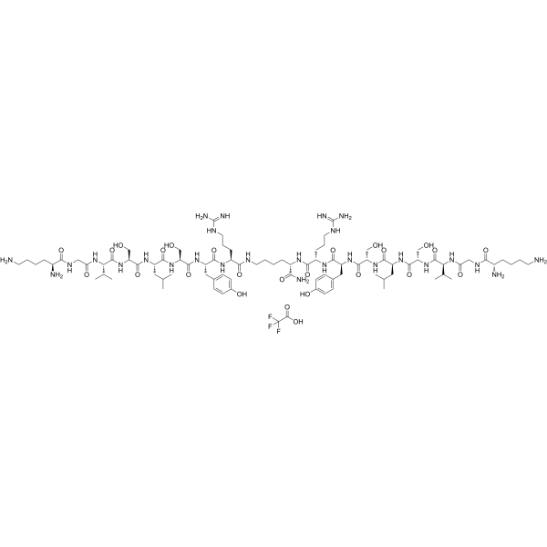
CTCE-9908 TFA
Based on 1 publication(s) in Google Scholar
CTCE-9908 TFA is a potent and selective CXCR4 antagonist. CTCE-9908 TFA induces mitotic catastrophe, cytotoxicity and inhibits migration in CXCR4-expressing ovarian cancer cells.
Nur für Forschungszwecke. Wir verkaufen nicht an Patienten.
- Formel: C88H148F3N27O25
- Molecular Weight:2041.28
-
Speicherung:
Please store the product under the recommended conditions in the Certificate of Analysis.

To place orders, for customer services and technical support, please contact: MedChemExpress USA
Tel: 609-228-6898 E-mail: [email protected] [email protected]


-
 Biologische Aktivität
-
Chemical Information
-
Lösungsmittel & Löslichkeit
- Protokoll
- Reinheit & Dokumentation
- Verweise
-
 Help & FAQs


Publications Citing Use of MedChemExpress (MCE) CTCE-9908 TFA
More

Biologische Aktivität
Beschreibung
IC50 & Target
[1]|
CXCR4 |
In Vitro
CTCE-9908 TFA (0-300 μg/mL; for 10 d) inhibits migration and growth in CXCR4-expressing in ovarian cancer cell lines (IGROV, TOV21G and SKOV3). CTCE-9908 TFA inhibits ovarian cancer cell migration to CXCL12. CTCE-9908 TFA does not cause apoptosis or cellular senescence, but induces multinucleation, G2-M arrest, and abnormal mitosis in ovarian cancer cells. CTCE-9908 TFA deregulates DNA damage checkpoint proteins and spindle assembly checkpoint proteins at G2-M phases of the cell cycle[1].
MedChemExpress (MCE) has not independently confirmed the accuracy of these methods. They are for reference only.
In Vivo
MedChemExpress (MCE) has not independently confirmed the accuracy of these methods. They are for reference only.


Chemical Information
-
Molecular Weight 2041.28
-
Formel C88H148F3N27O25
-
Sequence
Sequence 1:Lys-Gly-Val-Ser-Leu-Ser-Tyr-Arg-Lys-NH2;Sequence 1':Lys-Gly-Val-Ser-Leu-Ser-Tyr-Arg (Amide bridge:Lys9-Arg8’)
-
Sequence Shortening
Sequence 1:KGVSLSYRK-NH2;Sequence 1':KGVSLSYR (Amide bridge:Lys9-Arg8’)
-
Versand
Room temperature in continental US; may vary elsewhere.
-
Speicherung
Please store the product under the recommended conditions in the Certificate of Analysis.


Publications (1)
-
Journal Impact Factor
-
Most Recent


Lösungsmittel & Löslichkeit
In Vitro
H2O
Peptide Solubility and Storage Guidelines:
1. Calculate the length of the peptide.
2. Calculate the overall charge of the entire peptide according to the following table:
| Contents | Assign value | |
|---|---|---|
| Acidic amino acid | Asp (D), Glu (E), and the C-terminal -COOH. | -1 |
| Basic amino acid | Arg (R), Lys (K), His (H), and the N-terminal -NH2 | +1 |
| Neutral amino acid | Gly (G), Ala (A), Leu (L), Ile (I), Val (V), Cys (C), Met (M), Thr (T), Ser (S), Phe (F), Tyr (Y), Trp (W), Pro (P), Asn (N), Gln (Q) | 0 |
3. Recommended solution:
| Overall charge of peptide | Details |
|---|---|
| Negative (<0) |
1. Try to dissolve the peptide in water first. 2. If water fails, add NH4OH (<50 μL). 3. If the peptide still does not dissolve, add DMSO (50-100 μL) to solubilize the peptide. |
| Positive (>0) |
1. Try to dissolve the peptide in water first. 2. If water fails, try dissolving the peptide in a 10%-30% acetic acid solution. 3. If the peptide still does not dissolve, try dissolving the peptide in a small amount of DMSO. |
| Zero (=0) |
1. Try to dissolve the peptide in organic solvent (acetonitrile, methanol, etc.) first. 2. For very hydrophobic peptides, try dissolving the peptide in a small amount of DMSO, and then dilute the solution with water to the desired concentration. |


Reinheit & Dokumentation
Verweise
[1]. Joseph Kwong, et al. An antagonist of the chemokine receptor CXCR4 induces mitotic catastrophe in ovarian cancer cells. Mol Cancer Ther. 2009 Jul;8(7):1893-905. [Content Brief]
[2]. Saima Hassan, et al. CXCR4 peptide antagonist inhibits primary breast tumor growth, metastasis and enhances the efficacy of anti-VEGF treatment or docetaxel in a transgenic mouse model. Int J Cancer. 2011 Jul 1;129(1):225-32. [Content Brief]

Calculators
Konzentration (Stammlösung) × Volumen (Stammlösung) = Konzentration (Ziellösung) × Volumen (Ziellösung)


CTCE-9908 TFA Related Classifications
HY-P1103A Related Classifications
 Powered by Bioz