



Cysteinylglycine

Based on 1 Customer Validation
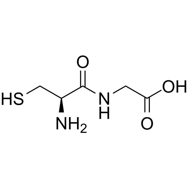
Cysteinylglycine (L-Cysteinylglycine; Cys-Gly) is a dipeptide formed by the peptide bond linkage between cysteine (Cysteine) and glycine (Glycine). Cysteinylglycine is an important metabolic intermediate in the human body, mainly derived from the degradation of glutathione (GSH). Cysteinylglycine reduces ferric iron to ferrous iron, drives the redox cycle of iron, generates reactive oxygen species (ROS), stimulates oxidative reactions, induces lipid peroxidation of human plasma LDL lipoproteins, and causes oxidative damage to DNA bases. Cysteinylglycine can be used as a biomarker to evaluate ischemic heart disease, breast cancer and other conditions.
For research use only. We do not sell to patients.
- Purity : 99.79%
- CAS No.: 19246-18-5
- Formula: C5H10N2O3S
- Molecular Weight:178.21
-
Storage:
Sealed storage, away from moisture and light, under nitrogen.
Powder -80°C, 2 years , -20°C, 1 year* In solvent : -80°C, 6 months; -20°C, 1 month (sealed storage, away from moisture and light, under nitrogen)

To place orders, for customer services and technical support, please contact: MedChemExpress USA
Tel: 609-228-6898 E-mail: [email protected] [email protected]


-
 Biological Activity
-
Chemical Information
-
Solvent & Solubility
- Protocols
- Purity & Documentation
- References
-
 Help & FAQs


-
HY-113402
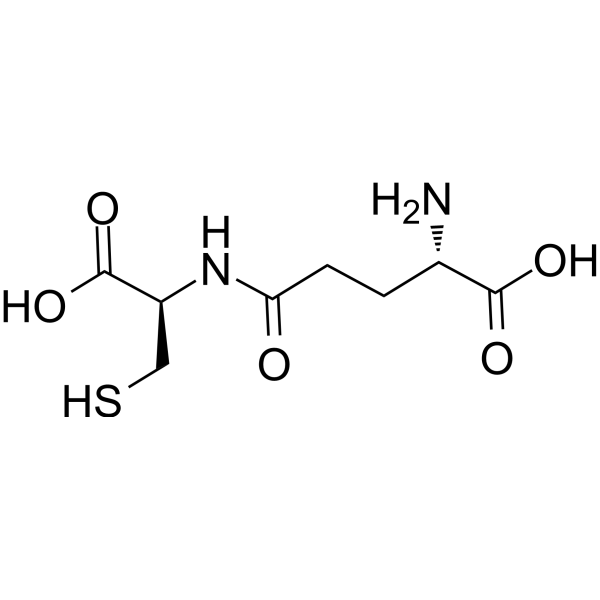
Gamma-glutamylcysteineDipeptide -
HY-D0187
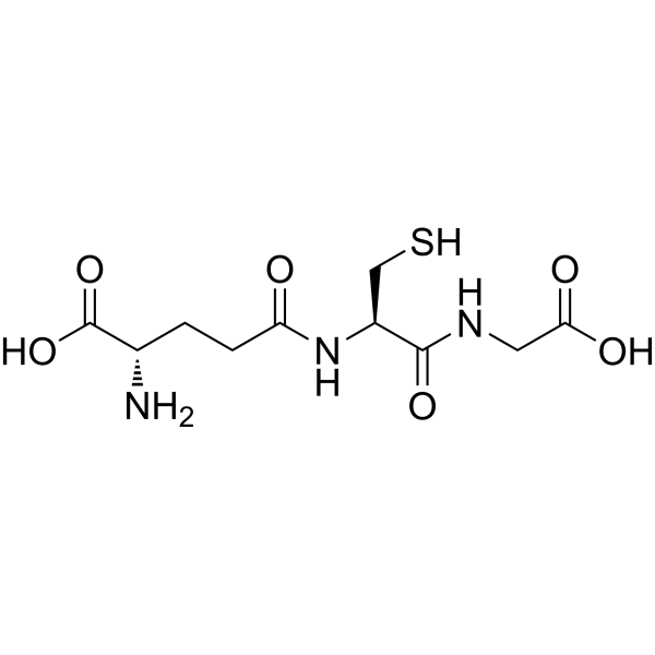
L-Glutathione reducedAntioxidant -
HY-Y0337
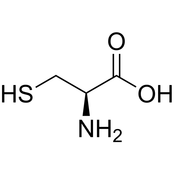
L-CysteineAnorectic Agent -

HY-113110R
Cysteinylglycine (Standard)Standard of Cysteinylglycine -
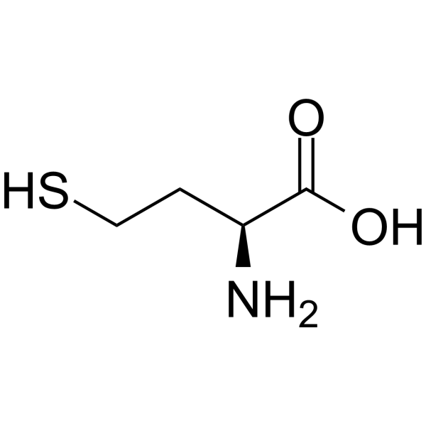
HY-W010347
L-HomocysteineAmino Acid -

HY-13259
MG-132Proteasome Inhibitor -
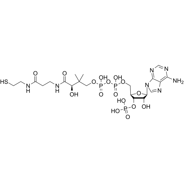
HY-128851
Coenzyme ACofactor -
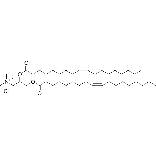
HY-112754A
DOTAP chlorideCationic Lipid -
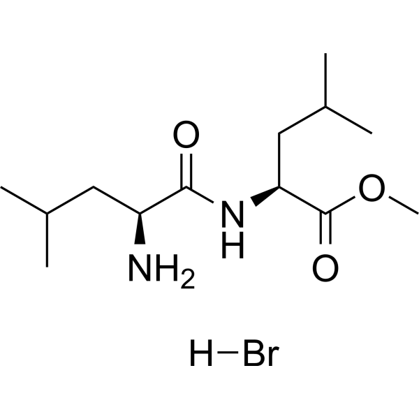
HY-129905A
L-Leucyl-L-Leucine methyl ester hydrobromideDipeptide condensation product -
HY-B1065
AceglutamideAutophagy Inducer -
HY-12056
BIX02189MEK5 Inhibitor -
HY-W053787
1-Methylhistamine dihydrochloride


Biological Activity
Description
IC50 & Target
|
Human Endogenous Metabolite |
Cellular Effect
|
Cell Line

|
Type | Value | Description | References |
|---|---|---|---|---|
| KB 3-1 | IC50 |
9.42 mM
Compound: Cys-Gly
|
Cytotoxicity against human KB-3-1 cells incubated for 72 hrs by MTT assay
Cytotoxicity against human KB-3-1 cells incubated for 72 hrs by MTT assay
|
[PMID: 21657271] |
| KB-V1 | IC50 |
4.23 mM
Compound: Cys-Gly
|
Cytotoxicity against drug-resistant human KBV1 cells expressing P-gp incubated for 72 hrs by MTT assay
Cytotoxicity against drug-resistant human KBV1 cells expressing P-gp incubated for 72 hrs by MTT assay
|
[PMID: 21657271] |
In Vitro
Cysteinylglycine (48 h; 10 μM-50 μM) shows a significant 1.2- to 1.7-fold increase in human astrocytoma U373-MG cells after 48 h of treatment with various Nrf2 activators, compared with control cells[1].
Cysteinylglycine (0.78-25 μM; incubated at 20℃ in the dark for 10 min) can be accurately quantified by this reversed-phase ion-pair liquid chromatography method, with a linear response range of 0.78 to 25 μM, a limit of detection < 2 pmol, and the adduct can be stably stored at -80℃ for 14 days[2].
MedChemExpress (MCE) has not independently confirmed the accuracy of these methods. They are for reference only. Further protocols information, click here.


Chemical Information
-
CAS No. 19246-18-5
-
Appearance Solid
-
Molecular Weight 178.21
-
Formula C5H10N2O3S
-
Color White to off-white
-
Synonyms
L-Cysteinylglycine; Cys-Gly; H-Cys-Gly-OH
-
Structure Classification
-
Shipping
Room temperature in continental US; may vary elsewhere.
-
Storage
Sealed storage, away from moisture and light, under nitrogen
Powder -80°C 2 years -20°C 1 year * In solvent : -80°C, 6 months; -20°C, 1 month (sealed storage, away from moisture and light, under nitrogen)


Solvent & Solubility
In Vitro:
H2O : 175 mg/mL (981.99 mM; Need ultrasonic)
Please refer to the solubility information to select the appropriate solvent. Once prepared, please aliquot and store the solution to prevent product inactivation from repeated freeze-thaw cycles.
Storage method and period of stock solution: -80°C, 6 months; -20°C, 1 month (sealed storage, away from moisture and light, under nitrogen). When stored at -80°C, please use it within 6 months. When stored at -20°C, please use it within 1 month.
* Note: If you choose water as the stock solution, please dilute it to the working solution, then filter and sterilize it with a 0.22 μm filter before use.
Please refer to the solubility information to select the appropriate solvent. Once prepared, please aliquot and store the solution to prevent product inactivation from repeated freeze-thaw cycles.
Storage method and period of stock solution: -80°C, 6 months; -20°C, 1 month (sealed storage, away from moisture and light, under nitrogen). When stored at -80°C, please use it within 6 months. When stored at -20°C, please use it within 1 month.
* Note: If you choose water as the stock solution, please dilute it to the working solution, then filter and sterilize it with a 0.22 μm filter before use.
Concentration (start) × Volume (start) = Concentration (final) × Volume (final)

Protocols
-
ROS/oxidative-stress fluorescent staining
ROS/oxidative-stress fluorescent staining uses cell-permeant fluorogenic probes that become fluorescent after oxidation inside cells or tissues; commonly used examples include DCFH-DA/DCFDA for broad cellular oxidant detection, DHE for superoxide-related signal detection, MitoSOX for mitochondrial superoxide-related signal detection, and CellROX probes for oxidative-stress-associated fluorescence readouts. The assay detects probe oxidation rather than a single ROS species unless the probe and analysis method have been chemically validated for that species. DCFH-DA enters cells, is deacetylated by intracellular esterases to DCFH, and produces fluorescent DCF after oxidation, so the readout is used as an operational measure of total cellular oxidative stress rather than a species-specific ROS measurement. DHE and MitoSOX can report superoxide-related oxidation, but red fluorescence alone can include non-specific ethidium-like oxidation products; HPLC or optimized spectral approaches are
-
Research Protocol for Cardiovascular Diseases
Cardiovascular disease can be modeled as maladaptive cardiac remodeling, where ischemic injury or pressure overload activates inflammatory signaling, fibroblast activation, extracellular-matrix deposition, cardiomyocyte hypertrophy, vascular remodeling, and progressive ventricular dysfunction. The TGF-β/SMAD axis is a central profibrotic pathway after myocardial injury and pressure overload, while innate immune and cytokine pathways regulate leukocyte recruitment, scar formation, and adverse remodeling. Key unresolved questions include which inflammatory signals are reparative versus harmful, when fibrosis is protective versus maladaptive, and whether pathway inhibition improves function without weakening necessary infarct healing or compensatory remodeling.
-
Ferroptosis Solutions
Ferroptosis is an iron-dependent, non-apoptotic form of regulated cell death characterized by lethal lipid peroxidation and sensitivity to suppression by iron chelators or lipophilic radical-trapping antioxidants. The core pathway links cystine uptake through system Xc−, glutathione availability, GPX4-dependent detoxification of phospholipid hydroperoxides, iron-dependent oxidative reactions, and polyunsaturated-phospholipid metabolism into a cell-death program that is biochemically and morphologically distinct from apoptosis, necrosis, and autophagy. The ferroptosis pathway is experimentally linked to phenotype through chemical and genetic perturbation. Erastin induces ferroptosis by inhibiting cystine uptake through system Xc− and weakening antioxidant defenses, while GPX4 inhibition or depletion causes lipid peroxide accumulation and ferroptotic cancer-cell death. ACSL4 and oxidizable arachidonoyl- or adrenoyl-containing phosphatidylethanolamines shape ferroptosis sensitivity by con
-
Perls' Prussian Blue Iron Staining
Perls' Prussian blue staining is a histochemical method used to detect non-heme ferric iron (Fe3+) in biological tissues by exploiting an acid-mediated release of loosely bound iron from storage complexes such as ferritin or hemosiderin, followed by its reaction with potassium ferrocyanide to form an insoluble blue ferric ferrocyanide (Prussian blue) precipitate that marks iron localization under light microscopy. The reaction is classically performed under acidic conditions, which liberate Fe3+ ions that subsequently bind ferrocyanide to generate the visible chromogen, enabling spatial visualization of iron deposits in tissues such as brain, liver, and spleen. Histochemical interpretations are limited to a reactive iron pool rather than total iron content, reflecting only histologically accessible iron species rather than tightly protein-bound iron.
-
Breast Cancer Modeling
Breast cancer is a heterogeneous cancer, and it has been distinguished into four subtypes: luminal A, luminal B, HER2-positive and basal-like. Molecular mutations, epigenetic alterations, hormone exposure and immune microenvironment are related to the progression of breast cancer.


Purity & Documentation
-
Data Sheet (271 KB)
-
SDS (701 KB)
- English - EN (701 KB)
- Français - FR (701 KB)
- Deutsch - DE (701 KB)
- Norwegian - NO (701 KB)
- Español - ES (701 KB)
- Swedish - SV (701 KB)
- Italian - IT (701 KB)
- Korean - KR (701 KB)
- Portuguese - PT (701 KB)
-
Handling Instructions (2659 KB)
References
[1]. Steele ML, et al. Effect of Nrf2 activators on release of glutathione, cysteinylglycine and homocysteine by human U373 astroglial cells. Redox Biol. 2013;1(1):441-445. Published 2013 Sep 12. [Content Brief]
[2]. Mansoor MA, et al. Determination of the in vivo redox status of cysteine, cysteinylglycine, homocysteine, and glutathione in human plasma. Anal Biochem. 1992;200(2):218-229. [Content Brief]
[3]. Lin J, et al. Plasma cysteinylglycine levels and breast cancer risk in women. Cancer Res. 2007;67(23):11123-11127. [Content Brief]
[4]. Houghton SC, et al. Plasma B-vitamin and one-carbon metabolites and risk of breast cancer before and after folic acid fortification in the United States. Int J Cancer. 2019;144(8):1929-1940. [Content Brief]


Complete Stock Solution Preparation Table
Please refer to the solubility information to select the appropriate solvent. Once prepared, please aliquot and store the solution to prevent product inactivation from repeated freeze-thaw cycles.
Storage method and period of stock solution: -80°C, 6 months; -20°C, 1 month (sealed storage, away from moisture and light, under nitrogen). When stored at -80°C, please use it within 6 months. When stored at -20°C, please use it within 1 month.
| Optional Solvent | Concentration Solvent Mass | 1 mg | 5 mg | 10 mg | 25 mg |
|---|---|---|---|---|---|
| H2O | 1 mM | 5.6114 mL | 28.0568 mL | 56.1136 mL | 140.2839 mL |
| 5 mM | 1.1223 mL | 5.6114 mL | 11.2227 mL | 28.0568 mL | |
| 10 mM | 0.5611 mL | 2.8057 mL | 5.6114 mL | 14.0284 mL | |
| 15 mM | 0.3741 mL | 1.8705 mL | 3.7409 mL | 9.3523 mL | |
| 20 mM | 0.2806 mL | 1.4028 mL | 2.8057 mL | 7.0142 mL | |
| 25 mM | 0.2245 mL | 1.1223 mL | 2.2445 mL | 5.6114 mL | |
| 30 mM | 0.1870 mL | 0.9352 mL | 1.8705 mL | 4.6761 mL | |
| 40 mM | 0.1403 mL | 0.7014 mL | 1.4028 mL | 3.5071 mL | |
| 50 mM | 0.1122 mL | 0.5611 mL | 1.1223 mL | 2.8057 mL | |
| 60 mM | 0.0935 mL | 0.4676 mL | 0.9352 mL | 2.3381 mL | |
| 80 mM | 0.0701 mL | 0.3507 mL | 0.7014 mL | 1.7535 mL | |
| 100 mM | 0.0561 mL | 0.2806 mL | 0.5611 mL | 1.4028 mL |
* Note: If you choose water as the stock solution, please dilute it to the working solution, then filter and sterilize it with a 0.22 μm filter before use.


Cysteinylglycine Related Classifications
HY-113110 Related Classifications
 Powered by Bioz
Keywords
- Cysteinylglycine
- 19246-18-5
- L-Cysteinylglycine
- Cys-Gly
- H-Cys-Gly-OH
- Endogenous Metabolite
- Reactive Oxygen Species (ROS)
- DNA/RNA Synthesis
- cysteine
- glycine
- human plasma LDL lipoproteins
- one-carbon metabolism
- U373-MG human astrocytoma cells
- Alzheimer’s disease
- neuronal ectopeptidase
- glutathione (GSH)
- astrocyte-derived γ-glutamyl transpeptidase
- Nrf2 activator
- Inhibitor
- inhibitor
- inhibit