



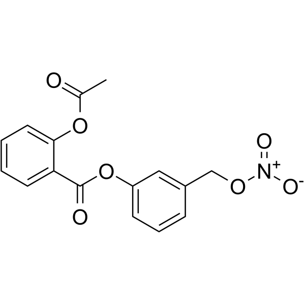
Nitroaspirin

Based on 1 Customer Validation
Nitroaspirin (NCX 4016) is an orally active inhibitor of ARG1 and NOS2. Nitroaspirin reduces ARG1 enzymatic activity via the STAT6-mediated signaling pathway, and feedback-inhibits the catalytic activity of NOS2 in a NO-releasing group-dependent manner. Nitroaspirin continuously releases intracellular NO, inducing redox-dependent loss of cell viability. Nitroaspirin also exerts anti-angiogenic effects by causing endothelial barrier dysfunction, and effectively inhibits the proliferation of Cisplatin (HY-17394)-resistant human ovarian cancer cells. Nitroaspirin can be used in research related to colon cancer, breast cancer and Cisplatin-resistant human ovarian cancer.
For research use only. We do not sell to patients.
- Purity : 98.57%
- CAS No.: 175033-36-0
- Formula: C16H13NO7
- Molecular Weight:331.28
-
Storage:Powder -20°C, 3 years , 4°C, 2 years ; In solvent -80°C, 6 months , -20°C, 1 month

To place orders, for customer services and technical support, please contact: MedChemExpress USA
Tel: 609-228-6898 E-mail: [email protected] [email protected]


-
 Biological Activity
-
Chemical Information
-
Solvent & Solubility
- Protocols
- Purity & Documentation
- References
-
 Help & FAQs




Biological Activity
Description
IC50 & Target
[1]|
STAT6 |
nNOS |
Cellular Effect
|
Cell Line

|
Type | Value | Description | References |
|---|---|---|---|---|
| HT-29 | EC50 |
1 mM
Compound: 2
|
Apoptosis induction in human HT29 cells after 24 hrs
Apoptosis induction in human HT29 cells after 24 hrs
|
[PMID: 17441704] |
In Vitro
Nitroaspirin (NCX 4016) (2 mM; 3 days) restores anti-CD3/anti-CD28-induced proliferation of BALB-c splenocytes inhibited by γ-irradiated MSC-2 myeloid suppressor cells[1].
Nitroaspirin (50-500 μM; 1-3 h) induces dose- and time-dependent loss of redox-dependent viability in BLMVECs, causing up to 74% viability loss at 100 μM after 3 h[2].
Nitroaspirin (100 μM; 2 h) induces a 4.7-fold increase in intracellular NO generation in BLMVECs after 2 h[2].
Nitroaspirin (100 μM; 1-2 h) induces time-dependent endothelial barrier dysfunction in BLMVEC monolayers, as measured by decreased TER after 1 and 2 h of treatment[2].
Nitroaspirin (100-500 μM; 2 h) induces dose-dependent actin cytoskeletal reorganization, including actin stress fiber formation and paracellular gap development, in BLMVECs after 2 h of treatment[2].
Nitroaspirin (25-100 μM; 10 h) inhibits in vitro angiogenesis in a dose-dependent manner in HUVECs and BLMVECs, with near-complete inhibition at 100 μM after 10 h of treatment[2].
MedChemExpress (MCE) has not independently confirmed the accuracy of these methods. They are for reference only.
-
Cell Line:bovine lung microvascular endothelial cells (BLMVECs)
-
Concentration:50-500 μM (2 h); 100 μM (1-3 h)
-
Incubation Time:1-3 h (100 μM); 2 h (50-500 μM)
-
Result:Induced a dose-dependent loss of cell viability: caused a 28% loss at 50 μM, 67% loss at 100 μM, and 70% loss at 500 μM relative to vehicle-treated controls after 2 h.
Caused a 19% loss of viability at 100 μM after 1 h, a 72% loss after 2 h, and a 74% loss after 3 h, with significantly greater viability loss at 2 and 3 h compared to 1 h.
-
Cell Line:bovine lung microvascular endothelial cells (BLMVECs)
-
Concentration:100 μM
-
Incubation Time:2 h (preceded by 5 mM NAC pretreatment for 2 h)
-
Result:Caused a significant loss of cell viability, which was completely attenuated by pretreatment with 5 mM NAC.
Resulted in viability slightly higher (12% increase) than vehicle-treated controls in NAC-pretreated cells.
-
Cell Line:bovine lung microvascular endothelial cells (BLMVECs)
-
Concentration:100-500 μM
-
Incubation Time:2 h
-
Result:Induced dose-dependent formation of actin stress fibers, along with changes in cell morphology and distinctive paracellular gaps indicative of endothelial barrier dysfunction.
In Vivo
MedChemExpress (MCE) has not independently confirmed the accuracy of these methods. They are for reference only.
-
Animal Model:BALB/c (8 weeks old)[1]
-
Dosage:6.25 mg/kg; 12.5 mg/kg; 25 mg/kg; 50 mg/kg
-
Administration:p.o.; twice daily; 9 days
-
Result:Restored compromised alloreactive CTL responses in tumor-bearing mice at 12.5, 25, and 50 mg/kg, with cytotoxicity levels approaching those of tumor-free mice; showed minimal activity at 6.25 mg/kg.
Reduced ARG activity in CD11b+ splenocytes from tumor-bearing mice from ~40 mU/106 cells to ~5 mU/106 cells, equivalent to tumor-free mice.
Reduced NOS activity in CD11b+ splenocytes from tumor-bearing mice from ~25 μM NO2-/NO3- to ~5 μM NO2-/NO3-, equivalent to tumor-free mice.
Completely abrogated intratumoral peroxynitrite production, as evidenced by the near disappearance of nitrotyrosine staining in tumor sections.
Reduced intratumoral CD11b+Gr-1+ myeloid suppressor cell content by ~41% (from 58.5 to 34.57 cells per ×400 field).
Caused only slight tumor growth retardation, with no significant difference in survival rates after treatment cessation.
- Cell Cytotoxicity Assay
- Mammalian live/dead viability and cytotoxicity staining
- ROS/oxidative-stress fluorescent staining
- Transepithelial/transendothelial electrical resistance assay
- CCK-8/WST-8 Cell Proliferation Assay
- MTT Cell Proliferation Assay
- Breast Cancer Modeling
- Cell Viability Determination by MTT Colorimetric Assay
Clinical Trial
| NCT Number | Sponsor | Condition | Start Date |
Phase
|
|---|---|---|---|---|
| NCT01329991 | Plexxikon| | 2011-05 | PHASE1 |


Chemical Information
-
CAS No. 175033-36-0
-
Appearance Solid
-
Molecular Weight 331.28
-
Formula C16H13NO7
-
Color White to off-white
-
SMILES
O=C(OC1=CC=CC(CO[N+]([O-])=O)=C1)C2=CC=CC=C2OC(C)=O
-
Synonyms
NCX 4016
-
Shipping
Room temperature in continental US; may vary elsewhere.
-
Storage
Powder -20°C 3 years 4°C 2 years In solvent -80°C 6 months -20°C 1 month


Solvent & Solubility
In Vitro:
DMSO : 200 mg/mL (603.72 mM; Need ultrasonic; Hygroscopic DMSO has a significant impact on the solubility of product, please use newly opened DMSO)
Please refer to the solubility information to select the appropriate solvent. Once prepared, please aliquot and store the solution to prevent product inactivation from repeated freeze-thaw cycles.
Storage method and period of stock solution: -80°C, 6 months; -20°C, 1 month. When stored at -80°C, please use it within 6 months. When stored at -20°C, please use it within 1 month.
Please refer to the solubility information to select the appropriate solvent. Once prepared, please aliquot and store the solution to prevent product inactivation from repeated freeze-thaw cycles.
Storage method and period of stock solution: -80°C, 6 months; -20°C, 1 month. When stored at -80°C, please use it within 6 months. When stored at -20°C, please use it within 1 month.
Concentration (start) × Volume (start) = Concentration (final) × Volume (final)
In Vivo:
Select the appropriate dissolution method based on your experimental animal and administration route.
- For the following dissolution methods, please ensure to first prepare a clear stock solution using an In Vitro approach and then sequentially add co-solvents:
- To ensure reliable experimental results, the clarified stock solution can be appropriately stored based on storage conditions. As for the working solution for In Vivo experiments, it is recommended to prepare freshly and use it on the same day.
- The percentages shown for the solvents indicate their volumetric ratio in the final prepared solution. If precipitation or phase separation occurs during preparation, heat and/or sonication can be used to aid dissolution.
In Vivo Dissolution Calculator
Please enter the basic information of animal experiments:
-
-
-
-
Recommended: Prepare an additional quantity of animals to account for potential losses during experiments.
Please enter your animal formula composition:
-
%DMSO +
Recommended: Keep the proportion of DMSO in working solution below 2% if your animal is weak.
-
%+
-
+%Tween-80 + +
-
%Saline +
The co-solvents required include: DMSO, . All of co-solvents are available by MedChemExpress (MCE). , Tween 80. All of co-solvents are available by MedChemExpress (MCE).
Working solution concentration: 0.22 mg/mL
Method for preparing stock solution: mg drug dissolved in μL DMSO. Stock solution concentration: mg/mL.
1. Take μL DMSO stock solution;
2. Add μL .
μL , mix evenly;
3. Then add μL Tween 80, mix evenly;
4. Then add μL
Please ensure that the stock solution in the first step is dissolved to a clear state, and add co-solvents in sequence. You can use ultrasonic heating (ultrasonic cleaner, recommended frequency 20-40 kHz), vortexing, etc. to assist dissolution.


Purity & Documentation
-
Data Sheet (289 KB)
-
SDS (254 KB)
- English - EN (254 KB)
- Français - FR (254 KB)
- Deutsch - DE (254 KB)
- Norwegian - NO (254 KB)
- Español - ES (254 KB)
- Swedish - SV (254 KB)
- Italian - IT (254 KB)
- Korean - KR (254 KB)
- Portuguese - PT (254 KB)
-
Handling Instructions (2659 KB)
References
[1]. De Santo C, et al. Nitroaspirin corrects immune dysfunction in tumor-bearing hosts and promotes tumor eradication by cancer vaccination. Proceedings of the National Academy of Sciences of the United States of America. 2005 Mar 15;102(11):4185-90. [Content Brief]
[2]. Parinandi NL, et al. Nitroaspirin (NCX-4016), an NO donor, is antiangiogenic through induction of loss of redox-dependent viability and cytoskeletal reorganization in endothelial cells. Antioxidants & redox signaling. 2007 Nov;9(11):1837-49. [Content Brief]


Complete Stock Solution Preparation Table
Please refer to the solubility information to select the appropriate solvent. Once prepared, please aliquot and store the solution to prevent product inactivation from repeated freeze-thaw cycles.
Storage method and period of stock solution: -80°C, 6 months; -20°C, 1 month. When stored at -80°C, please use it within 6 months. When stored at -20°C, please use it within 1 month.
| Optional Solvent | Concentration Solvent Mass | 1 mg | 5 mg | 10 mg | 25 mg |
|---|---|---|---|---|---|
| DMSO | 1 mM | 3.0186 mL | 15.0930 mL | 30.1859 mL | 75.4649 mL |
| 5 mM | 0.6037 mL | 3.0186 mL | 6.0372 mL | 15.0930 mL | |
| 10 mM | 0.3019 mL | 1.5093 mL | 3.0186 mL | 7.5465 mL | |
| 15 mM | 0.2012 mL | 1.0062 mL | 2.0124 mL | 5.0310 mL | |
| 20 mM | 0.1509 mL | 0.7546 mL | 1.5093 mL | 3.7732 mL | |
| 25 mM | 0.1207 mL | 0.6037 mL | 1.2074 mL | 3.0186 mL | |
| 30 mM | 0.1006 mL | 0.5031 mL | 1.0062 mL | 2.5155 mL | |
| 40 mM | 0.0755 mL | 0.3773 mL | 0.7546 mL | 1.8866 mL | |
| 50 mM | 0.0604 mL | 0.3019 mL | 0.6037 mL | 1.5093 mL | |
| 60 mM | 0.0503 mL | 0.2515 mL | 0.5031 mL | 1.2577 mL | |
| 80 mM | 0.0377 mL | 0.1887 mL | 0.3773 mL | 0.9433 mL | |
| 100 mM | 0.0302 mL | 0.1509 mL | 0.3019 mL | 0.7546 mL |


Nitroaspirin Related Classifications
HY-123823 Related Classifications
 Powered by Bioz