



TX2-121-1

Based on 1 Customer Validation
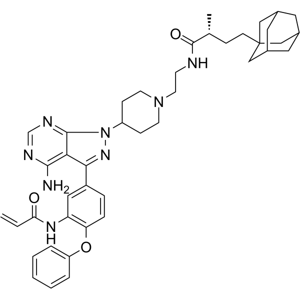
TX2-121-1 is a bifunctional hydrophobic tag Her3 degrader and also a Her3 inhibitor with an IC50 of 49 nM. TX2-121-1 covalently binds to Cys721 of Her3, induces Her3 degradation via the Hsp70/Hsp90-assisted proteasomal pathway, interferes with the heterodimerization of Her3 with Her2/c-Met, and attenuates downstream Erk/Akt signaling, thereby inhibiting Her3-dependent cancer cell growth. TX2-121-1 can be used for the research of non-small cell lung cancer, ovarian cancer and Her3-driven breast cancer.
(Pink: HER3 ligand (HY-164988); Blue: HyT ligand (HY-N2427); Black: linker).
Para uso exclusivo en investigación. No vendemos a pacientes.
- Pureza : 98.01%
- No. CAS: 1603845-42-6
- Fòrmula: C42H52N8O3
- Peso molecular:716.91
-
Almacenamiento:
4°C, sealed storage, away from moisture and light
* In solvent : -80°C, 6 months; -20°C, 1 month (sealed storage, away from moisture and light)

To place orders, for customer services and technical support, please contact: MedChemExpress USA
Tel: 609-228-6898 E-mail: [email protected] [email protected]


-
 Actividad biológica
-
Chemical Information
- Protocolo
- Pureza y Documentación
- Referencias
-
 Help & FAQs


Ver todos los productos específicos de isoformas EGFR
More

Actividad biológica
Descripciòn
IC50 & Target
[1]|
HER3 49 nM (IC50) |
Cellular Effect
|
Cell Line

|
Type | Value | Description | References |
|---|---|---|---|---|
| PC-9 | EC50 |
0.87 μM
|
Induction of preferential death of Her3-dependent PC9 GR4 cells via enhanced inhibition of Her3-dependent signals, measured by cell-based viability assay.
Induction of preferential death of Her3-dependent PC9 GR4 cells via enhanced inhibition of Her3-dependent signals, measured by cell-based viability assay.
|
34488823 |
| PC-9 | EC50 |
0.9 μM
|
Antiproliferative activity against PC9 GR4 lung cancer cells expressing wild-type Her3 assessed as reduction in cell viability incubated for 3 days by CellTiter-Glo® Luminescent Assay.
Antiproliferative activity against PC9 GR4 lung cancer cells expressing wild-type Her3 assessed as reduction in cell viability incubated for 3 days by CellTiter-Glo® Luminescent Assay.
|
25326665 |
| PC-9 | EC50 |
5.5 μM
|
Antiproliferative activity against PC9 GR4 lung cancer cells expressing Her3 C721S mutant assessed as reduction in cell viability incubated for 3 days by CellTiter-Glo® Luminescent Assay.
Antiproliferative activity against PC9 GR4 lung cancer cells expressing Her3 C721S mutant assessed as reduction in cell viability incubated for 3 days by CellTiter-Glo® Luminescent Assay.
|
25326665 |
| PC-9 | EC50 |
0.8 μM
|
Antiproliferative activity against Her3-dependent PC9 GR4 cancer cells assessed as reduction in cell viability incubated for 3 days by CellTiter-Glo® Luminescent Assay.
Antiproliferative activity against Her3-dependent PC9 GR4 cancer cells assessed as reduction in cell viability incubated for 3 days by CellTiter-Glo® Luminescent Assay.
|
25326665 |
| HCC827 | EC50 |
0.8 μM
|
Antiproliferative activity against Her3-dependent HCC827 GR6 cancer cells assessed as reduction in cell viability incubated for 3 days by CellTiter-Glo® Luminescent Assay.
Antiproliferative activity against Her3-dependent HCC827 GR6 cancer cells assessed as reduction in cell viability incubated for 3 days by CellTiter-Glo® Luminescent Assay.
|
25326665 |
| OVCAR-8 | EC50 |
0.8 μM
|
Antiproliferative activity against Her3-dependent Ovcar8 cancer cells assessed as reduction in cell viability incubated for 3 days by CellTiter-Glo® Luminescent Assay.
Antiproliferative activity against Her3-dependent Ovcar8 cancer cells assessed as reduction in cell viability incubated for 3 days by CellTiter-Glo® Luminescent Assay.
|
25326665 |
| PC-9 | EC50 |
0.8-1.4 μM
|
Antiproliferative activity against human PC9 GR4 cells.
Antiproliferative activity against human PC9 GR4 cells.
|
37535706 |
| HCC827 | EC50 |
0.8-1.4 μM
|
Antiproliferative activity against human HCC827 GR6 cells.
Antiproliferative activity against human HCC827 GR6 cells.
|
37535706 |
| OVCAR-8 | EC50 |
0.8-1.4 μM
|
Antiproliferative activity against human Ovcar8 cells.
Antiproliferative activity against human Ovcar8 cells.
|
37535706 |
In Vitro
TX2-121-1 (compound 2e) binds to purified Her3 with an IC50 of 49 nM[1].
TX2-121-1 (2 μM) acts as a selective binder for Her3 in live cells[1].
TX2-121-1 (1-5 μM; 12 h) induces partial degradation of Her3 in PC9 GR4 cells[1].
TX2-121-1 (0.5-2 μM; 12 h) reduces the phosphorylation levels of Erk and Akt, downstream effectors of Her3, in serum-starved PC9 GR4 cells stimulated with neuregulin[1].
TX2-121-1 (1 μM; 6 h) reduces the heterodimerization of Her3 with Her2 and c-Met in PC9 GR4 cells[1].
TX2-121-1 (3 h) potently binds to the purified human Her3 kinase domain with an IC50 of 49.2 nM, and exhibits Her3-centered kinome selectivity, showing no direct inhibitory activity against EGFR or Her2 enzymes at concentrations below 10 μM[3].
TX2-121-1 (1 μM; 6 h) inhibits the heterodimerization of Her3 with Her2 and c-Met in PC9 GR4 lung cancer cells, and exhibits stronger inhibitory activity against Her2/Her3 dimers[3].
TX2-121-1 potently binds to purified Her3 protein, with an IC50 value of 49.2 nM[4].
TX2-121-1 (500 nM) covalently binds to and degrades endogenously expressed ErbB3 in ErbB3-dependent cell lines, thereby inhibiting downstream signal transduction and cell proliferation[5].
TX2-121-1 inhibits HER3-dependent signaling pathways, induces partial degradation of HER3, and disrupts the heterodimerization of HER3 with HER2 and c-Met in cultured cells[6].
TX2-121-1 inhibits the proliferation of PC9 GR4 cells, and its potency is approximately 7-fold that of the non-covalent control TX2-135-2[1].
TX2-121-1 selectively induces PC9 GR4 cell death by enhancing the inhibition of Her3-dependent signaling, with an EC50 of 0.87 μM[2].
TX2-121-1-mediated intracellular Her3 degradation is enhanced upon combined treatment with the chaperone inhibitors 116-9e or Tanespimycin (17-AAG) (HY-10211)[2].
TX2-121-1 (0.5-2 μM; 12 h) induces partial degradation of Her3 in starved PC9 GR4 lung cancer cells and inhibits NRG-stimulated phosphorylation of Erk and Akt[3].
TX2-121-1 (administered consecutively for 3 days) inhibits the proliferation of PC9 GR4 lung cancer cells expressing wild-type Her3, with an EC50 of 0.9 μM; in cells expressing the Her3C721S mutant, the activity of this compound decreases (EC50 = 5.5 μM), indicating that its targeting activity depends on covalent binding to Cys721[3].
TX2-121-1 (administered for 3 consecutive days) selectively inhibits the proliferation of Her3-dependent cancer cell lines (PC9 GR4, HCC827 GR6, Ovcar8), with EC50 values ranging from 0.8 to 1.4 μM[3].
Degradation of Her3 in PC9 GR4 lung cancer cells induced by TX2-121-1 (0.5 μM) is enhanced by inhibition of Hsp70 or Hsp90, and is dependent on the proteasome[3].
TX2-121-1 (2 μM; 12 h) induces partial degradation of Her3 in starved PC9 GR4 cells, thereby inhibiting the downstream Her2 and c-Met signaling pathways[4].
TX2-121-1 inhibits the proliferation of Her3-dependent PC9 GR4, HCC827 GR6 and Ovcar8 cell lines, with EC50 values ranging from 0.8 to 1.4 μM[4].
MedChemExpress (MCE) has not independently confirmed the accuracy of these methods. They are for reference only.
-
Cell Line:PC9 GR4 cells
-
Concentration:1, 5 μM
-
Incubation Time:12 h
-
Result:Induced partial degradation of Her3 at both tested concentrations.
Showed more evident degradation compared to other adamantane conjugates and the non-adamantane control TX2-120-1.
-
Cell Line:serum-starved PC9 GR4 cells
-
Concentration:0.5, 2 μM
-
Incubation Time:12 h
-
Result:Attenuated phosphorylation of the downstream Her3 effectors Erk and Akt after NRG stimulation.
Outperformed non-covalent or non-adamantane control compounds, which did not show this effect.
-
Cell Line:starved PC9 GR4 (EGFR E746_A750/T790M) lung cancer cells
-
Concentration:0.5, 2 μM
-
Incubation Time:12 h
-
Result:Induced partial degradation of Her3 protein.
Inhibited the phosphorylation of Erk and Akt following NRG stimulation.
-
Cell Line:PC9 GR4 (EGFR E746_A750/T790M) lung cancer cells
-
Concentration:1 μM
-
Incubation Time:6 h
-
Result:Effectively reduced the association of Her3 with both Her2 and c-Met.
Showed a greater inhibitory effect on Her2/Her3 heterodimerization than on c-Met/Her3 heterodimerization.


Chemical Information
-
No. CAS 1603845-42-6
-
Appearance Solid
-
Peso molecular 716.91
-
Fòrmula C42H52N8O3
-
Color White to off-white
-
SMILES
C[C@@H](C(NCCN1CCC(N2C3=NC=NC(N)=C3C(C(C=C4)=CC(NC(C=C)=O)=C4OC5=CC=CC=C5)=N2)CC1)=O)CCC6(C[C@H](C7)C8)C[C@H]8C[C@H]7C6
-
Envío
Room temperature in continental US; may vary elsewhere.
-
Almacenamiento
4°C, sealed storage, away from moisture and light
* In solvent : -80°C, 6 months; -20°C, 1 month (sealed storage, away from moisture and light)


Pureza y Documentación
-
Ficha de datos (288 KB)
-
SDS (252 KB)
- English - EN (252 KB)
- Français - FR (252 KB)
- Deutsch - DE (252 KB)
- Norwegian - NO (252 KB)
- Español - ES (252 KB)
- Swedish - SV (252 KB)
- Italian - IT (252 KB)
- Korean - KR (252 KB)
- Portuguese - PT (252 KB)
-
Instrucciones de manejo (2659 KB)
Referencias
[1]. Lim SM, et al. Development of small molecules targeting the pseudokinase Her3. Bioorganic & medicinal chemistry letters. 2015 Aug 15;25(16):3382-9. [Content Brief]
[3]. Xie T, et al. Pharmacological targeting of the pseudokinase Her3. Nature chemical biology. 2014 Dec;10(12):1006-12. [Content Brief]
[4]. Xie S, et al. Small-Molecule Hydrophobic Tagging: A Promising Strategy of Druglike Technology for Targeted Protein Degradation. Journal of medicinal chemistry. 2023 Aug 24;66(16):10917-10933. [Content Brief]

Calculators
Concentration (start) × Volume (start) = Concentration (final) × Volume (final)


TX2-121-1 Related Classifications
HY-118998 Related Classifications
 Powered by Bioz