



Gramicidin S
Based on 2 publication(s) in Google Scholar
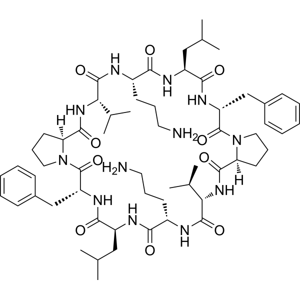
Gramicidin S (Gramicidin soviet) is a cationic cyclic peptide antibiotic that selectively targets bacterial cell membranes and has anticancer activity. Gramicidin S also exerts antibacterial activity by destroying membrane integrity and interfering with membrane protein function. Gramicidin S inserts into the phospholipid bilayer through hydrophobic amino acid residues, specifically binds to negatively charged membrane lipids and disrupts membrane structure, thereby inhibiting cell division and cell wall synthesis, and ultimately causing bacterial death. Gramicidin S also inhibits ion channels, with IC50s of 41 μM, 24 μM, and 3 μM for Na+/K+-ATPase, tobacco leaf plasma membrane Mg2+/K+-ATPase, and rat heart plasma membrane Ca2+-ATPase, respectively.
For research use only. We do not sell to patients.
- Purity : 99.89%
- CAS No.: 113-73-5
- Formula: C60H92N12O10
- Molecular Weight:1141.45
-
Storage:
Sealed storage, away from moisture and light.
Powder -80°C, 2 years , -20°C, 1 year* In solvent : -80°C, 6 months; -20°C, 1 month (sealed storage, away from moisture and light)

To place orders, for customer services and technical support, please contact: MedChemExpress USA
Tel: 609-228-6898 E-mail: [email protected] [email protected]


-
 Biological Activity
-
Chemical Information
-
Solvent & Solubility
- Protocols
- Purity & Documentation
- References
-
 Help & FAQs


-

HY-107193
BacitracinPolypeptide Antibiotic -
HY-B0108
DaptomycinBacterial Inhibitor -

HY-P0163
GramicidinAntimicrobial Peptide -

HY-P3914
Cecropin A (1-7)-Melittin A (2-9)Antibacterial Peptide -
HY-105088
PexigananAntimicrobial Peptide -

HY-P0092
Cecropin BCytochrome P450 Inhibitor -

HY-120435
TyrothricinAntibiotic Agent -

HY-P0269
Magainin 1Antimicrobial Peptide -

HY-P1629
Temporin AAntimicrobial Peptide -
HY-105048
OmigananCationic antimicrobial peptide
Publications Citing Use of MedChemExpress (MCE) Gramicidin S
MoreAll Antibiotic Isoforms
More

Biological Activity
Description
Cellular Effect
|
Cell Line

|
Type | Value | Description | References |
|---|---|---|---|---|
| Erythrocyte | EC50 |
11.7 μM
Compound: GS
|
Cytotoxicity against human erythrocytes assessed as hemolysis after 4 hrs by twofold dilution assay
Cytotoxicity against human erythrocytes assessed as hemolysis after 4 hrs by twofold dilution assay
|
[PMID: 19674904] |
| Erythrocyte | ED50 |
5 μM
Compound: GS
|
Hemolytic activity against human erythrocytes
Hemolytic activity against human erythrocytes
|
10.1039/C1MD00081K |
In Vitro
In the membrane protein localization experiment, Gramicidin S (1 μM; 10 min) causes the localization disorder of the peripheral membrane proteins MinD, DivIVA and PlsX of Bacillus subtilis, among which PlsX completely lost its membrane binding ability[1].
In the cell viability experiment, Gramicidin S (1 μM; 30 min) significantly reduces the viability of Escherichia coli (E. coli) cells, causing about 70% cell death by destroying the cell membrane integrity and inducing lipid phase separation[1].
Gramicidin S has nematicidal activity with IC50=0.08 μM (Bursaphelenchus lignicolus) and inhibits Escherichia coli with IC50=3.5 μM[1].
Gramicidin S (1 μg/mL, 2 μg/mL; 9 h) can induce fluid lipid domains independently of the MreB cytoskeleton, leading to the delocalization of peripheral membrane proteins (such as MurG involved in cell wall synthesis)[2].
MedChemExpress (MCE) has not independently confirmed the accuracy of these methods. They are for reference only. Further protocols information, click here.
-
Cell Line:Escherichia coli
-
Concentration:1 μM
-
Incubation Time:30 min
-
Result:Significantly reduced cell viability, with a 70% decrease in viable cell count compared to the control group.
Disrupted the bacterial membrane by inserting hydrophobic residues into the phospholipid bilayer, inducing lipid phase separation and fluid membrane microdomain formation, which ultimately led to membrane permeability loss and cell death.
In Vivo
Gramicidin S (20-50 μg/mouse/day; intraperitoneal injection; daily; 16 days) shows a 60% tumor inhibition rate in BALB/c mice subcutaneously implanted with Meth A fibrosarcoma at the high dose (50 μg/mouse/day) after 16 days, while low-dose treatment exhibits weaker inhibition[3].
MedChemExpress (MCE) has not independently confirmed the accuracy of these methods. They are for reference only.
-
Animal Model:Female ICR mice (8 weeks old,) subcutaneously implanted with Sarcoma 180 (S180)[3]
-
Dosage:0 (control), 20 μg/mouse/day, 200 μg/mouse/day (dissolved in ethanol and diluted with Dulbecco's PBS)
-
Administration:Intraperitoneal injection daily for 21 days starting from day 1 after tumor inoculation
-
Result:On day 21, the high-dose group (200 μg/mouse/day) showed a 60% tumor growth inhibition rate, with a slight increase in body weight, similar to the control group.
No significant difference in survival rate was observed among groups, with 6/8 mice surviving in both the high-dose and control groups.


Chemical Information
-
CAS No. 113-73-5
-
Appearance Solid
-
Molecular Weight 1141.45
-
Formula C60H92N12O10
-
Color White to off-white
-
Synonyms
Gramicidin soviet
-
Sequence
cyclo(Val-Orn-Leu-D-Phe-Pro)2
-
Sequence Shortening
cyclo(VOLDFP)2
-
Shipping
Room temperature in continental US; may vary elsewhere.
-
Storage
Sealed storage, away from moisture and light
Powder -80°C 2 years -20°C 1 year * In solvent : -80°C, 6 months; -20°C, 1 month (sealed storage, away from moisture and light)


Publications (2)
-
Journal Impact Factor
-
Most Recent
-
-
ACS Synth Biol
Combinatorial Nonribosomal Peptide Synthetase Libraries Using the SEAM-Combi-OGAB Method. [Abstract]2025 Feb 21;14(2):520-530. PMID: 39907600


Solvent & Solubility
In Vitro:
DMSO : 100 mg/mL (87.61 mM; Need ultrasonic; Hygroscopic DMSO has a significant impact on the solubility of product, please use newly opened DMSO)
Please refer to the solubility information to select the appropriate solvent. Once prepared, please aliquot and store the solution to prevent product inactivation from repeated freeze-thaw cycles.
Storage method and period of stock solution: -80°C, 6 months; -20°C, 1 month (sealed storage, away from moisture and light). When stored at -80°C, please use it within 6 months. When stored at -20°C, please use it within 1 month.
Please refer to the solubility information to select the appropriate solvent. Once prepared, please aliquot and store the solution to prevent product inactivation from repeated freeze-thaw cycles.
Storage method and period of stock solution: -80°C, 6 months; -20°C, 1 month (sealed storage, away from moisture and light). When stored at -80°C, please use it within 6 months. When stored at -20°C, please use it within 1 month.
Concentration (start) × Volume (start) = Concentration (final) × Volume (final)

Protocols
-
Research Protocol for Infectious Diseases
Infectious-disease experiments test how pathogens interact with host barriers, innate immune receptors, inflammatory signaling, pathogen replication, and tissue injury; pattern-recognition receptors such as TLRs, RIG-I-like receptors, NOD-like receptors, and inflammasomes detect microbial molecules and activate NF-κB, interferon, and cytokine responses. The central hypothesis is that infection severity reflects the balance between pathogen burden and host response: protective inflammation restricts pathogen growth, whereas excessive or mislocalized inflammation contributes to tissue damage and disease phenotype. Unresolved questions include which host pathways are protective versus pathogenic, why some infection models fail to translate to human disease, and which combined readouts best predict clinically relevant infection outcomes.
-
Membrane Protein Extraction Using Detergents and Chaotropes
Membrane protein extraction with detergents and chaotropes solubilizes lipid-bilayer-associated proteins by disrupting protein-lipid and protein-protein interactions while maintaining proteins in a soluble state for downstream electrophoresis, purification, or mass spectrometry. Chaotropes such as urea and thiourea improve solubilization of difficult proteins, while nonionic and zwitterionic detergents such as CHAPS, ASB-14, SB 3-10, MEGA-10, dodecyl maltoside, and Triton X-100 differ in extraction efficiency depending on sample type and membrane protein properties.
-
CFSE Dye Dilution Proliferation Assay
The CFSE (carboxyfluorescein diacetate succinimidyl ester) dye dilution proliferation assay is based on the covalent labeling of intracellular proteins by a cell-permeant fluorescent dye that becomes fluorescent upon intracellular ester cleavage and then is stably retained within cells. As labeled cells divide, the dye is partitioned equally between daughter cells, resulting in a stepwise halving of fluorescence intensity that can be quantified by flow cytometry to determine the number of cell divisions undergone by each cell population. This fluorescence dilution approach enables quantitative tracking of lymphocyte proliferation at the single-cell level over multiple rounds of division. CFSE-based proliferation analysis has been widely applied to measure antigen-driven lymphocyte expansion in vitro, where discrete fluorescence peaks correspond to successive cell divisions and allow reconstruction of proliferative history within heterogeneous populations.
-
Dye-dilution cell tracking and proliferation staining
Dye-dilution cell tracking assays quantify cell proliferation by covalently labeling intracellular proteins with a stable fluorescent dye that is equally partitioned between daughter cells during mitosis, resulting in stepwise halving of fluorescence intensity with each cell division as measured by flow cytometry histograms. Carboxyfluorescein diacetate succinimidyl ester (CFSE) is a prototypical dye that diffuses into cells, is enzymatically converted into a fluorescent compound, and then covalently binds intracellular amine groups, producing long-lived fluorescence suitable for tracking multiple rounds of division in vitro and in vivo. Successive generations of dividing cells form discrete peaks of decreasing fluorescence intensity, enabling estimation of proliferation history, precursor frequency, and division index within heterogeneous populations. Alternative dyes such as CellTrace Violet (CTV) and far-red membrane dyes (e. g. , PKH26) follow the same dilution principle but differ
-
Ca2+ Staining Technique
Ca2+ staining is an experimental technique that utilizes specific fluorescent probes (such as Fluo-4 AM, Fura-2, etc.) to qualitatively or quantitatively detect dynamic changes in intracellular Ca2+ concentrations; this is achieved by monitoring the changes in fluorescent signals generated when these probes bind to free intracellular calcium ions. The underlying principle relies primarily on the presence of chelating groups within the probe's molecular structure that possess high affinity for calcium ions.
-
Bacterial live/dead nucleic-acid viability staining
The LIVE/DEAD bacterial viability staining method is based on differential permeability of nucleic-acid-binding fluorescent dyes, most commonly SYTO 9 and propidium iodide (PI), which enables discrimination of bacterial populations with intact versus compromised cytoplasmic membranes. SYTO 9 penetrates both intact and damaged bacterial membranes and binds nucleic acids to produce green fluorescence, whereas propidium iodide penetrates only cells with compromised membranes and fluoresces red while also reducing SYTO 9 signal through competitive binding and fluorescence interactions. The resulting fluorescence pattern is interpreted as a proxy for membrane integrity, which is widely used as an indicator of bacterial viability in microscopy, flow cytometry, and spectroscopic platforms. However, mechanistic studies show that SYTO 9 and PI interactions involve displacement and fluorescence resonance energy transfer effects, which can influence signal interpretation depending on dye ratios a


Purity & Documentation
-
Data Sheet (289 KB)
-
SDS (252 KB)
- English - EN (252 KB)
- Français - FR (252 KB)
- Deutsch - DE (252 KB)
- Norwegian - NO (252 KB)
- Español - ES (252 KB)
- Swedish - SV (252 KB)
- Italian - IT (252 KB)
- Korean - KR (252 KB)
- Portuguese - PT (252 KB)
-
Handling Instructions (2659 KB)
References
[1]. Mogi T, et al. Gramicidin S and polymyxins: the revival of cationic cyclic peptide antibiotics. Cell Mol Life Sci. 2009 Dec;66(23):3821-6. [Content Brief]
[2]. Wenzel M, et al. The Multifaceted Antibacterial Mechanisms of the Pioneering Peptide Antibiotics Tyrocidine and Gramicidin S. mBio. 2018 Oct 9;9(5):e00802-18. [Content Brief]
[3]. Okamoto K, et al. Inhibitory effect of gramicidin S on the growth of murine tumor cells in vitro and in vivo. Oncology. 1984;41(1):43-8. [Content Brief]


Complete Stock Solution Preparation Table
Please refer to the solubility information to select the appropriate solvent. Once prepared, please aliquot and store the solution to prevent product inactivation from repeated freeze-thaw cycles.
Storage method and period of stock solution: -80°C, 6 months; -20°C, 1 month (sealed storage, away from moisture and light). When stored at -80°C, please use it within 6 months. When stored at -20°C, please use it within 1 month.
| Optional Solvent | Concentration Solvent Mass | 1 mg | 5 mg | 10 mg | 25 mg |
|---|---|---|---|---|---|
| DMSO | 1 mM | 0.8761 mL | 4.3804 mL | 8.7608 mL | 21.9020 mL |
| 5 mM | 0.1752 mL | 0.8761 mL | 1.7522 mL | 4.3804 mL | |
| 10 mM | 0.0876 mL | 0.4380 mL | 0.8761 mL | 2.1902 mL | |
| 15 mM | 0.0584 mL | 0.2920 mL | 0.5841 mL | 1.4601 mL | |
| 20 mM | 0.0438 mL | 0.2190 mL | 0.4380 mL | 1.0951 mL | |
| 25 mM | 0.0350 mL | 0.1752 mL | 0.3504 mL | 0.8761 mL | |
| 30 mM | 0.0292 mL | 0.1460 mL | 0.2920 mL | 0.7301 mL | |
| 40 mM | 0.0219 mL | 0.1095 mL | 0.2190 mL | 0.5475 mL | |
| 50 mM | 0.0175 mL | 0.0876 mL | 0.1752 mL | 0.4380 mL | |
| 60 mM | 0.0146 mL | 0.0730 mL | 0.1460 mL | 0.3650 mL | |
| 80 mM | 0.0110 mL | 0.0548 mL | 0.1095 mL | 0.2738 mL |


Gramicidin S Related Classifications
HY-P5712 Related Classifications
 Powered by Bioz