



hAChE-IN-5
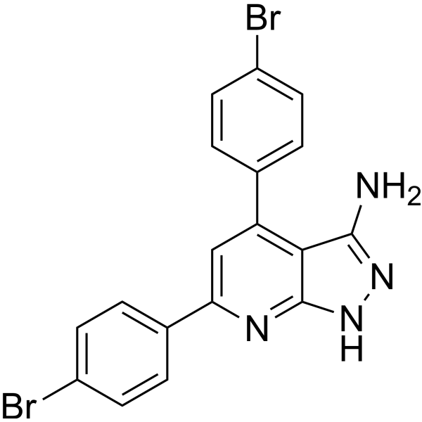
hAChE-IN-5 (compound 49) is a potent hAChE and hBuChE inhibitor with IC50 values of 0.17 μM and 0.17 μM, respectively. hAChE-IN-5 shows potent GSK3β inhibition with an IC50 value of 0.21 μM. hAChE-IN-5 is used as tau protein aggregation and Aβ1-42 self-aggregation inhibitor. hAChE-IN-5 can bind virtually with the PAS affecting Aβ aggregation, thus preventing Aβ-dependent neurotoxicity. hAChE-IN-5 can penetrate BBB and has the potential for multi-targeted anti-Alzheimer's agents research.
商品は「研究用試薬」です。人や動物の医療用・臨床診断用・食品用の製品ではありません。
研究用途以外に使用した場合、当社は一切の責任を負いかねます。
- CAS 番号: 3007667-15-1
- 分子式: C18H12Br2N4
- 分子量:444.12
-
保管条件:
Please store the product under the recommended conditions in the Certificate of Analysis.

To place orders, for customer services and technical support, please contact: MedChemExpress USA
Tel: 609-228-6898 E-mail: [email protected] [email protected]


-
 生物活性
生物活性
-
化学情報
- プロトコル
- 純度とドキュメンテーション
- 参考文献
-
 Help & FAQs




生物活性
製品説明
Cellular Effect
|
Cell Line

|
Type | Value | Description | References |
|---|---|---|---|---|
| WI-38 | IC50 |
58.28 μM
Compound: 49
|
Cytotoxicity against human WI-38 cells assessed as inhibition of cell growth incubated for 72 hrs by MTT assay
Cytotoxicity against human WI-38 cells assessed as inhibition of cell growth incubated for 72 hrs by MTT assay
|
[PMID: 37871406] |


化学情報
-
CAS 番号 3007667-15-1
-
分子量 444.12
-
分子式 C18H12Br2N4
-
SMILES
BrC1=CC=C(C2=C3C(NN=C3N)=NC(C4=CC=C(Br)C=C4)=C2)C=C1
-
輸送条件
Room temperature in continental US; may vary elsewhere.
-
保管条件
Please store the product under the recommended conditions in the Certificate of Analysis.


純度とドキュメンテーション
参考文献

Calculators
濃度 (開始) × 体積 (開始) = 濃度 (終了) × 体積 (終了)


hAChE-IN-5 関連分類
HY-155365 関連分類
 Powered by Bioz